
Pathogenesis of asthama
asthma pathogenesis mechanism diagram airway inflammation
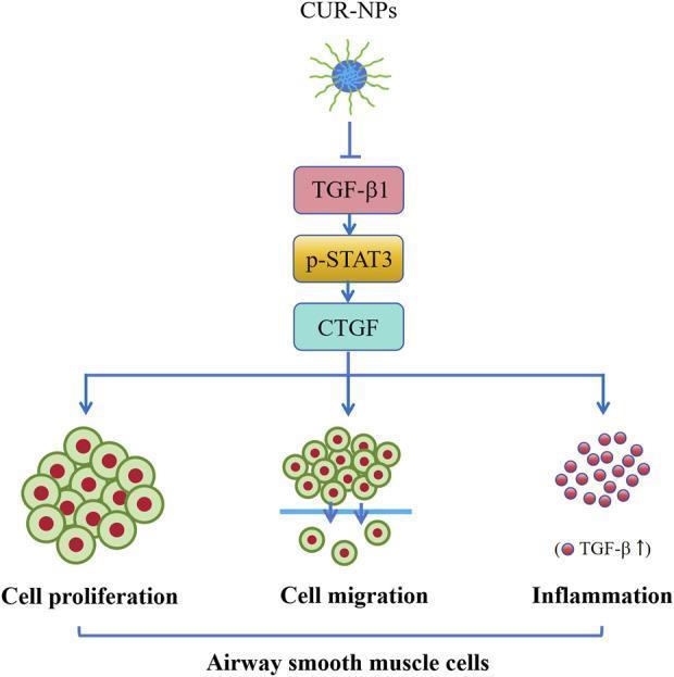
This pathophysiology diagram illustrates the molecular mechanism by which curcumin nanoparticles (CUR-NPs) inhibit asthma-related changes in airway smooth muscle cells (ASMCs). The diagram shows a vertical signaling cascade starting with CUR-NPs exerting an inhibitory effect (indicated by a T-bar arrow) on Transforming Growth Factor-beta 1 (TGF-̢1). This suppresses the downstream signaling pathway consisting of phosphorylated Signal Transducer and Activator of Transcription 3 (p-STAT3) and Connective Tissue Growth Factor (CTGF). The activation of CTGF is linked via branching arrows to three key pathological processes in ASMCs: cell proliferation (depicted as a dense cluster of nucleated cells), cell migration (shown as cells passing through a basement membrane), and inflammation (represented by small red circular markers labeled with 'TGF-̢1 ↑'). The flow indicates that by targeting the TGF-̢1/p-STAT3/CTGF axis, CUR-NPs can mitigate airway remodeling and inflammatory infiltration in conditions like asthma. This pedagogical visual is designed for intermediate to advanced medical education regarding pharmacology and respiratory pathophysiology.
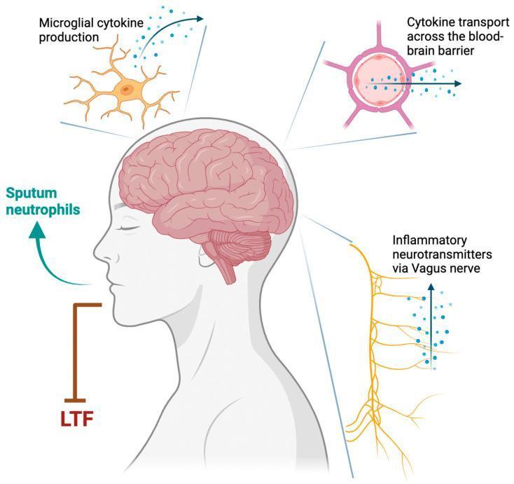
This pathophysiology diagram illustrates the mechanisms by which Type 1 inflammation in asthma may destabilize respiratory control, potentially contributing to obstructive sleep apnea (OSA). A sagittal profile of a human head and neck highlights the brain, brainstem, and central nervous system as the primary regulatory sites. Three key neural-immune pathways are detailed via callouts: 1) Microglial cytokine production, depicting an activated microglial cell with branching processes releasing signaling molecules; 2) Cytokine transport across the blood-brain barrier (BBB), shown as molecules moving from a peripheral blood vessel into the central nervous system parenchyma; and 3) Inflammatory neurotransmitters via the Vagus nerve, represented by an ascending neural pathway from the lungs/airways carrying afferent signals. Additional elements include 'Sputum neutrophils' indicating airway inflammation and 'LTF' (Long-Term Facilitation), representing a compensatory mechanism for respiratory stability that becomes compromised. The diagram serves as an educational tool for pulmonologists and neurologists to understand the neuro-inflammatory cross-talk involving central chemoreceptors and peripheral lung injury.
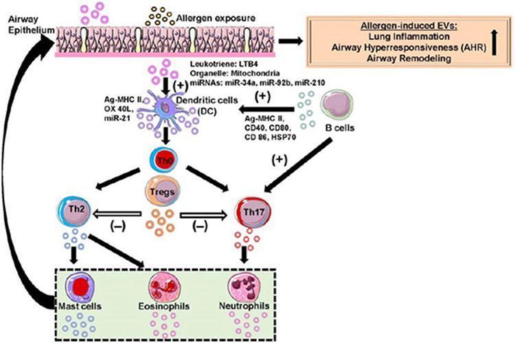
This pathophysiology diagram illustrates extracellular vesicle (EV)-mediated signaling within the lung microenvironment during allergic asthma. The schematic shows how allergen exposure triggers the Airway Epithelium to release EVs containing Leukotriene LTB4, mitochondria, and miRNAs (miR-34a, miR-92b, miR-210) to stimulate Dendritic Cells (DCs). These DCs, alongside B cell-derived EVs (carrying Ag-MHC II, CD40, CD80, CD86, and HSP70), promote the activation of naïve T cells (Th0). The Th0 cells differentiate into Th2, Th17, and Regulatory T cells (Tregs). A key regulatory pathway is shown where Treg-derived EVs inhibit Th2 and Th17 responses (indicated by '-' symbols). Th2 and Th17 cells release EVs that activate effector cells, including Mast cells, Eosinophils, and Neutrophils. The resulting phenotypic changes culminate in increased lung inflammation, airway hyperresponsiveness (AHR), and airway remodeling. The diagram utilizes arrows to denote communication flow, with (+) indicating promotion/maturation and (-) indicating inhibition, highlighting the complex immune crosstalk in asthma pathogenesis.
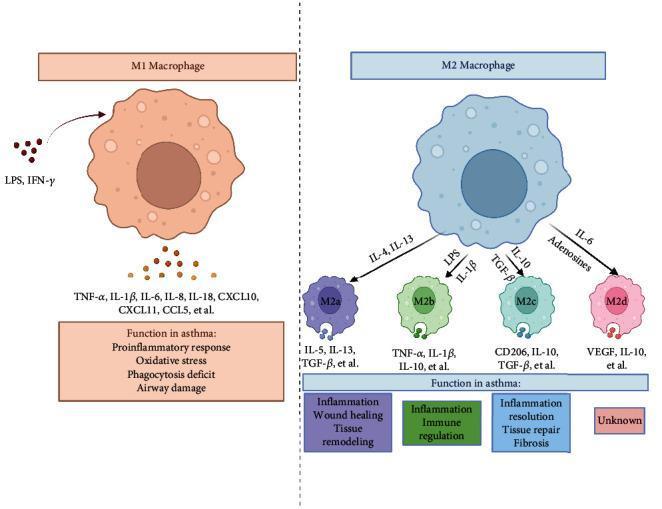
This medical illustration depicts the polarization and functional diversity of M1 and M2 macrophage phenotypes in the context of asthma pathogenesis. The diagram is divided into two main sections. On the left, an M1 macrophage (classically activated) is shown responding to LPS and IFN-γ. It releases proinflammatory mediators including TNF-α, IL-1β, IL-6, IL-8, IL-18, CXCL10, and CCL5, contributing to oxidative stress and airway damage. On the right, the M2 macrophage (alternatively activated) is shown as a central hub differentiating into four distinct subtypes: M2a (activated by IL-4/IL-13), M2b (by LPS/IL-1β), M2c (by IL-10/TGF-β), and M2d (by IL-6/adenosines). Each M2 subtype is associated with specific effector molecules and clinical roles: M2a is linked to wound healing and tissue remodeling; M2b to immune regulation; M2c to inflammation resolution and fibrosis; and M2d has an unknown function in this context. The diagram illustrates the complex immunological balance between pro-inflammatory M1 responses and the immunomodulatory or tissue-repairing roles of M2 subtypes in asthmatic airway disease.
"asthma"[MeSH Terms] AND "pathogenesis"[MeSH Terms]
Pathogenesis of Asthma
1. Overview and Classification
| Feature | Atopic (Extrinsic) | Nonatopic (Intrinsic) |
|---|---|---|
| Allergen sensitization | Yes | No |
| Skin test | Positive | Usually negative |
| Family history | Common | Less common |
| IgE levels | Elevated | Normal |
| Onset | Childhood | Adulthood |
| Triggers | Allergens | Infections, irritants |
2. Genetic Predisposition
- Asthma shows strong familial clustering. Genome-wide association studies (GWAS) have identified multiple susceptibility loci, including genes encoding the IL-4 receptor and other mediators directly involved in airway inflammation.
- The tendency to produce IgE (atopy) is at least partly genetically determined, and asthma clusters with allergic rhinitis, atopic dermatitis, and food allergy in family groups.
- The hygiene hypothesis proposes that lack of early microbial/allergen exposure leads to immune hyperreactivity later in life, though a precise mechanistic basis remains unproven.
3. Atopic Asthma - Immunopathogenesis
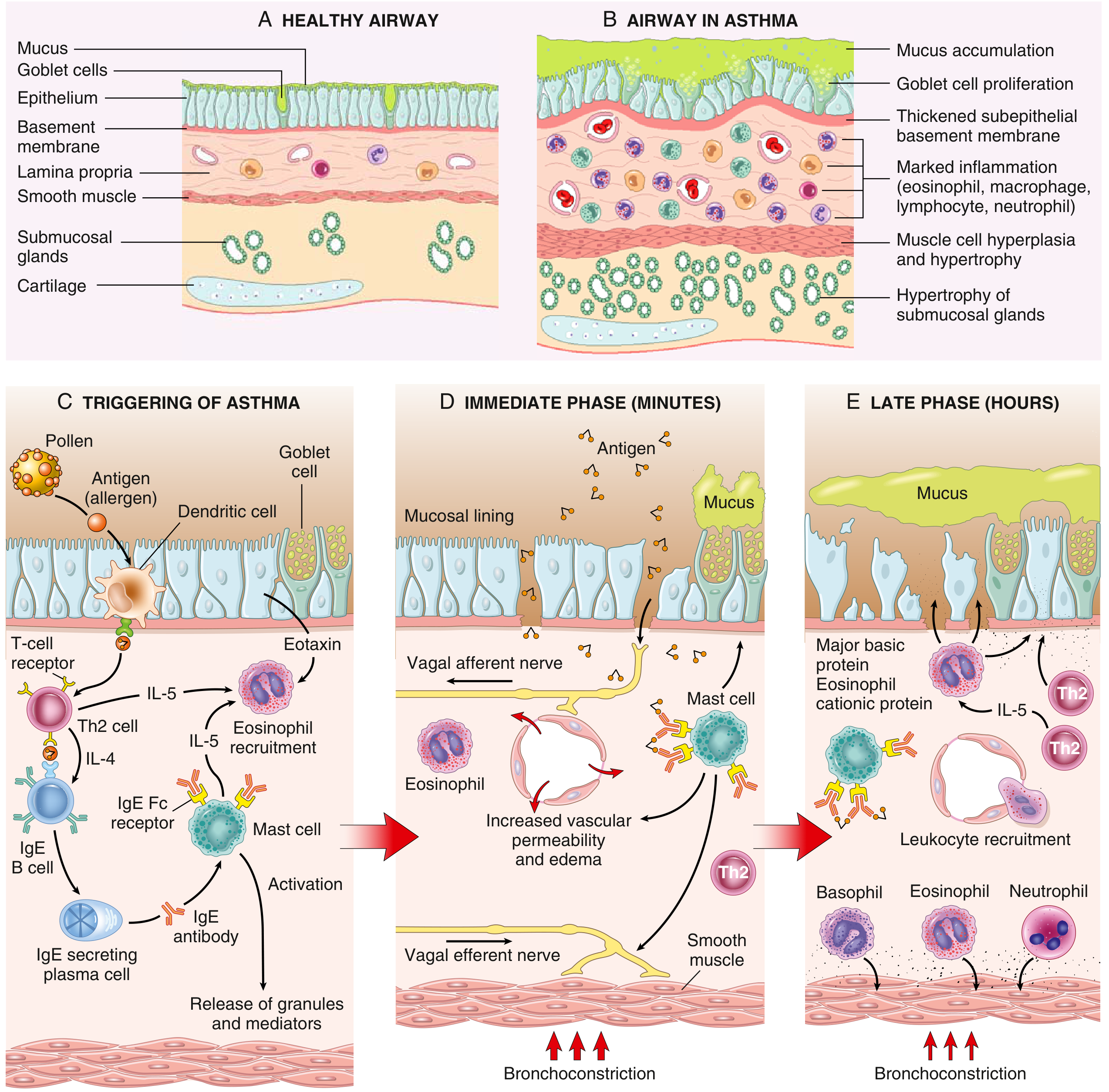
Step 1: Sensitization Phase
- Inhaled allergens (house dust mite, cockroach, pollen, animal dander, molds) are processed by dendritic cells in the airway mucosa.
- Dendritic cells present antigen to naive T cells, driving differentiation into Th2 lymphocytes.
- Th2 cells secrete key cytokines:
- IL-4 and IL-13 → stimulate B cells to undergo class switching and produce IgE
- IL-5 → recruits and activates eosinophils
- IL-13 → stimulates mucus hypersecretion by goblet cells
- IgE binds to high-affinity FcεRI receptors on submucosal mast cells, "sensitizing" them.
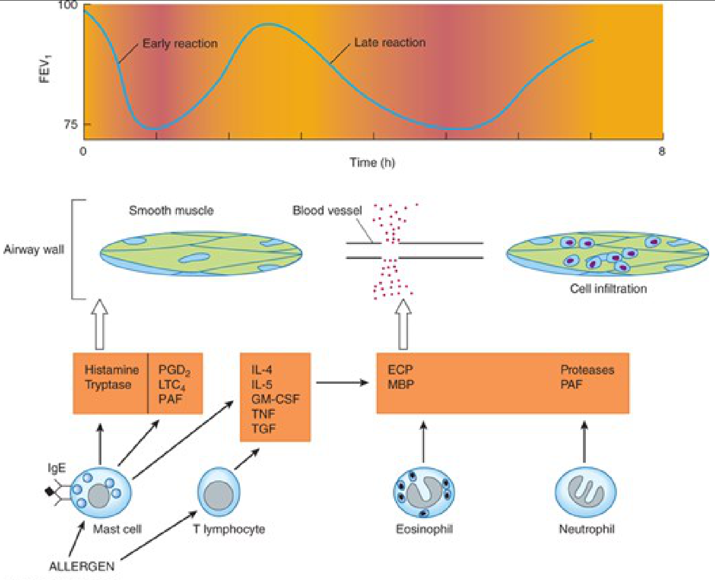
Step 2: Early (Immediate) Phase Reaction - Minutes
- Allergen cross-links IgE on mast cell surfaces → mast cell degranulation
- Released mediators cause:
- Histamine → bronchoconstriction, vascular dilation
- Tryptase → airway inflammation
- Leukotriene C4 and D4 (LTC4, LTD4) → potent bronchoconstrictors
- Prostaglandin D2 (PGD2) → bronchoconstriction
- Platelet-activating factor (PAF) → inflammation
Step 3: Late Phase Reaction - 3-6 Hours
- Mast cells and T lymphocytes release cytokines: IL-4, IL-5, GM-CSF, TNF, TGF
- These attract and activate eosinophils and neutrophils into the bronchial mucosa
- Eosinophils release:
- Eosinophil cationic protein (ECP)
- Major basic protein (MBP)
- Proteases and PAF
- These cause edema, mucus hypersecretion, smooth muscle contraction, and increased bronchial reactivity (second FEV1 fall at ~3-6 hours)
- Epithelial cells produce eotaxin (a potent eosinophil chemoattractant) which amplifies Th2 recruitment, creating a self-sustaining inflammatory loop
4. Complete Airway Pathology Diagram
5. Nonatopic Asthma
- No allergen sensitization - skin tests are negative, IgE is not elevated
- Common triggers: viral respiratory infections (rhinovirus, parainfluenza), inhaled air pollutants (SO2, ozone, NO2), cold air, exercise, stress
- Mechanism: virus-induced inflammation lowers the threshold of subepithelial vagal receptors to irritants
- Despite different triggers, the downstream effectors are the same - eosinophils, mast cells, and their mediators - hence treatment is similar to atopic asthma
6. Drug-Induced and Occupational Asthma
- Aspirin-sensitive asthma: Aspirin inhibits cyclo-oxygenase (COX), diverting arachidonic acid toward the lipoxygenase pathway, generating excess leukotrienes. Presents with rhinitis, nasal polyps, urticaria, and bronchospasm.
- Occupational asthma: Triggered by epoxy resins, organic dusts (wood, cotton), platinum salts, toluene; develops after repeated exposure. Can be immunologic (IgE-mediated) or nonimmunologic (irritant).
7. Key Inflammatory Cells Summary
| Cell | Role in Asthma |
|---|---|
| Mast cells | Early phase; release histamine, LTs, PGD2 on IgE cross-linking |
| Eosinophils | Key effector cells; release MBP, ECP, proteases causing epithelial damage |
| Th2 lymphocytes | Orchestrate immune response via IL-4, IL-5, IL-13 |
| Neutrophils | Late phase and severe/steroid-resistant asthma |
| Dendritic cells | Antigen presentation; initiate Th2 polarization |
| Basophils | Late phase inflammation |
| Epithelial cells | Produce eotaxin; barrier dysfunction |
8. Airway Remodeling (Chronic Changes)
- Subbasement membrane fibrosis (thickened subepithelial collagen)
- Goblet cell metaplasia and hyperplasia → mucus hypersecretion
- Hypertrophy and hyperplasia of bronchial smooth muscle
- Increased submucosal vascularity (angiogenesis)
- Hypertrophy of submucosal glands
- These changes add an irreversible obstructive component to what was initially a reversible disease
9. Morphological Findings in Fatal Asthma
- Lungs distended with air trapping (overinflation)
- Bronchi occluded by thick, tenacious mucus plugs
- Curschmann spirals (whorls of shed epithelium in mucus)
- Charcot-Leyden crystals (composed of galectin-10 from eosinophils - also pro-inflammatory)
- Eosinophilic infiltration of the bronchial wall
- Subbasement membrane thickening (Fig. 11.11 in Robbins)
10. Patterns of Inflammation
- Eosinophilic - most common; type 2 driven; steroid-responsive
- Neutrophilic - associated with severe/steroid-resistant asthma and occupational triggers
- Mixed eosinophilic/neutrophilic
- Pauci-granulocytic - minimal inflammatory cells despite symptoms
Summary of Key Mediators
| Mediator | Source | Effect |
|---|---|---|
| IgE | B cells (IL-4/IL-13 driven) | Sensitizes mast cells |
| IL-4, IL-13 | Th2 cells | IgE production, mucus secretion |
| IL-5 | Th2 cells | Eosinophil recruitment/activation |
| Histamine | Mast cells | Bronchoconstriction, vasodilation |
| LTC4, LTD4 | Mast cells | Potent bronchoconstriction |
| PGD2 | Mast cells | Bronchoconstriction |
| Eotaxin | Epithelial cells | Eosinophil chemotaxis |
| MBP, ECP | Eosinophils | Epithelial damage |
| TGF-beta | T cells, mast cells | Airway remodeling/fibrosis |