

A 45 yr old female present with pus filled lesions on back of body since one week . Associated with itching.whats the diagnosis and management for this condition?
folliculitis back pustules papules skin
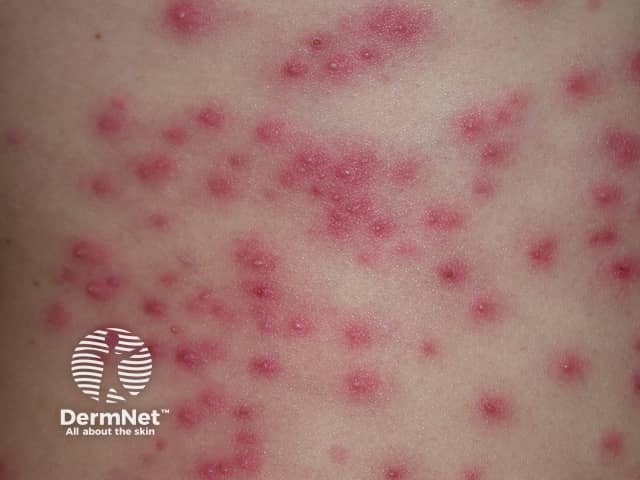
Clinical skin photograph showing numerous small erythematous papules and pustules distributed across trunk/back. The lesions are centered on hair follicles, 2–5 mm in diameter, with surrounding perifollicular erythema and occasional crusting. The overall pattern is diffuse and crops of lesions are evident, consistent with folliculitis. This presentation is classically associated with Pseudomonas aeruginosa infection, often termed hot tub folliculitis, though other gram-negative organisms can cause a similar eruption. Incubation after warm water exposure is typically 24–48 hours. The image demonstrates superficial involvement of the follicular unit without deeper necrosis; no comedones are evident; there is mild edema of the surrounding skin. The phototype appears light to moderate; however, color fidelity is preserved, aiding assessment of vascular dilation and inflammation. Clinically, a history of recent hot tub, pool, or whirlpool exposure should be elicited; pustule cultures or Gram stain can confirm Pseudomonas. Management is usually supportive, with spontaneous resolution in days to weeks; antibiotics with anti-pseudomonal activity are reserved for persistent or extensive disease or immunocompromised patients. These images are suitable for medical education, case repositories, and digital dermatology teaching materials.
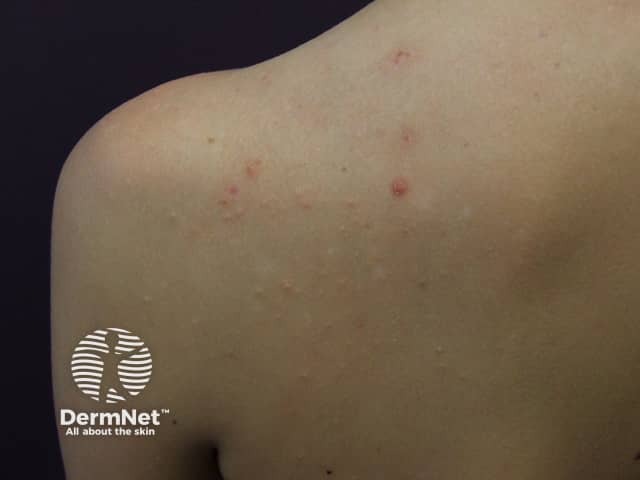
Clinical photography of the upper back and left shoulder/scapular region illustrating a mild inflammatory dermatosis. Modality: clinical photography; perspective: posterior dorsal view. The skin shows multiple small erythematous papules, several with central pustules, clustered along the upper thoracic/scapular zone. Lesions measure approximately 2–4 mm in diameter and are discrete but in close proximity, consistent with a follicular pattern. No significant crusting or scarring is evident; surrounding skin is otherwise uninvolved. The appearance is suggestive of an acneiform eruption or folliculitis, with inflammatory insult centered on hair follicles. Differential diagnoses include acne vulgaris of the back (bacne), folliculitis (bacterial or fungal), keratosis pilaris-like follicular papules, insect bites, or contact dermatitis. The lesions' symmetry and distribution favor follicular inflammation in regions with higher sebaceous gland density and tolerated occlusion. Clinically, this presentation may respond to topical anti-inflammatory agents, benzoyl peroxide, or topical retinoids if cosmetic concerns persist; antibiotic therapy may be considered for secondary infection. Correlation with patient history (pubertal status, hormonal fluctuations, sweating, clothing friction, recent hygiene products) and possible Gram stain or culture if lesions enlarge or fail to improve can guide management. This image highlights common back acne patterns and serves educational purposes for recognition and differential diagnosis. education.
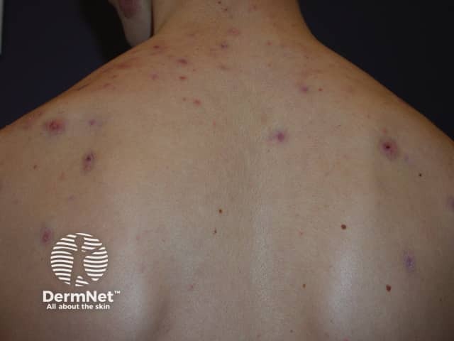
Imaging modality and technique: Clinical photography in color, dorsal posterior trunk view, standard ambient lighting with minimal glare; no dermatoscopic enhancement or chemical staining. The specimen is human skin surface. The primary subject is the back of an adolescent or adult individual presenting an acneiform eruption. The image shows multiple erythematous papules scattered across the upper back and shoulder regions, with a subset displaying central pustulation and a follicular distribution pattern. Lesions vary from small target-like pustules to larger inflamed papules, some with slight crusts, and there is minimal surrounding edema. The overall appearance is consistent with inflammatory acne involving the trunk, with folliculitis-like features at several sites. No open comedones or nodules are clearly evident in this view, though clinically these may be present elsewhere. The background is clear, allowing visualization of lesion morphology, distribution, and degree of surface inflammation. This photographic documentation is suitable for tracking lesion response to therapy, counseling patients, and aiding dermatologic differential diagnosis, including acne vulgaris of the trunk, folliculitis, and less likely keratosis pilaris sparing the trunk. The image lacks histopathology data and laboratory correlation; a clinical diagnosis should be confirmed with history, examination, and, if needed, culture or biopsy.
Diagnosis: Bacterial Folliculitis (Staphylococcal Folliculitis)
Image Analysis
- Multiple small erythematous papules (1-5 mm), scattered across the posterior shoulder/upper back
- Pinpoint dark crusts at lesion centers - indicating excoriation from itching
- Follicular distribution pattern (lesions centred on hair follicles)
- No large abscesses or carbuncles visible
Pathophysiology
- Andrews' Diseases of the Skin, p. 14.5
- Excessive sweating / friction from clothing (blouse/saree border visible)
- Obesity
- Diabetes mellitus (must be screened)
- Immunocompromised state
- Poor hygiene or occlusive clothing
Differential Diagnoses
| Condition | Key Distinguishing Feature |
|---|---|
| Staphylococcal folliculitis (most likely) | Pustules at follicle openings, back/trunk, pruritic |
| Pityrosporum (Malassezia) folliculitis | Intensely pruritic, monomorphic, upper back/chest, worsens with antibiotics |
| Gram-negative / Hot tub folliculitis | History of hot tub/pool exposure, Pseudomonas |
| Acne vulgaris | Usually has comedones, predominantly sebaceous areas |
| Insect bite reaction | Lesions not follicle-centred, often linear/grouped |
| Miliaria pustulosa | Heat exposure, not follicle-centred |
Management
Step 1 - General/Hygiene Measures
- Keep skin clean and dry; avoid occlusive/tight-fitting clothing
- Antibacterial washes: chlorhexidine or benzoyl peroxide wash
- Dilute sodium hypochlorite baths (bleach baths: ½ cup bleach in 40-gallon tub) to decolonize skin
- Avoid shaving the affected area
Step 2 - Topical Antibiotics (first-line for mild-moderate disease)
-
Mupirocin 2% ointment applied to lesions twice daily x 7-10 days
-
Alternative: Topical clindamycin, bacitracin, or retapamulin
-
Topical mupirocin also applied to anterior nares (5 days) if recurrent, to eradicate nasal carriage
-
Andrews' Diseases of the Skin, p. 14.6; Schwartz's Surgery p. 2330
Step 3 - Systemic Antibiotics (for extensive or recurrent disease)
- Dicloxacillin or cephalexin 500 mg orally twice daily x 7-10 days (empiric anti-staphylococcal)
- If MRSA suspected: Trimethoprim-sulfamethoxazole (TMP-SMX) or doxycycline
- If Pseudomonas/hot tub folliculitis suspected: Ciprofloxacin orally
- Textbook of Family Medicine 9e, p. 3885
Step 4 - Symptomatic Relief
- Antihistamines (cetirizine or loratadine) for pruritus control
- Warm compresses to individual lesions
Step 5 - Investigations to Order
- Blood glucose / HbA1c - to rule out diabetes mellitus as a predisposing factor
- Swab culture and sensitivity from an intact pustule - if not responding to empiric therapy
- KOH preparation - if fungal folliculitis (Pityrosporum) is suspected (especially if worsening on antibiotics)
Red Flags / When to Escalate
- Development of furuncles (deep boils) or carbuncles - may need incision and drainage
- Recurrent folliculitis despite treatment - consider MRSA, diabetes, or immunosuppression workup
- Failure to respond to topical antibiotics within 5-7 days - escalate to systemic therapy
Note: In a 45-year-old female with pruritic folliculitis on the back, diabetes screening (FBS/HbA1c) is strongly recommended as folliculitis in middle-aged patients frequently heralds undiagnosed diabetes mellitus.