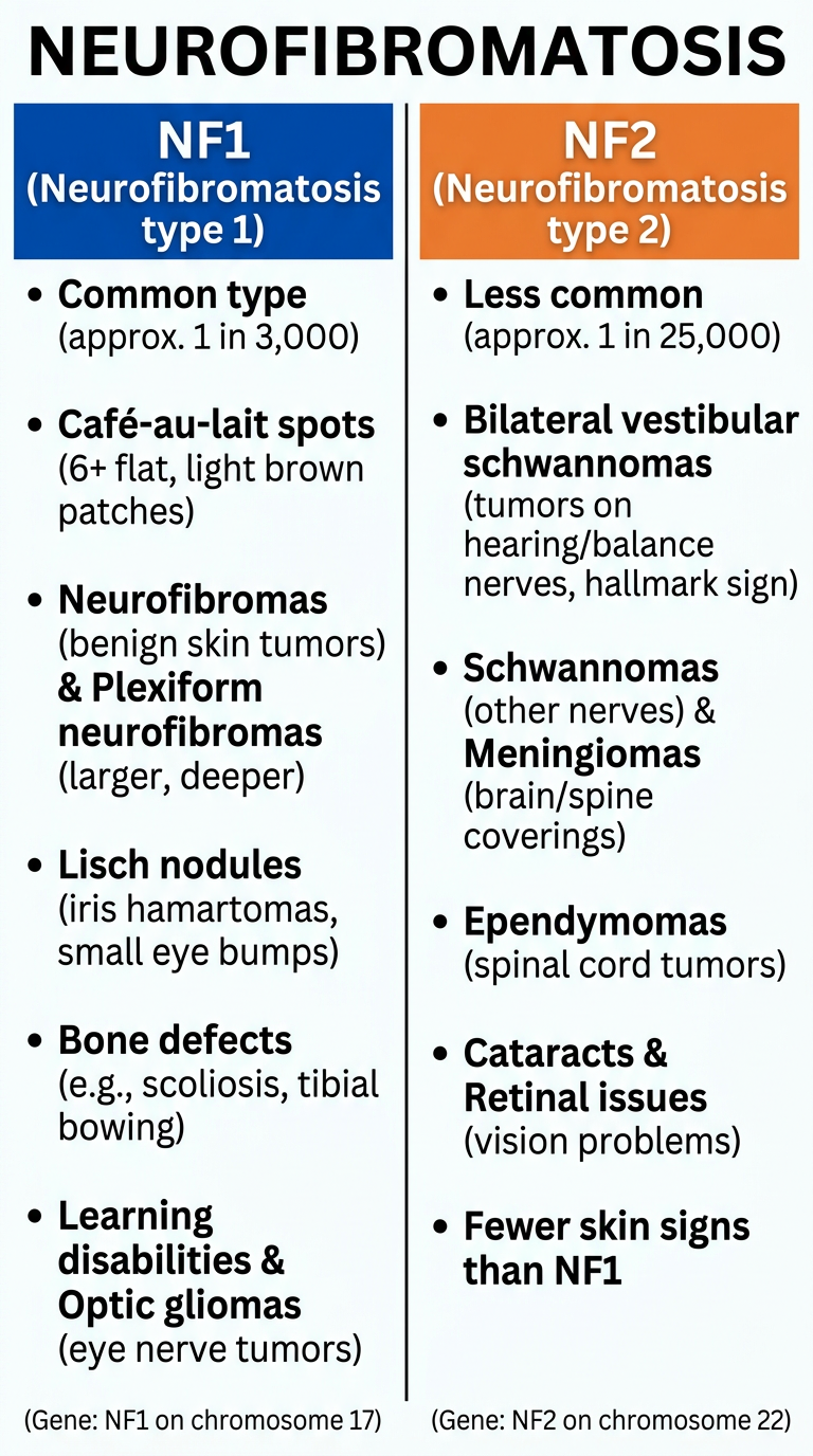
DVT Detailed notes As per Harrison latest edition + latest official guidelines
DVT deep vein thrombosis 2024 2025 guidelines ASH CHEST treatment diagnosis
"deep vein thrombosis" AND "anticoagulation"
DEEP VEIN THROMBOSIS (DVT) - Comprehensive Notes
Source: Harrison's 22E (2025) + Major Textbooks + ASH 2020 / ACCP 2022 Guidelines
1. DEFINITION & OVERVIEW
- DVT = thrombosis (clot formation) within the deep venous system, most commonly the lower extremities
- DVT and pulmonary embolism (PE) are together termed venous thromboembolism (VTE)
- Estimated incidence: 1-2 per 1000 persons/year in the US
- 300,000-600,000 new VTE cases/year in the US; 60,000-80,000 deaths/year
- Up to 30% of patients die within 30 days; 1 in 5 suffer sudden death as the first manifestation
- Leg DVT is ~10 times more common than upper extremity DVT
- Patients present with DVT symptoms about twice as frequently as with PE symptoms
2. ANATOMY & CLASSIFICATION
Lower Extremity DVT
- Distal (calf) DVT: Below the popliteal vein (posterior tibial, peroneal, anterior tibial, muscular veins)
- Proximal DVT: Involves popliteal vein and above (popliteal, femoral, common femoral, iliac veins)
- More likely to embolize to lungs
- DVT usually begins in the calf and propagates proximally to popliteal → femoral → iliac veins
Classification by Location
| Type | Veins Involved | Significance |
|---|---|---|
| Isolated calf (distal) DVT | Tibial, peroneal veins | Low PE risk; controversial treatment |
| Popliteal DVT | Popliteal vein | Moderate-high PE risk |
| Femoral DVT | Femoral/common femoral vein | High PE risk |
| Iliofemoral DVT | Iliac + femoral veins | Highest PE risk, massive leg swelling |
Upper Extremity DVT
- Increasingly common due to:
- Central venous catheters (CVC), PICC lines, pacemakers, ICDs
- Chemotherapy access devices
- Effort thrombosis (Paget-Schroetter syndrome) in athletes
- Risk increases with larger catheter diameter and more lumens
- COVID-19 associated with catheter-based upper extremity DVT
- Complications: PE, SVC syndrome, loss of vascular access, central venous stenosis
Special Forms
- Phlegmasia cerulea dolens: Most severe DVT; diffuse edema of entire limb; limb-threatening
- May-Thurner syndrome: Right proximal iliac artery compresses left proximal iliac vein → recurrent left thigh edema, especially in young women → predisposes to DVT
3. PATHOPHYSIOLOGY - VIRCHOW'S TRIAD
| Component | Examples |
|---|---|
| Hypercoagulability | Inherited thrombophilia, cancer, pregnancy, OCP, antiphospholipid syndrome |
| Venous stasis | Immobility, heart failure, prolonged travel, obesity |
| Endothelial injury | Surgery, trauma, indwelling catheters, chemotherapy |
Coagulation Cascade (Harrison's Ch. 122)
- Vascular injury → tissue factor exposure → TF binds Factor VII → activates downstream
- TF-FVIIa complex → activates FX (via FIX-FVIIIa pathway) → FXa + FVa → prothrombin → thrombin
- Thrombin → converts fibrinogen → insoluble fibrin + activates FXIII → cross-linked stable clot
- Antithrombotic regulators: antithrombin, TFPI, heparin cofactor II, protein C/protein S
- Additional contributors: procoagulant microparticles, inflammatory cells, microvesicles, fibrin structure
4. RISK FACTORS
Acquired Risk Factors
- Surgery (highest risk): neurosurgery, major abdominal/pelvic surgery, orthopedic (hip/knee replacement)
- Cancer: ~4-fold increased VTE risk; annual risk on chemotherapy ~11% (6x general population)
- Especially: lung, pancreatic, GI, breast, ovarian, GU cancers; lymphomas; brain tumors
- Trousseau's syndrome = migratory thrombophlebitis + visceral carcinoma (especially pancreatic)
- Immobilization: prolonged bed rest, paralysis, plaster cast, long-distance travel (>4-6 hrs)
- Prior VTE: strongest predictor of recurrence
- Pregnancy and postpartum period
- Oral contraceptives / hormone replacement therapy (estrogen-containing)
- Obesity (BMI >30)
- Increasing age
- Infections: community- or hospital-acquired
- Inflammatory conditions: IBD, rheumatologic diseases
- Renal disease, heart failure
- Central venous catheters
- COVID-19
Inherited Thrombophilias (Genetic Risk Factors)
- 5-8% of US population has a known genetic predisposition to venous thrombosis
- Factor V Leiden mutation (most common): resistance to activated protein C - autosomal dominant
- Prothrombin G20210A mutation: elevated prothrombin levels
- Protein C deficiency
- Protein S deficiency
- Antithrombin deficiency (most severe thrombophilia)
- Hyperhomocysteinemia (MTHFR mutation)
- Elevated Factor VIII, IX, XI levels
Independent Predictors of Recurrence
- Increasing age
- Obesity
- Malignancy
- Acute extremity paresis
5. CLINICAL FEATURES
Symptoms
- Unilateral leg pain (especially calf)
- Leg swelling - unilateral, progressive
- Skin erythema and warmth over affected area
- Symptoms may be acute to subacute in onset
Signs
- Calf tenderness on palpation along deep vein distribution
- Pitting edema of the affected limb
- Unilateral calf swelling >3 cm compared to opposite leg
- Erythema and warmth
- Collateral superficial non-varicose veins (visible)
- Homan's sign (calf pain on dorsiflexion of foot) - historically taught but insensitive and non-specific; NOT recommended for diagnosis
Clinical Pearls (Harrison's 22E)
- Not all leg pain is DVT: sudden severe calf pain → ruptured Baker's cyst
- Fever + chills → more likely cellulitis than DVT
- Mild cases: only mild palpation discomfort in lower calf
- Massive DVT: marked thigh swelling, tenderness, erythema
- If diffusely edematous leg → DVT unlikely (except phlegmasia cerulea dolens)
- Upper extremity DVT: asymmetry in supraclavicular fossa or upper arm circumference
Superficial Venous Thrombosis (SVT)
- Erythema, tenderness, palpable "cord" along superficial vein
- Most common: lower extremity (varicose veins), upper extremity (IV catheters)
- Urschel's sign: superficial veins visible in chest wall
- Risk: extension to deep venous system
6. DIAGNOSIS
Step 1: Pre-test Clinical Probability - Wells DVT Score
| Clinical Variable | Score |
|---|---|
| Active cancer (treatment ongoing or within 6 months, or palliative) | +1 |
| Paralysis, paresis, or recent plaster immobilization of lower extremity | +1 |
| Recently bedridden >3 days or major surgery within 12 weeks | +1 |
| Tenderness along distribution of deep veins | +1 |
| Entire leg swollen | +1 |
| Calf swelling >3 cm compared to asymptomatic leg | +1 |
| Pitting edema (greater in symptomatic leg) | +1 |
| Collateral superficial non-varicose veins | +1 |
| Strong family history of DVT (≥2 first-degree relatives) | +1 (in some versions) |
| Alternative diagnosis at least as likely as DVT | -2 |
- ≤0: Low probability (DVT can be effectively ruled out with negative D-dimer)
- 1-2: Moderate probability
- ≥3: High probability (proceed to imaging)
Step 2: D-Dimer
- High sensitivity, low specificity (many causes of elevated D-dimer)
- Standard threshold: 500 ng/mL (or 500 mcg/L FEU)
- Age-adjusted D-dimer (for PE evaluation, not DVT): Age × 10 ng/mL for patients >50 years
- Note: Age-adjusted D-dimer does NOT apply to suspected acute DVT
- Negative D-dimer + low/moderate pre-test probability = DVT ruled out (no imaging needed)
- Causes of false positive D-dimer: cancer, pregnancy, infection, post-surgical, age, liver disease, heart failure
- D-dimer is not reliably predictive in cancer patients (baseline elevation common)
Step 3: Imaging
Duplex Compression Ultrasonography (CUS) - PRIMARY TEST
- Gold standard non-invasive test for DVT
- Primary criterion: loss of vein compressibility (non-compressible segment)
- Normal vein collapses with gentle pressure → creates "wink" appearance
- DVT: vein remains open/distended under pressure
- Additional features: direct thrombus visualization, altered Doppler flow dynamics
- Loss of respiratory variation = obstruction at/proximal to measurement site
- Loss of flow augmentation with calf compression = distal obstruction
- Sensitivity: 96.5% for symptomatic proximal DVT; 71.2% for distal (calf) DVT
- Specificity: ~94%
- Technically poor views: consider CT or MRI venography
CT Venography (CTV)
- Role: evaluation of pelvic veins, assessing for extrinsic compression, when US non-diagnostic
- Challenge: appropriate contrast timing
MR Venography (MRV)
- Superior for pelvic/IVC evaluation, no contrast issue
- Limited in patients with metallic implants
Contrast (Catheter) Phlebography/Venography
- Gold standard (invasive) - largely replaced by CUS
- Used when interventional procedure planned (thrombolysis, stenting)
- Definitive: direct visualization of intraluminal filling defect
- Allows intraluminal pressure measurements, IVUS
Integrated Diagnostic Algorithm (Harrison's 22E)
Suspected DVT
      ↓
Clinical Pre-test Probability (Wells Score)
      ↓                          ↓
 Low/Moderate                   High
      ↓                          ↓
  D-Dimer                   Compression Ultrasound
  Negative → DVT excluded    Non-compressible → DVT confirmed
  Positive → CUS             Compressible → D-dimer / repeat CUS in 7 days
Differential Diagnosis of DVT
- Ruptured Baker's cyst
- Muscle strain/injury (especially gastrocnemius tear)
- Cellulitis (fever + chills suggest this)
- Acute postthrombotic syndrome/venous insufficiency
- Lymphedema
- Hematoma
- Superficial venous thrombosis
- Popliteal artery aneurysm
7. THROMBOPHILIA WORKUP
- DVT in unusual sites (portal, hepatic, mesenteric, cerebral veins)
- DVT in young patients (<45 years) without provoking factor
- Recurrent unprovoked VTE
- Strong family history
- DVT during pregnancy or OCP use
- Factor V Leiden mutation (PCR)
- Prothrombin G20210A mutation (PCR)
- Protein C activity
- Protein S antigen/activity (free and total)
- Antithrombin activity
- Antiphospholipid antibodies: lupus anticoagulant, anticardiolipin Ab, anti-β2-glycoprotein I Ab
- Homocysteine level
- Factor VIII level
8. TREATMENT
Phase Classification (ASH 2020 Guidelines)
- Initial management: First 5-21 days
- Primary treatment: First 3-6 months
- Secondary prevention: Beyond 3-6 months
8A. Anticoagulation - Drug Options
DOACs (Direct Oral Anticoagulants) - FIRST-LINE (ASH & ACCP 2022)
| Drug | Mechanism | DVT Dosing Regimen |
|---|---|---|
| Rivaroxaban (Xarelto) | Factor Xa inhibitor | 15 mg BID × 21 days, then 20 mg OD with evening meal |
| Apixaban (Eliquis) | Factor Xa inhibitor | 10 mg BID × 7 days, then 5 mg BID |
| Edoxaban (Savaysa/Lixiana) | Factor Xa inhibitor | Requires 5-10 days LMWH first, then 60 mg OD |
| Dabigatran (Pradaxa) | Direct thrombin inhibitor | Requires 5-10 days LMWH first, then 150 mg BID |
- Rivaroxaban and Apixaban: Can be initiated orally without initial parenteral therapy ("DOAC-only" regimens)
- DOACs vs. warfarin: Lower risk of recurrent VTE, lower major bleeding, no routine monitoring required
Low Molecular Weight Heparin (LMWH)
- Enoxaparin: 1 mg/kg SC q12h OR 1.5 mg/kg SC OD (therapeutic)
- More effective than UFH for DVT prophylaxis in high-risk patients (orthopedic surgery)
- Lower incidence of HIT than UFH
- Preferred in pregnancy (does not cross placenta), cancer patients (historically)
- Limited in severe renal dysfunction (CrCl <30 mL/min) - use anti-Xa monitoring
- Cancer-associated DVT: LMWH was traditional standard; DOACs (edoxaban, rivaroxaban) now found non-inferior in select cancer patients (avoid in GI/GU cancer due to bleeding risk)
Unfractionated Heparin (UFH)
- IV: 80 units/kg bolus (or 5000 units), then 18 units/kg/hr infusion
- Target PTT: 60-80 seconds (corresponds to anti-Xa level 0.3-0.7 units/mL)
- Adjust every 6 hours based on PTT
- Must reach therapeutic levels within 24 hours (delays increase recurrent VTE risk)
- Role: ICU patients, renal failure, when rapid reversal needed (protamine)
- Risk: HIT (heparin-induced thrombocytopenia)
Vitamin K Antagonist (VKA) - Warfarin
- Target INR: 2.0-3.0 (2.5 target)
- Requires bridging with heparin/LMWH for at least 5 days AND until INR therapeutic for 24h
- Warfarin is teratogenic → contraindicated in pregnancy
- Drug and food interactions; requires regular INR monitoring
- Transient hypercoagulable state on initiation (protein C & S fall before other VK-dependent factors)
Fondaparinux
- Factor Xa inhibitor (indirect, parenteral)
- Alternative to LMWH; once-daily SC injection
- Renally cleared; avoid if CrCl <30 mL/min
8B. Duration of Anticoagulation (ASH 2020 + ACCP)
| Clinical Scenario | Recommended Duration |
|---|---|
| Provoked DVT (reversible risk factor: surgery, trauma, immobility) | 3 months |
| First unprovoked proximal DVT or PE | Minimum 3-6 months; consider indefinite if low-moderate bleeding risk |
| Distal (calf) DVT, symptomatic | 3 months (anticoagulation) |
| Distal DVT, asymptomatic or low risk | Surveillance vs. 3 months (controversial) |
| Recurrent unprovoked DVT | Indefinite anticoagulation |
| DVT with known thrombophilia (antiphospholipid syndrome, antithrombin deficiency) | Indefinite |
| Cancer-associated DVT | Indefinite or until cancer resolved |
| DVT in pregnancy | LMWH throughout pregnancy + at least 6 weeks postpartum (minimum 3 months total) |
- Apixaban 2.5 mg BID (AMPLIFY-EXT trial): reduces recurrent VTE vs. placebo, lower bleeding than full dose
- Rivaroxaban 10 mg OD (EINSTEIN-CHOICE trial): superior to aspirin for secondary prevention
8C. Isolated Calf (Distal) DVT
- ASH 2020 suggests anticoagulation for patients with symptomatic isolated distal DVT
- Randomized trial: 6 weeks anticoagulation did NOT reduce adverse outcomes vs. no treatment in low-risk patients with isolated calf DVT (3% vs 5%, p=0.54), but DID cause bleeding (4% vs 0%)
- Meta-analysis: anticoagulation → 50% reduction in recurrent VTE (RR 0.50; 95% CI 0.31-0.79) without increasing major bleeding
- 2025 meta-analysis (PMID 40400471): supports anticoagulation for isolated distal DVT in symptomatic patients
- Alternative: serial ultrasound at 5-7 days to detect proximal extension; anticoagulate only if extension occurs
8D. Advanced/Interventional Therapy
Catheter-Directed Thrombolysis (CDT)
- Indications: Extensive proximal (iliofemoral) DVT with high clot burden, phlegmasia, limb-threatening ischemia, young patients with good functional status
- ASH 2020: Suggests CDT over systemic thrombolysis for extensive DVT (if thrombolysis appropriate)
- Agents: tPA (alteplase), urokinase
- Goal: reduce clot burden, prevent post-thrombotic syndrome (PTS)
- ATTRACT trial: CDT did NOT prevent PTS at 24 months vs. anticoagulation alone; did reduce severe PTS
Inferior Vena Cava (IVC) Filter
- Indications:
- Acute DVT/PE + absolute contraindication to anticoagulation
- Recurrent PE despite adequate anticoagulation
- ASH 2020 + ACCP: Suggest against routine IVC filter placement; anticoagulation alone preferred when feasible
- Retrievable filters: Preferred; must be removed when anticoagulation resumed
- Complications: filter thrombosis, IVC occlusion, penetration, migration
Surgical Thrombectomy
- Rarely performed; reserved for cases of massive phlegmasia cerulea dolens with arterial compromise
Pharmacomechanical CDT (PCDT)
- Combines CDT with mechanical thrombus disruption/aspiration
- May accelerate thrombus clearance
8E. Ancillary Measures
- Compression stockings: For symptom relief and edema management
- Historically used for prevention of PTS; SOX trial showed graduated elastic stockings did NOT prevent PTS vs. placebo
- ASH 2020: Suggests against routine use of compression stockings for PTS prevention after DVT
- Early ambulation: Encouraged (does not increase PE risk; may reduce symptoms)
- Leg elevation: During acute phase
- Avoid prolonged immobility
9. COMPLICATIONS
Pulmonary Embolism (PE)
- Most feared acute complication
- Occurs in ~30% of untreated proximal DVT
- Massive PE: systemic hypotension, >50% pulmonary vascular obstruction, cardiogenic shock
- Small PE: pleuritic chest pain (peripheral, near pleural innervation = pulmonary infarction)
Post-Thrombotic Syndrome (PTS)
- Chronic venous insufficiency after DVT
- Incidence: 20-50% after proximal DVT (within 2 years)
- Features: chronic leg pain, swelling, heaviness, skin changes (lipodermatosclerosis), venous ulcers (Villalta scale)
- Mechanism: valvular incompetence + venous obstruction → ambulatory venous hypertension
- Prevention: effective anticoagulation, possibly compression stockings (benefit disputed)
- Treatment: graduated compression, wound care for ulcers, venoactive drugs (diosmin), endovascular intervention for obstructive lesions
Recurrent VTE
- Rate: ~30% over 10 years after a first episode
- Higher in: unprovoked VTE, cancer, antiphospholipid syndrome, thrombophilia
Chronic Thromboembolic Pulmonary Hypertension (CTEPH)
- Occurs in 2-4% of patients after PE
- Results from persistent pulmonary arterial obstruction + secondary small-vessel arteriopathy
- Features: progressive dyspnea, functional limitation, RV failure
- Treatment: pulmonary thromboendarterectomy (most effective), riociguat, balloon pulmonary angioplasty
10. DVT IN SPECIAL POPULATIONS
Cancer-Associated DVT
- LMWH traditionally preferred (showed superiority over warfarin)
- DOACs (edoxaban, rivaroxaban): non-inferior to LMWH for most cancers
- Avoid edoxaban/rivaroxaban in GI or GU malignancies (higher luminal bleeding risk) - use LMWH
- ASH 2021 Cancer VTE guidelines: DOAC or LMWH both acceptable depending on bleeding risk
- Duration: Indefinite (as long as cancer active)
- Elevated D-dimer not reliably predictive in cancer (baseline elevation); venous US or venography preferred
Pregnancy-Associated DVT
- LMWH = treatment of choice throughout pregnancy (does not cross placenta; safe)
- Warfarin: teratogenic (especially 6-12 weeks) - contraindicated in 1st trimester
- DOACs: not recommended in pregnancy (insufficient safety data)
- Duration: Continue LMWH through labor/delivery + ≥6 weeks postpartum (minimum 3-6 months total)
- Heparin-associated osteoporosis risk with long-term LMWH
HIT (Heparin-Induced Thrombocytopenia)
- Immune-mediated: IgG antibodies to heparin-PF4 complex
- Paradoxically pro-thrombotic (both arterial and venous)
- Onset: 5-14 days after heparin initiation
- Diagnosis: 4T score + anti-PF4/heparin antibodies + SRA (serotonin release assay)
- Management: stop ALL heparin immediately; switch to argatroban (direct thrombin inhibitor) or fondaparinux; avoid warfarin until platelets recover
- LMWH has lower HIT incidence than UFH
ICU Patients
- High DVT risk due to immobility
- Prophylaxis: subcutaneous UFH or LMWH + sequential compression devices (SCDs)
- LMWH more effective than UFH for DVT prophylaxis in high-risk patients
- Renal dysfunction limits LMWH use → consider UFH with monitoring
Upper Extremity DVT
- Complications: PE (risk similar to leg DVT), SVC syndrome, loss of vascular access, central venous stenosis
- Treatment: anticoagulation × 3 months (same principles as lower extremity)
- Consider CDT for Paget-Schroetter syndrome (effort thrombosis) in young, active patients
- If catheter-related: catheter removal if no longer needed + anticoagulation
- If catheter must remain: anticoagulate with catheter in place
11. DVT PROPHYLAXIS
Risk Stratification (Caprini Score commonly used)
- Low risk: Early ambulation sufficient
- Moderate risk: Mechanical (SCDs) or pharmacologic prophylaxis
- High risk: Pharmacologic prophylaxis preferred (LMWH or UFH)
Pharmacologic Options
| Agent | Dose |
|---|---|
| Enoxaparin (LMWH) | 40 mg SC OD (general surgery); 30 mg SC BID (orthopedic) |
| Heparin (UFH) | 5000 units SC q8-12h |
| Fondaparinux | 2.5 mg SC OD |
| Rivaroxaban | 10 mg OD (post-orthopedic surgery) |
| Apixaban | 2.5 mg BID (post-orthopedic surgery) |
| Aspirin | Limited role; ASH: use only when GCS/LMWH not feasible (high-risk travelers) |
Mechanical Methods
- Sequential compression devices (SCDs) / intermittent pneumatic compression (IPC)
- Graduated compression stockings (GCS): 15-30 mmHg
Long-Distance Travel (>4-6 hours)
- Without risk factors: Ambulation, hydration, calf exercises; no pharmacologic prophylaxis
- With risk factors (prior VTE, recent surgery, cancer, pregnancy): ASH suggests GCS or prophylactic LMWH; aspirin if those not feasible
- ACCP recommends GCS for high-risk travelers on flights >6 hours
12. GUIDELINE SUMMARY TABLE
| Topic | Recommendation | Source |
|---|---|---|
| First-line anticoagulant | DOAC (rivaroxaban, apixaban, edoxaban, dabigatran) over VKA | ASH 2020, ACCP 2022 |
| Cancer DVT: anticoagulant | DOAC or LMWH (avoid DOAC in GI/GU cancers) | ASH 2021 |
| Calf DVT: anticoagulation | Yes for symptomatic; serial US acceptable in low-risk asymptomatic | ASH 2020 |
| CDT vs systemic thrombolysis for DVT | CDT preferred (if thrombolysis chosen) | ASH 2020 |
| IVC filter | Only when anticoagulation contraindicated | ASH 2020, ACCP 2022 |
| Compression stockings for PTS | Against routine use | ASH 2020 |
| Duration: provoked DVT | 3 months | ASH 2020, ACCP 2022 |
| Duration: unprovoked DVT | ≥3 months; consider indefinite | ASH 2020 |
| D-dimer to guide duration | Against routine use | ASH 2020 |
| Home treatment for low-risk DVT | Yes, preferred | ASH 2020 |
13. KEY CLINICAL FACTS TO REMEMBER
- Wells Score is the validated pre-test probability tool for DVT; score ≤0 = low probability
- Compression ultrasonography is the primary diagnostic test; non-compressibility = DVT
- D-dimer: Highly sensitive but non-specific; negative value in low-risk patient rules out DVT
- DOACs are first-line anticoagulants for most DVT patients (ASH 2020, ACCP 2022)
- Rivaroxaban and apixaban do NOT require initial parenteral therapy ("oral-only" approach)
- Dabigatran and edoxaban require 5-10 days of LMWH/UFH first
- LMWH preferred in pregnancy (warfarin is teratogenic; DOACs not recommended)
- 3 months anticoagulation for provoked DVT; indefinite for recurrent/unprovoked/cancer-associated
- May-Thurner syndrome: Right iliac artery compresses left iliac vein → recurrent left leg DVT in young women
- Phlegmasia cerulea dolens: Most severe DVT - entire limb swollen, potential limb loss
- HIT: Stop all heparin immediately; switch to argatroban or fondaparinux (NOT warfarin initially)
- CTEPH occurs in 2-4% after PE; treat with pulmonary endarterectomy ± riociguat
- IVC filter: Only when absolute contraindication to anticoagulation; prefer retrievable type
- Age-adjusted D-dimer applies to suspected PE, NOT suspected DVT
- Trousseau syndrome = migratory thrombophlebitis + occult cancer (especially pancreatic)
- Harrison's Principles of Internal Medicine 22nd Edition (2025), Chapters 122, 290, and 74 (Oncologic complications)
- Fuster & Hurst's The Heart, 15th Edition, Chapter 56
- Sabiston Textbook of Surgery, Chapter on Venous Thromboembolism
- Braunwald's Heart Disease, Chapter 87
- ASH 2020 VTE Guidelines: Treatment of DVT and PE
- ACCP/CHEST 2022 Antithrombotic Therapy for VTE
Give summary
DVT - Quick Summary
What is it?
Pathophysiology
- Hypercoagulability (cancer, thrombophilia, OCP, pregnancy)
- Stasis (immobility, heart failure, long travel)
- Endothelial injury (surgery, catheters, trauma)
Risk Factors
| High Risk | Moderate Risk |
|---|---|
| Surgery (ortho/neuro/abdominal) | OCP / HRT |
| Active cancer (4x risk) | Obesity |
| Prior VTE | Long-distance travel |
| Thrombophilia (Factor V Leiden most common) | Pregnancy/postpartum |
Clinical Features
- Unilateral leg pain, swelling, erythema, warmth
- Homan's sign - historically taught; NOT reliable
- Massive DVT: entire limb swollen (phlegmasia cerulea dolens = limb-threatening)
- May-Thurner: right iliac artery compresses left iliac vein → recurrent left leg DVT in young women
Diagnosis - 3 Steps
- ≤0 = Low | 1-2 = Moderate | ≥3 = High
- Negative + low/moderate probability = DVT excluded (no imaging needed)
- Positive = proceed to ultrasound
- Not reliable in cancer patients
- Non-compressible vein = DVT confirmed
- Sensitivity: 96.5% proximal, 71.2% distal DVT
Treatment
Drug of Choice
| Drug | Initiation |
|---|---|
| Rivaroxaban | 15 mg BID × 21 days → 20 mg OD |
| Apixaban | 10 mg BID × 7 days → 5 mg BID |
| Edoxaban / Dabigatran | LMWH × 5-10 days first, then oral |
Duration
| Scenario | Duration |
|---|---|
| Provoked DVT (surgery/trauma) | 3 months |
| Unprovoked DVT | ≥3-6 months → consider indefinite |
| Cancer-associated | Indefinite |
| Recurrent unprovoked | Indefinite |
| Pregnancy | LMWH throughout + 6 weeks postpartum |
Special Situations
- Pregnancy: LMWH only (warfarin = teratogenic; DOACs = not recommended)
- Cancer: LMWH or DOAC (avoid DOAC in GI/GU cancer - bleeding risk)
- HIT: Stop ALL heparin → switch to argatroban/fondaparinux
- IVC filter: Only if absolute contraindication to anticoagulation
Complications
| Complication | Key Point |
|---|---|
| PE | 30% of untreated proximal DVT |
| Post-thrombotic syndrome (PTS) | 20-50%; chronic swelling, ulcers, venous insufficiency |
| Recurrent VTE | ~30% over 10 years |
| CTEPH | 2-4% after PE; treat with pulmonary endarterectomy |
Top 5 Exam Points
- Wells Score stratifies pre-test probability; score ≤0 + negative D-dimer = DVT excluded
- DOACs are first-line; rivaroxaban/apixaban need no initial heparin
- Provoked = 3 months; unprovoked = indefinite consideration
- LMWH in pregnancy; never warfarin (teratogenic), never DOACs
- Non-compressible vein on US = DVT confirmed
Give poster
Medical education poster titled "DEEP VEIN THROMBOSIS (DVT)" with a clean, professional clinical layout on a deep navy blue background with white and teal accents. Include the following sections clearly organized in boxes/panels: TOP HEADER: "DEEP VEIN THROMBOSIS (DVT)" in large bold white text, subtitle "Harrison's 22E + ASH 2020 + ACCP 2022 Guidelines" SECTION 1 - VIRCHOW'S TRIAD (red triangle icon): Three boxes: Hypercoagulability (cancer, thrombophilia, OCP), Stasis (immobility, HF, travel), Endothelial Injury (surgery, catheters, trauma) SECTION 2 - WELLS DVT SCORE (scoring table): +1 each: Active cancer, Paralysis/cast, Bedridden >3 days, Deep vein tenderness, Entire leg swollen, Calf >3cm larger, Pitting edema, Collateral veins. MINUS 2: Alternative diagnosis equally likely. Score: ≤0=Low, 1-2=Moderate, ≥3=High SECTION 3 - DIAGNOSIS ALGORITHM (flowchart): Wells Score → Low/Mod: D-Dimer (Negative=Excluded, Positive=CUS) → High: Compression Ultrasound → Non-compressible=DVT CONFIRMED SECTION 4 - TREATMENT (green section): FIRST-LINE: DOACs. Table showing Rivaroxaban 15mg BD×21d→20mg OD, Apixaban 10mg BD×7d→5mg BD, Edoxaban/Dabigatran need LMWH bridge first. DURATION: Provoked=3 months, Unprovoked=≥6 months/indefinite, Cancer=Indefinite, Pregnancy=LMWH only SECTION 5 - COMPLICATIONS (orange boxes): PE (30% untreated), Post-thrombotic Syndrome (20-50%), Recurrent VTE (30%/10yr), CTEPH (2-4%) SECTION 6 - KEY PEARLS (yellow highlight box): 5 bullet points: DOACs first-line; Rivo/Apixaban no heparin bridge; LMWH in pregnancy; HIT→stop heparin→argatroban; IVC filter only if anticoagulation contraindicated Small anatomical leg diagram showing DVT location. Professional medical poster style, high contrast, readable fonts.
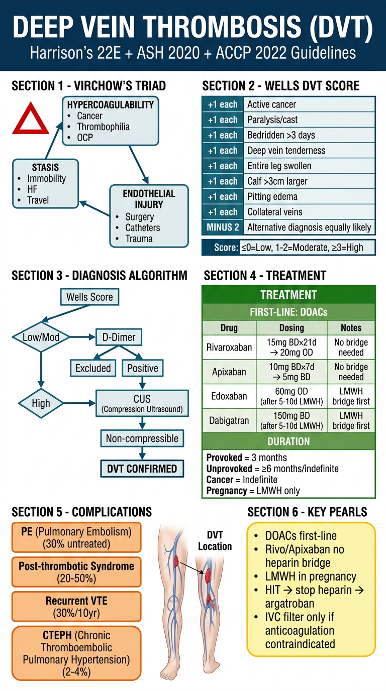
Neurofibromatosis Detailed notes Harrison latest edition Official latest guidelines
neurofibromatosis NF1 NF2 2024 2025 guidelines management selumetinib
"neurofibromatosis" AND "management"
neurofibromatosis NF1 2024 NIH revised diagnostic criteria surveillance guidelines mirdametinib
NEUROFIBROMATOSIS - Comprehensive Notes
Source: Harrison's 22E (2025) + Goldman-Cecil + Fitzpatrick's + Latest Guidelines
1. OVERVIEW & CLASSIFICATION
| Feature | NF Type 1 (NF1) | NF Type 2 (NF2) |
|---|---|---|
| Old name | von Recklinghausen's disease | Central NF / Bilateral acoustic NF |
| Gene | NF1 (chromosome 17q11.2) | NF2 (chromosome 22q) |
| Protein | Neurofibromin | Merlin (schwannomin) |
| Prevalence | 1 in 2,500-3,000 births | 1 in 40,000-50,000 births |
| Inheritance | Autosomal dominant | Autosomal dominant |
| Penetrance | High; but variable expressivity | High |
| De novo mutations | ~50% of cases | ~50% of cases |
| Hallmark | Café-au-lait macules + neurofibromas | Bilateral vestibular schwannomas |
2. NEUROFIBROMATOSIS TYPE 1 (NF1)
2A. Genetics & Molecular Pathology
- Gene: NF1 on chromosome 17q11.2 - one of the largest human genes
- 60 exons spanning >350 kb of genomic DNA
- Protein: Neurofibromin - a GTPase-activating protein (GAP)
- Normally converts p21-Ras-GTP (active) → p21-Ras-GDP (inactive)
- Acts as a tumor suppressor by downregulating the Ras/MAPK/MEK signaling pathway
- Mutations in NF1 impair this downregulation → uncontrolled cell proliferation
- Mutations are diverse, scattered throughout exons - no consistent genotype-phenotype correlation
- Exception: complete NF1 gene deletion → early-onset multiple neurofibromas, developmental delay, facial dysmorphism
- Derived from neural crest cells: Schwann cells, melanocytes, and related cells most affected
2B. Clinical Features
Cutaneous/Dermatologic
- Café-au-lait macules (CALMs)
- Most frequent early manifestation; present in virtually all NF1 patients
- Well-defined, uniformly pigmented, light-to-dark brown macules
- Size threshold: >5 mm in prepubertal individuals; >15 mm in postpubertal individuals
- NF1-associated CALMs contain significantly increased numbers of melanocytes (vs. isolated CALMs)
- Appear in infancy; nearly all patients have them by age 5 years
- Axillary/inguinal freckling (Crowe's sign)
- Smaller café-au-lait spots in skin folds (axilla, groin, inframammary, neck)
- Pathognomonic for NF1 (rarely occurs in other conditions)
- Also contain increased epidermal melanocytes (distinguishes from ordinary freckles)
- Cutaneous neurofibromas
- Benign peripheral nerve sheath tumors; arise within skin
- Soft, flesh-colored, sessile or pedunculated papules/nodules
- Begin appearing in late childhood/adolescence; increase with age and pregnancy
- Not present at birth; progressively increase in number throughout life
- May become numerous (hundreds to thousands in severe cases)
- Subcutaneous neurofibromas
- Firm, palpable nodules along peripheral nerves; may be painful
- Can be disfiguring
- Plexiform neurofibromas (PNF)
- Involve multiple nerve fascicles; described as "bag of worms" on palpation
- Often large; may involve deep tissues and organs
- Present at birth (congenital) or appear in early childhood
- Can cause disfigurement, pain, and functional impairment
- Risk of malignant transformation to MPNST (malignant peripheral nerve sheath tumor): 8-13% lifetime risk
- Overlying skin often hyperpigmented and hypertrichotic
Ophthalmologic
- Lisch nodules (iris hamartomas)
- Dome-shaped, melanocytic hamartomas on the iris surface
- Asymptomatic; detected by slit-lamp examination
- Found in >90% of adults with NF1 but rare in unaffected individuals
- Pathognomonic for NF1; increase with age
- NOT found in NF2
- Optic pathway gliomas (OPG)
- Most common CNS tumor in NF1; occur in ~15% of patients
- Usually low-grade pilocytic astrocytomas (WHO Grade I)
- Most occur in first decade of life (peak age 3-5 years)
- Most are asymptomatic and discovered incidentally on screening MRI
- Symptomatic: visual loss, proptosis, precocious puberty (if hypothalamic extension)
- May spontaneously stabilize or regress
Neurologic
- Spinal neurofibromas: multiple, along spinal nerve roots
- Learning disabilities: ~50% of NF1 patients; most common cognitive complication
- ADHD/attention deficit disorder: common
- Macrocephaly: common
- Epilepsy/seizures: increased risk
- Cognitive impairment: variable; rarely severe
- T2 hyperintensities on MRI: "unidentified bright objects" (UBOs) in basal ganglia, brainstem, cerebellum; significance unclear, may correlate with cognitive issues
- Intracranial gliomas: beyond optic pathway; brainstem gliomas, striatothalamic gliomas
Skeletal
- Scoliosis: dystrophic or non-dystrophic; common
- Short stature: frequent (even without GH deficiency)
- Macrocephaly: head circumference above 97th percentile
- Tibial pseudarthrosis/dysplasia: anterolateral bowing of the tibia; pathognomonic skeletal finding; may progress to fracture and nonunion
- Sphenoid bone dysplasia: characteristic skull base abnormality; may cause pulsating exophthalmos
- Vertebral scalloping, rib penciling
Vascular
- Renal artery stenosis: leading to hypertension in children with NF1
- Intracranial arterial stenoses/moyamoya pattern
- Cerebrovascular disease: increased stroke risk
- Vascular lesions are distinct from atherosclerosis
Endocrine
- Pheochromocytoma: ~1% of NF1 patients (3% per Goldman-Cecil)
- Usually solitary benign adrenal tumor
- 90% within adrenal medulla, 10% extra-adrenal (para-aortic)
- Presentation similar to sporadic pheochromocytoma
- Carcinoid tumors: ~1%; often periampullary or ileal
- Hepatic metastases → carcinoid syndrome (flushing, diarrhea, bronchoconstriction, tricuspid valve disease)
- Precocious puberty: associated with optic glioma extending to hypothalamus → early GnRH activation
- Growth hormone deficiency: reported in some NF1 patients
Gastrointestinal
- GI neurofibromas: throughout GI tract; may cause obstruction, bleeding
- GIST (gastrointestinal stromal tumors): increased frequency in NF1; usually small intestinal, often multiple
- Carcinoid tumors: periampullary region most common
Malignancies (Lifetime Risk ~60%)
- MPNST (Malignant Peripheral Nerve Sheath Tumors): most important malignancy
- Risk: 8-15% lifetime; highest malignancy risk in NF1
- Arise from plexiform neurofibromas
- Age 20-50 years typical presentation
- Poor prognosis: 5-year survival ~20-50%
- CNS gliomas: optic pathway, brainstem, high-grade gliomas
- Leukemia: especially juvenile myelomonocytic leukemia (JMML) in children
- Rhabdomyosarcoma: in young children
- Breast cancer: increased risk in women <50 years (2-4x general population)
- Overall: 3-15% additional lifetime cancer risk vs. general population
2C. NIH Diagnostic Criteria for NF1
- ≥6 café-au-lait macules: >5 mm (prepubertal) OR >15 mm (postpubertal)
- ≥2 neurofibromas of any type OR ≥1 plexiform neurofibroma
- Axillary or inguinal freckling (Crowe's sign)
- Optic nerve/pathway glioma
- ≥2 Lisch nodules (iris hamartomas on slit-lamp examination)
- Sphenoid bone dysplasia OR tibial dysplasia/pseudarthrosis
- First-degree relative with NF1 (diagnosed by above criteria)
- Heterozygous pathogenic NF1 variant identified by molecular testing (genetic diagnosis)
- Choroidal freckling/choroidal abnormalities on MRI
- Spinal neurofibromas as a qualifying criterion in appropriate context
3. NEUROFIBROMATOSIS TYPE 2 (NF2)
3A. Genetics & Molecular Pathology
- Gene: NF2 on chromosome 22q11-q12
- Protein: Merlin (also called schwannomin) - a membrane-cytoskeletal linking protein
- Merlin is a tumor suppressor that downregulates cellular growth
- Mutations of NF2 → loss of merlin → uncontrolled proliferation of Schwann cells, meningeal cells
- Diverse mutations; truncating mutations tend to produce more severe disease
- Mosaic NF2 (somatic mosaicism) can present with milder or unilateral disease
3B. Clinical Features
- Bilateral vestibular schwannomas (acoustic neuromas): HALLMARK
- Benign tumors of cranial nerve VIII (vestibulocochlear nerve)
- Tumors often begin unilaterally and become bilateral
- ~60% of patients present with hearing loss, tinnitus, or loss of balance
- Age of presentation: usually young adulthood (mean ~20s)
- Younger age of onset = greater ultimate disease severity
- Children more likely to present with non-CN VIII tumors
- Meningiomas: multiple; intracranial, intraspinal, optic nerve sheath
- Spinal schwannomas: dorsal roots of spinal cord, peripheral nerves, cranial nerves
- Ependymomas and gliomas of CNS (less frequent)
- Juvenile posterior subcapsular cataracts (60-80% of patients) - early marker
- Retinal hamartomas and combined hamartomas of retina and RPE
- Optic nerve sheath meningiomas (especially in children)
- Cutaneous lesions:
- Café-au-lait spots: present but rarely >6; NO intertriginous freckling
- Cutaneous schwannomas: plaques with slightly raised, faint violaceous hue, sometimes with hair - characteristic of NF2
- Occasional cutaneous neurofibromas (indistinguishable from NF1)
- Morbidity: HIGH - patients frequently become paralyzed and deaf
- NO endocrine abnormalities (endocrine tumors are NF1-specific)
3C. Diagnostic Criteria for NF2 (Fitzpatrick's / Manchester Criteria)
- Bilateral vestibular schwannomas (on CT or MRI)
- First-degree relative with NF2 + unilateral vestibular schwannoma OR any 2 of: meningioma, schwannoma, glioma, neurofibroma, posterior subcapsular lens opacities
- Unilateral vestibular schwannoma + any 2 of: meningioma, schwannoma, glioma, neurofibroma, posterior subcapsular lens opacities
- Multiple meningiomas + unilateral vestibular schwannoma OR any 2 of: schwannoma, glioma, neurofibroma, cataract
4. COMPARISON TABLE: NF1 vs NF2
| Feature | NF1 | NF2 |
|---|---|---|
| Chromosome | 17q11.2 | 22q |
| Gene/Protein | NF1/Neurofibromin | NF2/Merlin |
| Prevalence | 1 in 2,500-3,000 | 1 in 40,000-50,000 |
| Café-au-lait macules | ≥6 required (diagnostic) | Present but <6; not diagnostic |
| Axillary freckling | Yes - pathognomonic | No |
| Lisch nodules | Yes - pathognomonic | No |
| Neurofibromas | Multiple cutaneous, subcutaneous, plexiform | Rare; occasional cutaneous |
| Vestibular schwannomas | Rare/occasional | Bilateral - HALLMARK |
| Meningiomas | Occasional | Multiple - common |
| Spinal schwannomas | No | Yes |
| Cataracts | No | Posterior subcapsular (60-80%) |
| Endocrine tumors | Yes (pheo, carcinoid) | No |
| MPNST risk | 8-15% | No |
| Cognitive issues | 50% have learning disabilities | No |
| Skeletal | Scoliosis, tibial pseudarthrosis | Spinal cord compression |
| Skin lesions | Prominent | Minimal |
| Onset | Infancy/childhood | Young adulthood |
| Prognosis | Variable; significant cancer risk | Higher morbidity; deafness, paralysis |
5. OTHER NF SPECTRUM DISORDERS
Schwannomatosis (NF Type 3)
- Third distinct type; NOT allelic to NF1 or NF2
- Gene: SMARCB1 (chromosome 22q11) or LZTR1 mutations
- Features: multiple schwannomas WITHOUT bilateral CN VIII tumors; chronic pain; no neurofibromas or café-au-lait macules
- Sporadic or familial; adult onset typical
- No risk of MPNST
Mosaic/Segmental NF1
- Postconceptional somatic NF1 mutation → mosaicism
- Manifestations (CALMs, neurofibromas, freckling) limited to one body segment
- Risk for NF1 complications in affected area
- Gonadal mosaicism possible: affected children with apparently unaffected parent
- Often misdiagnosed as full NF1
NF1-Noonan Syndrome
- Patients meet NF1 criteria but also have Noonan syndrome features (short stature, pulmonic stenosis, webbed neck, hypertelorism)
- Caused by NF1 mutation with Noonan-like phenotype
6. SURVEILLANCE / MONITORING GUIDELINES
NF1 Surveillance Schedule
| Age/Interval | Assessment |
|---|---|
| Every visit | Blood pressure, height/weight, skin exam, neurologic exam |
| Annual | Ophthalmologic exam (visual acuity, slit lamp) until age 8; then as indicated |
| Annual | Developmental/cognitive/behavioral assessment |
| Annual | Scoliosis screening |
| Annual | Whole-body MRI in adults (for plexiform NF surveillance, MPNST detection) |
| Baseline MRI brain + spine | At diagnosis or when neurological symptoms arise |
| Optic pathway MRI | Every 1-2 years up to age 7-8 in symptomatic patients |
| Blood pressure monitoring | Screen for renal artery stenosis / pheochromocytoma |
| Genetic counseling | All patients and families |
| Breast cancer screening | Women with NF1: annual MRI from age 30 (earlier/more intensive than general population) |
NF2 Surveillance
- Annual MRI brain + spine from diagnosis
- Audiology testing (baseline and annual)
- Ophthalmology (slit lamp for cataracts, visual fields)
- Genetic counseling
- Early family screening (MRI at ~age 10)
7. TREATMENT
7A. NF1 - General Principles
- No curative treatment exists for NF1 itself
- Many patients with mild disease require only surveillance and symptomatic management
- Genetic counseling recommended for all patients and families
- Treatment directed at specific complications
7B. Plexiform Neurofibromas - MEK Inhibitors (LANDMARK THERAPY)
Selumetinib (Koselugo) - MEK1/2 Inhibitor
- FDA approved 2020: Pediatric patients ≥2 years with NF1 and symptomatic inoperable PN
- FDA approved November 2025: EXPANDED to adult patients (≥18 years) with NF1 and symptomatic inoperable PN (based on KOMET trial)
- FDA approved September 2025: Expanded to pediatric patients ≥1 year (granule formulation - SPRINKLE trial)
- Dose: 25 mg/m² orally twice daily in 28-day cycles
- Mechanism: Inhibits MEK1/2 → blocks RAS-MAPK pathway → reduces tumor proliferation
- Efficacy: Reduces tumor volume in ~70% of pediatric patients; sustained responses
- Side effects (key warnings): Cardiomyopathy, ocular toxicity (retinopathy), GI toxicity (nausea, diarrhea, vomiting), skin toxicity (acneiform rash, paronychia), elevated CPK, embryo-fetal toxicity; capsule formulation → elevated vitamin E + bleeding risk
- Also shows promise in: low-grade astrocytoma, spinal neurofibromas, cognitive dysfunction
Mirdametinib (Gomekli) - Highly Selective MEK Inhibitor (NEW 2025)
- FDA approved February 2025: Adult and pediatric patients ≥2 years with NF1 and symptomatic PN not amenable to complete resection (ReNeu trial - Phase IIb)
- Dose: 2 mg/m² orally twice daily for first 21 days of each 28-day cycle
- Efficacy: ORR ~45-47% in adults; responses deepen over time
- Second MEK inhibitor option for NF1-PN; provides option for patients who failed or cannot tolerate selumetinib
- Key point: Two MEK inhibitors now FDA-approved for NF1 plexiform neurofibromas
7C. Optic Pathway Gliomas (OPG)
- Asymptomatic OPG: usually observe with regular ophthalmologic monitoring
- Symptomatic OPG (visual decline, progression): chemotherapy
- First-line: Carboplatin + vincristine OR cisplatin + temozolomide
- Selumetinib: showing promise for NF1-associated OPG and low-grade astrocytoma
- Radiation NOT recommended (risk of secondary malignancies, cerebrovascular injury, and cognitive decline in NF1 patients)
- MEK inhibitors are now being evaluated as first-line therapy for OPG
7D. Surgical Management
- Surgical excision: effective for subcutaneous, intraspinal, intracranial lesions when technically feasible
- Plexiform neurofibromas: surgical debulking possible but complete resection rarely achievable; high recurrence rate; MEK inhibitors preferred for inoperable lesions
- Spinal neurofibromas: surgical decompression if causing cord compression
- Scoliosis: orthopedic management; bracing or spinal fusion if progressive
- Tibial pseudarthrosis: orthopedic management (challenging; high nonunion rate)
- Pheochromocytoma: surgical resection after appropriate alpha-blockade (as for sporadic tumors)
7E. Other Pharmacologic Approaches
- Epilepsy: standard antiepileptics
- ADHD: standard management (methylphenidate, etc.)
- Short stature + GH deficiency: GH replacement if confirmed deficient (note: GH use requires caution given tumor risk)
- Pain management for painful neurofibromas
7F. NF2 Treatment
Vestibular Schwannomas
- Surgery: mainstay; but hearing preservation is challenging even with early intervention
- Options: total resection (sacrifices hearing), hearing preservation surgery, stereotactic radiosurgery (Gamma Knife)
- Timing and approach depend on tumor size, hearing status, and patient age
- Stereotactic radiosurgery (SRS/Gamma Knife):
- For small to medium tumors; slows growth
- May ultimately cause hearing loss
- Bevacizumab (Avastin): anti-VEGF monoclonal antibody
- 5 mg/kg IV every 2 weeks
- Can improve hearing in some patients with vestibular schwannomas
- Used for patients where surgery/radiation not suitable
- Role remains uncertain; not uniformly effective
- Annual MRI monitoring for known schwannomas
Other NF2 Tumors
- Meningiomas: surgical resection when symptomatic or growing; SRS an option
- Spinal schwannomas/ependymomas: surgical decompression when causing symptoms
- Cataracts: surgical extraction when visually significant
8. MALIGNANT PERIPHERAL NERVE SHEATH TUMOR (MPNST)
- Most serious complication of NF1
- ~8-15% lifetime risk in NF1; >50-fold increased risk vs. general population
- Most arise from transformation of plexiform neurofibromas
- Typical age: 20-50 years
- Histologically: spindle cell sarcoma with fascicular growth pattern
- Markers of malignant transformation in a plexiform NF:
- Rapid growth
- New or worsening pain (especially nocturnal)
- Hardening of a previously soft mass
- Neurologic deficit in distribution of affected nerve
- Diagnosis: MRI (heterogeneous signal, perilesional edema, invasion); FDG-PET (high SUV); biopsy confirms
- Treatment: Wide surgical excision (primary); adjuvant radiation; chemotherapy (limited efficacy)
- Prognosis: Poor - 5-year survival ~20-50%; NF1-associated MPNST has worse prognosis than sporadic MPNST
9. KEY FACTS FOR EXAMS
| Fact | Detail |
|---|---|
| NF1 gene location | Chromosome 17q11.2 |
| NF2 gene location | Chromosome 22q |
| NF1 protein | Neurofibromin (RAS-GAP; tumor suppressor) |
| NF2 protein | Merlin/schwannomin (membrane-cytoskeletal protein; tumor suppressor) |
| De novo mutations | ~50% in both NF1 and NF2 |
| Most common tumor NF1 | Cutaneous neurofibroma (cosmetic); OPG (clinical) |
| Most dangerous tumor NF1 | MPNST (8-15% risk) |
| NF2 hallmark | Bilateral vestibular schwannomas |
| Pathognomonic NF1 skin | Axillary/inguinal freckling (Crowe's sign) |
| Pathognomonic NF1 eye | Lisch nodules (slit lamp) |
| NF2 eye finding | Posterior subcapsular cataracts (60-80%) |
| NF1 CALM threshold | ≥6 macules; >5mm pre-pubertal, >15mm post-pubertal |
| First-line drug for inoperable PN | Selumetinib (MEK inhibitor) |
| Newest approved drug (2025) | Mirdametinib (Gomekli) - Feb 2025 |
| NF2 drug | Bevacizumab (for vestibular schwannomas; uncertain role) |
| Radiation in NF1 | CONTRAINDICATED for OPG (secondary malignancy risk) |
| Why MEK inhibitors work | NF1 loss → uncontrolled RAS → MEK → cell proliferation; MEK inhibition corrects this |
| Cancer lifetime risk NF1 | ~60% (Goldman-Cecil) |
| Learning disabilities NF1 | ~50% |
| Precocious puberty in NF1 | Due to optic glioma extending to hypothalamus → early GnRH activation |
10. PATHOLOGY MNEMONICS
- C - Café-au-lait macules (≥6)
- A - Axillary/inguinal freckling
- F - Fibrous neurofibromas (≥2 or 1 plexiform)
- E - Eye: Lisch nodules (≥2)
- PLUS: Optic glioma, Sphenoid dysplasia, Family history
- M - Multiple meningiomas
- I - Intracranial schwannomas
- S - Subcutaneous masses (schwannomas)
- M - Mixed tumors (gliomas, ependymomas)
- E - Eighth nerve tumors (bilateral vestibular schwannomas = hallmark)
- Harrison's Principles of Internal Medicine 22nd Edition (2025), Chapter 400 (Endocrine Tumors - NF section) and Chapter 74
- Goldman-Cecil Medicine, Chapter 385 (Neurocutaneous Syndromes)
- Fitzpatrick's Dermatology, Chapter 135 (NF1 and NF2)
- Adams & Victor's Principles of Neurology, 12th Edition, Chapter on Phakomatoses
- FDA approval: Mirdametinib (Gomekli) Feb 11, 2025
- FDA approval: Selumetinib expanded to adults Nov 2025 - KOMET trial
- 2021 International Consensus Revised NF1 Diagnostic Criteria
Give summary
Neurofibromatosis - Quick Summary
Two Distinct Disorders
| NF1 | NF2 | |
|---|---|---|
| Chromosome | 17q11.2 | 22q |
| Protein | Neurofibromin (RAS-GAP) | Merlin (cytoskeletal) |
| Prevalence | 1 in 2,500-3,000 | 1 in 40,000-50,000 |
| Hallmark | Café-au-lait + neurofibromas | Bilateral vestibular schwannomas |
| De novo | ~50% | ~50% |
NF1 - Clinical Features
- ≥6 café-au-lait macules (>5mm pre-pubertal; >15mm post-pubertal)
- Axillary/inguinal freckling - pathognomonic
- Cutaneous + subcutaneous neurofibromas
- Plexiform neurofibromas (congenital; "bag of worms")
NF1 Diagnostic Criteria (NIH 1987, Revised 2021)
- ≥6 café-au-lait macules (size threshold above)
- ≥2 neurofibromas OR ≥1 plexiform neurofibroma
- Axillary/inguinal freckling
- Optic pathway glioma
- ≥2 Lisch nodules
- Sphenoid dysplasia OR tibial dysplasia
- First-degree relative with NF1
- (2021 update) Pathogenic NF1 variant on genetic testing
NF2 - Clinical Features
- Bilateral CN VIII schwannomas → hearing loss, tinnitus, vertigo (onset young adulthood)
- Multiple meningiomas (intracranial + spinal)
- Spinal + cranial nerve schwannomas
- Posterior subcapsular cataracts (60-80%) - early marker
- NO Lisch nodules, NO axillary freckling, NO endocrine tumors
- Café-au-lait spots present but rarely >6
- Very high morbidity: deafness + paralysis common
NF2 Diagnostic Criteria
- Bilateral vestibular schwannomas on MRI OR
- FDR with NF2 + unilateral schwannoma OR 2 of (meningioma, schwannoma, glioma, cataract) OR
- Unilateral vestibular schwannoma + 2 of above features
Treatment
NF1 - Plexiform Neurofibromas (MEK Inhibitors - KEY)
| Drug | Approval | Dose |
|---|---|---|
| Selumetinib (Koselugo) | Peds ≥1 yr (2020/2025); Adults 2025 | 25 mg/m² PO BID (28-day cycles) |
| Mirdametinib (Gomekli) | Peds ≥2 yr + adults Feb 2025 | 2 mg/m² PO BID × 21 days/cycle |
- Both are MEK1/2 inhibitors - block RAS-MAPK pathway
- Used for symptomatic, inoperable plexiform neurofibromas
- ORR ~45-70%; tumor shrinkage; not curative
NF1 - Other
- Optic glioma: Carboplatin + vincristine; selumetinib (emerging); NO radiation
- Surgery: For accessible subcutaneous/intraspinal/intracranial lesions
- MPNST: Wide resection ± radiation; poor prognosis
- Pheochromocytoma: Alpha-blockade then surgery
NF2
- Vestibular schwannomas: Surgery (hearing preservation challenging), stereotactic radiosurgery
- Bevacizumab 5 mg/kg IV q2 weeks - may improve hearing; role uncertain
- Meningiomas/spinal tumors: Surgery when symptomatic
Surveillance (NF1)
- Annual: BP, skin, ophthalmology (until age 8), scoliosis, developmental assessment
- Annual whole-body MRI (adults) for MPNST surveillance
- Breast MRI from age 30 in women with NF1
- Genetic counseling for all
5 Key Exam Points
- NF1 = chr 17, neurofibromin (RAS-GAP); NF2 = chr 22, merlin
- Axillary freckling + Lisch nodules = pathognomonic for NF1 (not NF2)
- Selumetinib and mirdametinib (both MEK inhibitors) FDA-approved for NF1 plexiform neurofibromas
- No radiation for NF1 optic gliomas (risk of secondary malignancy + vascular injury)
- MPNST = most dangerous NF1 complication; rapid growth + new pain in a plexiform NF = red flag
Give poster
Medical education poster titled "NEUROFIBROMATOSIS (NF1 vs NF2)" with a professional clinical layout on a deep dark purple/navy background with white, gold, and teal accents. Clean, organized, readable medical poster style. TOP HEADER: "NEUROFIBROMATOSIS" in large bold white text, subtitle "NF1 vs NF2 | Harrison's 22E + NIH 2021 Guidelines" SECTION 1 - TWO COLUMNS SIDE BY SIDE comparing NF1 and NF2: NF1 COLUMN (teal accent): - "NF1 - Von Recklinghausen's" - Chromosome 17q11.2 | Gene: NF1 | Protein: Neurofibromin (RAS-GAP) - Prevalence: 1 in 2,500-3,000 - De novo: ~50% NF2 COLUMN (gold accent): - "NF2 - Bilateral Acoustic NF" - Chromosome 22q | Gene: NF2 | Protein: Merlin - Prevalence: 1 in 40,000-50,000 - De novo: ~50% SECTION 2 - NF1 DIAGNOSTIC CRITERIA BOX (teal border): Title: "NIH Diagnostic Criteria (2 or more):" 1. ≥6 café-au-lait macules (>5mm pre-pubertal; >15mm post-pubertal) 2. ≥2 neurofibromas OR ≥1 plexiform neurofibroma 3. Axillary/inguinal freckling ★PATHOGNOMONIC 4. Optic pathway glioma 5. ≥2 Lisch nodules ★PATHOGNOMONIC 6. Sphenoid dysplasia OR tibial dysplasia 7. First-degree relative with NF1 8. Pathogenic NF1 variant (2021 update) SECTION 3 - NF1 CLINICAL FEATURES (small icons, bullet points): Skin: CALMs, freckling, neurofibromas, plexiform NF Eyes: Lisch nodules (>90% adults) Neuro: Learning disability 50%, ADHD, seizures, OPG 15% Skeletal: Scoliosis, tibial pseudarthrosis Vascular: Renal artery stenosis Endocrine: Pheochromocytoma 3%, carcinoid Cancer: MPNST 8-15% lifetime risk, GIST, leukemia SECTION 4 - NF2 FEATURES (gold box): • Bilateral vestibular schwannomas (HALLMARK) • Multiple meningiomas • Posterior subcapsular cataracts 60-80% • NO Lisch nodules | NO freckling | NO endocrine tumors • Deafness + paralysis = major morbidity SECTION 5 - TREATMENT (green/teal box): NF1 PLEXIFORM NEUROFIBROMAS - MEK INHIBITORS: • Selumetinib (Koselugo) 25mg/m² BID - FDA 2020/2025 (peds+adults) • Mirdametinib (Gomekli) 2mg/m² BID - FDA Feb 2025 (peds+adults) NF1 OPTIC GLIOMA: Carboplatin + Vincristine | NO RADIATION NF2: Surgery for schwannomas | Bevacizumab 5mg/kg q2wk (hearing) SECTION 6 - RED FLAG BOX (red/orange): "MPNST WARNING: Rapid growth + NEW PAIN in plexiform NF = malignant transformation" BOTTOM: Small human figure silhouettes showing café-au-lait spots and neurofibromas distribution on skin
400, message: Got more than 4194304 bytes when reading: b'data: {"candidates": [{"content": {"role": "model","parts": [{"inlineData": {"mimeType": "image/png"...'.
We encountered an error while processing your request. Please try again or reach out to us for help.
Give poster
Medical education poster on Neurofibromatosis NF1 vs NF2. Dark navy background, white and teal text. Six clearly labeled sections: HEADER: NEUROFIBROMATOSIS - NF1 vs NF2 BOX 1 - GENETICS: NF1: Chr 17q11.2, Neurofibromin, 1 in 3000. NF2: Chr 22q, Merlin, 1 in 50000 BOX 2 - NF1 DIAGNOSIS (2 or more): 6+ cafe-au-lait macules, 2+ neurofibromas or 1 plexiform, Axillary freckling PATHOGNOMONIC, Lisch nodules PATHOGNOMONIC, Optic glioma, Sphenoid or tibial dysplasia, Family history BOX 3 - NF1 FEATURES: Skin: cafe-au-lait, freckling, neurofibromas. Eyes: Lisch nodules. Brain: learning disability 50%, OPG 15%. Bone: scoliosis, tibial pseudarthrosis. Endocrine: pheo 3%, carcinoid. Cancer: MPNST 8-15% lifetime BOX 4 - NF2 FEATURES: Bilateral vestibular schwannomas HALLMARK. Multiple meningiomas. Posterior subcapsular cataracts 60-80%. NO Lisch nodules. NO freckling. NO endocrine tumors. BOX 5 - TREATMENT: MEK inhibitors for NF1 plexiform: Selumetinib 25mg/m2 BID FDA 2020/2025. Mirdametinib 2mg/m2 BID FDA Feb 2025. OPG: carboplatin plus vincristine NO RADIATION. NF2: Surgery plus Bevacizumab 5mg/kg q2wk BOX 6 - RED FLAG: Rapid growth plus new pain in plexiform neurofibroma = MPNST malignant transformation
400, message: Got more than 4194304 bytes when reading: b'data: {"candidates": [{"content": {"role": "model","parts": [{"inlineData": {"mimeType": "image/png"...'.
We encountered an error while processing your request. Please try again or reach out to us for help.
Give summary poster
Clean medical summary poster for Neurofibromatosis. White background, simple bold text, minimal design, key facts only. Title: NEUROFIBROMATOSIS at top. Two main columns NF1 and NF2 with colored headers. Short bullet points. Small font where needed.