
What to do if we suspect snake bite
snakebite first aid management guidelines 2024 WHO
snakebite first aid management steps
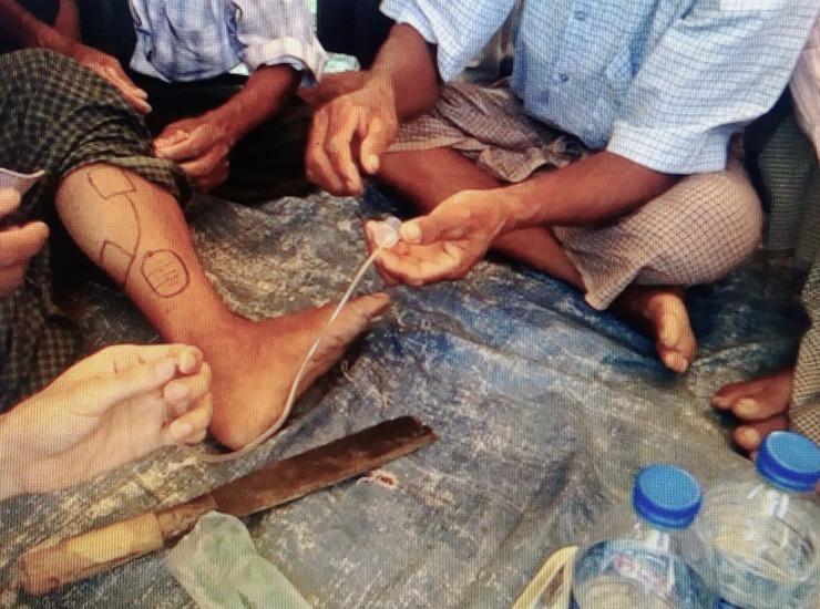
This clinical photograph illustrates a community-based demonstration of traditional healing practices for snakebite management. The central focus is a person's lower leg and foot, which feature black ink demarcations consisting of geometric outlines and a circular zone, likely indicating surgical or extraction boundaries. Parallel skin incisions are visible within these marked areas. An improvised suction device is depicted, consisting of a 20cc plastic syringe barrel (with the distal end removed) applied directly to the skin, connected via transparent tubing to a second syringe held by a practitioner to create negative pressure. On the ground adjacent to the limb is a large, weathered blade used for making the incisions. The scene highlights traditional first aid interventions, specifically the 'cut and suck' method, which is medically discouraged in modern toxicology due to the risks of secondary infection, tissue damage, and lack of efficacy in removing systemic venom. This image serves as educational material for global health and toxicology to illustrate cultural health practices and the challenges of pre-hospital snakebite care.
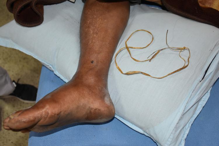
This clinical photograph displays a human lower limb resting on a pillow, illustrating complications related to snakebite management. The anatomical focus is the lower leg and foot, which exhibits significant edema (swelling) and skin tension. A small, dark, circular puncture or lesion is visible on the anterior aspect of the lower leg, suggestive of a bite site. The skin shows areas of mottled pigmentation and localized dryness, particularly around the heel and ankle. Adjacent to the limb is a discarded makeshift tourniquet made of yellow plastic rope, which was reportedly used as a tight ligature. This image serves as a clinical example of traditional first aid practices for snakebites, highlighting the physiological changes in the affected limb, such as swelling and potential tissue ischemia, as well as the use of non-medical interventions like mechanical constriction and localized topical applications.
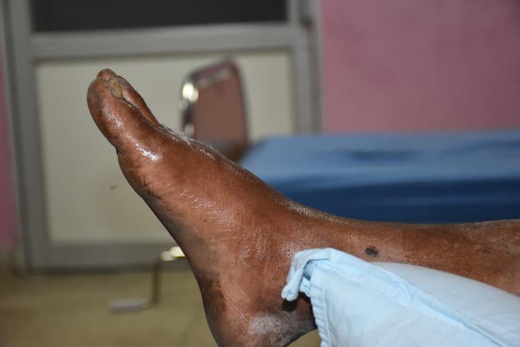
This clinical photograph displays the right foot and ankle of a patient in a medical setting, following a reported snakebite. The foot exhibits moderate diffuse swelling (edema). The skin is coated in a thick, shiny, and viscous amber-colored substance, identified as honey, which was applied as a traditional first-aid remedy prior to hospital arrival. A high specular light reflection is visible across the dorsal and lateral surfaces of the foot due to the topical application. There is a dark, circular lesion or puncture mark on the lateral aspect of the distal lower leg, near the ankle joint, potentially representing the bite site. The ankle is partially covered by a light blue medical cloth. This image illustrates traditional pre-hospital practices and local wound management for snakebite envenomation in a clinical context, highlighting the intersection of traditional medicine and emergency medical care.
Suspected Snakebite: What to Do
1. Recognize the Situation
- Local: pain, swelling, bruising, blistering, fang marks at the bite site
- Systemic: nausea/vomiting, dizziness, blurred vision, difficulty swallowing, drooling, drooping eyelids (ptosis)
- Neurological: numbness/tingling, muscle weakness, difficulty breathing
- Hematological: bleeding from gums or wounds, dark/bloody urine
2. Immediate First Aid - What TO DO
At the Scene
- Stay calm and keep the victim still - Movement accelerates venom absorption. Immobilize the entire patient and especially the bitten limb.
- Call emergency services immediately (or arrange fastest transport to a hospital). Do not wait for symptoms to develop.
- Immobilize the bitten limb at or below heart level - keep it still and lower than the heart to slow systemic spread.
- Remove constrictive items - Remove jewelry (rings, bracelets, watches), tight clothing, and footwear from the affected limb before swelling makes this impossible.
- Note the time of the bite - This helps doctors calculate envenomation progression.
- Try to safely identify the snake - Do NOT try to catch or handle it. A photograph taken at a safe distance is useful. Remember: even a recently killed snake can still bite reflexively.
- Keep the victim warm and positioned comfortably (lying down is ideal).
- If venom is sprayed into the eyes (spitting cobras), irrigate immediately with large volumes of water.
In Australia Specifically (Elapid snakes - neurotoxic)
3. What NOT to Do - Dangerous Myths
| Action | Why It's Harmful |
|---|---|
| Cutting or incising the bite | Causes tissue damage, introduces infection, does not remove venom |
| Sucking out venom | Does not remove significant venom; contaminates wound with oral bacteria |
| Applying a tourniquet | Causes ischemia, nerve injury, and gangrene within 20 minutes to 2 hours |
| Applying ice or cold packs | Can worsen tissue damage from cytotoxic venom |
| Applying electric shock | No evidence of benefit, can cause injury |
| Drinking alcohol | Vasodilates and accelerates absorption |
| Traditional remedies / "black stones" | No proven benefit; waste critical time |
| Letting the victim walk | Increases venom spread |
4. Hospital Management
Rapid Assessment
- Full vital signs, airway/breathing/circulation (ABC) assessment
- IV access and blood draws (CBC, coagulation panel, renal/liver function, urinalysis)
- Mark the edge of swelling with a pen and measure limb circumference every 30 minutes to track progression
Species Identification + Envenomation Grading
- Mild: local swelling only, no systemic signs
- Moderate: swelling extending beyond bite area, mild systemic signs
- Severe: rapidly advancing swelling, coagulopathy, neurotoxicity, hemodynamic instability
Antivenom - The Only Definitive Treatment
- Children require the same dose as adults (dose is based on venom load, not body weight)
- Must be given in a setting with resuscitation equipment available (epinephrine must be immediately available for anaphylaxis)
- If an acute allergic reaction occurs, stop infusion, give antihistamines (H1 + H2 blockers)
- Repeat dosing may be required if symptoms progress
- Rosen's Emergency Medicine notes: CroFab and Anavip F(ab')2 are the antivenoms of choice for North American pit viper (crotalid) bites
Supportive Care
- IV fluids + vasopressors for hypotension/shock
- Blood products if active bleeding (after antivenom)
- Compartment syndrome management: if compartment pressure >30 mmHg, elevate limb, give mannitol (1-2 g/kg IV), additional antivenom; fasciotomy only as a last resort
- Wound care: debridement of necrotic tissue as needed
- Tetanus prophylaxis
- Dialysis support if acute kidney injury develops (kidneys can fail in viper envenomation)
5. When to Suspect Envenomation vs. Dry Bite
- No pain or swelling beyond the bite site
- No systemic symptoms
- Normal lab values
Summary Pocket Guide
SUSPECT SNAKEBITE?
  ✓ Stay calm, keep victim still
  ✓ Immobilize limb below heart level
  ✓ Remove rings/watches/tight clothing
  ✓ Call ambulance / go to ER IMMEDIATELY
  ✓ Note the time, try to photograph snake safely

  ✗ Do NOT cut/suck wound
  ✗ Do NOT apply tourniquet
  ✗ Do NOT apply ice
  ✗ Do NOT let patient walk
  ✗ Do NOT use traditional remedies
Important: This information is for general education. In an actual emergency, call your local emergency number immediately (911 in USA, 102/112 in India, 999 in UK, 000 in Australia, etc.).
In hospital
In-Hospital Management of Snakebite
Step 1: Rapid Assessment & Stabilization (on arrival)
Primary Survey - ABCs First
- Airway: Elapid (cobra/krait/coral snake) bites can cause respiratory paralysis - assess immediately. Intubate early if there is any sign of bulbar weakness, drooling, dysphagia, or worsening respiratory effort.
- Breathing: Baseline and serial pulmonary function measurements (inspiratory pressure, vital capacity) in suspected elapid bites - respiratory failure is the main cause of death.
- Circulation: Establish IV access (in an unaffected limb), assess for hypotension and tachycardia (signs of hypovolemia from venom-induced plasma/blood extravasation into tissues).
Immediate Actions
- Remove any pre-hospital tourniquets or constricting bands only when antivenom is available and IV access is secured - be prepared for rapid systemic envenomation after removal
- Immobilize the bitten limb in a neutral position, below heart level
- Administer supplemental oxygen
- Tetanus prophylaxis - check immunization status and update as needed
- Clean and document the wound (distance between fang marks estimates snake size)
Step 2: Investigations / Labs
| Test | What to Look For |
|---|---|
| CBC | Thrombocytopenia (platelet consumption), hemolysis, hemoconcentration |
| Coagulation screen (PT/INR, aPTT, fibrinogen) | Prolonged times, hypofibrinogenemia - sign of DIC/coagulopathy |
| Blood group + crossmatch | In case transfusion needed |
| Renal function (BUN, creatinine, electrolytes) | AKI from viper envenomation, myoglobinuria |
| LFTs | Hepatotoxicity with some species |
| Urinalysis | Hemoglobinuria, myoglobinuria (dark urine = poor sign) |
| CK | Rhabdomyolysis (sea snakes, some kraits, Australian elapids) |
| Blood glucose | Hypoglycemia possible |
| ECG | Arrhythmias rare but documented |
Step 3: Grading Envenomation Severity
| Grade | Features |
|---|---|
| Dry bite | Fang marks only, no swelling, no systemic signs, normal labs after 8-12 hours |
| Mild | Local swelling and pain confined to bite site, minimal/no systemic signs |
| Moderate | Swelling progressing beyond bite site, mild systemic signs, mild lab abnormalities |
| Severe | Rapidly advancing swelling/ecchymosis, significant coagulopathy, systemic effects (hypotension, altered sensorium, respiratory compromise) |
Step 4: Antivenom - The Cornerstone of Treatment
Principles
- Antivenoms consist of heterologous antibodies (from immunized sheep or horses) that bind and neutralize venom molecules
- Children require the same dose as adults - dosing is based on venom load, not body weight
- Must be given in a critical care setting (ED or ICU) with resuscitation equipment and epinephrine immediately available
- Give IV, never IM
When to Give Antivenom
North American Pit Viper (Crotalid) Antivenoms
| Product | Details |
|---|---|
| CroFab (Crotalidae Polyvalent Immune Fab, Ovine) | Fab fragments from sheep; shorter half-life; first line |
| Anavip (Crotalidae Immune F(ab')2, Equine) | F(ab')2 fragments; longer half-life; lower incidence of recurrent coagulopathy; caution in horse allergy |
Elapid Antivenoms (Coral snakes, Cobras, Kraits, Mambas)
- Admit all confirmed/suspected elapid bites - neurological effects can be delayed 12+ hours and are not easily reversed
- Coral snake (US): 3-5 vials of Anti-venin (M. fulvius) IV; reserve additional doses if symptoms appear; monitor for respiratory failure
- Exotic elapids (cobras, kraits, mambas): Contact regional poison control center for specific antivenom location and dosing
Managing Antivenom Reactions
- Acute allergic reaction: Stop infusion immediately, give antihistamines (H1 + H2 blockers). Have epinephrine drawn and ready.
- Anaphylaxis: Epinephrine immediately
- Serum sickness (delayed, 1-3 weeks): Fever, rash, arthralgias - treat with prednisone 1 mg/kg/day PO tapered over 1-2 weeks
Step 5: Managing Specific Complications
Coagulopathy / DIC
- Best treated with antivenom (which neutralizes the procoagulant venom enzymes)
- Blood component replacement (FFP, platelets, cryoprecipitate) only if active bleeding and AFTER antivenom has been given
- Do not transfuse first - venom will continue consuming clotting factors until neutralized
Hypotension / Shock
- IV isotonic fluid resuscitation (crotalid venom causes plasma extravasation into tissues)
- Vasopressors if fluid-refractory
Compartment Syndrome
- Measure intracompartmental pressure
- If pressure >30 mmHg: elevate limb + mannitol 1-2 g/kg IV over 30 min + additional antivenom concurrently
- Reassess after 60 minutes
- Fasciotomy only as a last resort if pressure remains elevated - antivenom is more effective than fasciotomy for venom-induced compartment syndrome, and fasciotomy may actually worsen local tissue loss
Neurotoxicity (Elapid bites)
- Watch for: ptosis, drooling, dysarthria, diplopia, dysphagia, respiratory muscle weakness
- Onset can be delayed 12+ hours (some species)
- Serial vital capacity and inspiratory pressure measurements
- Early intubation and ventilatory support may be required for prolonged periods
- For coral snake: cholinergic agonists at first sign of symptoms; calcium infusion may reduce seizure onset
Acute Kidney Injury
- IV fluids to maintain urine output
- Supportive dialysis if needed (same approach as AKI from any other cause)
- Early antivenom administration reduces AKI severity
Rhabdomyolysis
- Especially with sea snakes, some kraits, some Australian elapids
- Aggressive IV hydration, urine alkalinization, monitor CK
Wound Care
- Clean wound, debride necrotic tissue as it demarcates
- Prophylactic antibiotics: not routinely recommended - infection after snakebite is surprisingly uncommon and most apparent "infections" (erythema, edema, necrosis) are sterile from venom effects
- Treat culture-proven infection with appropriate antibiotics (potential organisms: Pseudomonas, Enterobacteriaceae, Staphylococcus, Clostridia)
- Steroids: not effective for envenomation; use only for allergic reactions or serum sickness
Step 6: Disposition
| Situation | Disposition |
|---|---|
| Dry bite (8-12 hrs observation, no signs) | Discharge with return precautions |
| Mild-moderate, antivenom completed | General ward |
| Severe, life-threatening, receiving antivenom | ICU |
| All confirmed elapid bites | Admit (ICU preferred) - delayed neurological effects |
Quick Reference: In-Hospital Flowchart
ARRIVAL
  → ABCs (airway priority for elapid bites)
  → IV access (unaffected limb)
  → Remove prehospital tourniquets (with antivenom ready)
  → Labs: CBC, coag, renal, LFTs, UA, CK, type & screen
  → ECG
  → Mark edema border, measure limb circumference
  ↓
GRADE ENVENOMATION
  → Dry bite? → Observe 8-12 hrs → discharge if stable
  → Signs of progression? → ANTIVENOM
  ↓
ANTIVENOM (IV, in ICU/ED, epinephrine drawn)
  → Establish "initial control" (stop progression)
  → Reassess q30 min, repeat labs q4h
  → Re-dose if progression continues
  ↓
MANAGE COMPLICATIONS
  → Coagulopathy → more antivenom → blood products if active bleed
  → Compartment syndrome → mannitol + antivenom → fasciotomy last resort
  → Respiratory failure → intubate early in elapid bites
  → Hypotension → fluids → vasopressors
  → AKI → fluids → dialysis if needed
  ↓
DISPOSITION
  → ICU (severe) / ward (moderate) / discharge (dry bite)