
Clinical examination of respiratory system in detail and associated concept
chest inspection respiratory examination patterns breathing
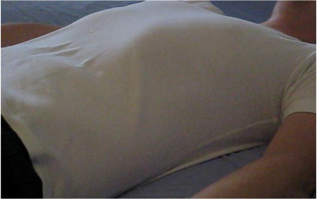
A clinical photograph of a human subject in a supine position, focused on the thorax and neck. The individual is wearing a loose-fitting white t-shirt against a dark background. This visual serves as a standardized pedagogical tool for assessing respiratory patterns and calculating respiratory rate (RR). The image captures the rise and fall of the chest wall and abdominal excursions, which are critical visual cues for clinical observation in emergency medicine and nursing education. The setup is designed to teach healthcare professionals how to identify tachypnea, bradypnea, or normal breathing rhythms (eupnea) through non-invasive visual inspection of the thoracic region. By highlighting the contour of the chest against the fabric, the image facilitates the identification of respiratory effort and chest wall symmetry.
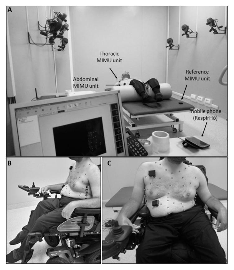
Clinical and procedural photographs illustrating a research setup for respiratory monitoring using wearable inertial sensors and Optoelectronic Plethysmography (OEP). Panel A shows a subject in the supine position on an elevated examination table within a laboratory equipped with wall-mounted infrared cameras. The setup identifies Thoracic and Abdominal Magnetic Inertial Measurement Units (MIMU) placed on the subject's trunk, a Reference MIMU on the table, a monitoring smartphone ('RespirHó'), and a data display monitor. Panels B (lateral view) and C (frontal view) depict a male subject seated in a power wheelchair. The subject's torso is shirtless, revealing multiple small, spherical retro-reflective markers and rectangular MIMU sensors adhered to the chest and abdominal wall. This clinical setup is designed to evaluate chest wall movement and breathing patterns in static conditions (supine vs. seated), often used in the context of neuromuscular disease monitoring to validate wearable sensor accuracy against the OEP gold standard.
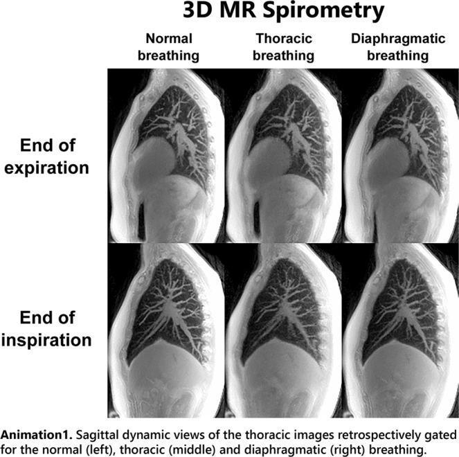
This diagnostic imaging panel presents six sagittal views of the human thorax obtained using 3D ultrashort echo time (UTE) Magnetic Resonance Imaging (MRI). The images compare three respiratory patterns: normal breathing (left), thoracic breathing (middle), and diaphragmatic breathing (right), captured at two distinct phases: end-expiration (top row) and end-inspiration (bottom row). Red overlay curves on the inspiration images mark the lung boundaries from the corresponding expiratory state to highlight anatomical displacement. Red arrows indicate the primary direction of motion, emphasizing diaphragmatic descent and anterior-posterior chest wall expansion. The 'normal' column shows a combination of diaphragmatic and thoracic movement, while 'thoracic' breathing exhibits more pronounced anterior chest wall protrusion, and 'diaphragmatic' breathing shows maximal inferior displacement of the diaphragm. This visualization is part of a 3D MR Spirometry study used to analyze respiratory mechanics, pulmonary dynamics, and regional volume expansion, demonstrating how different breathing maneuvers affect lung parenchyma motion and thoracic cavity volume.
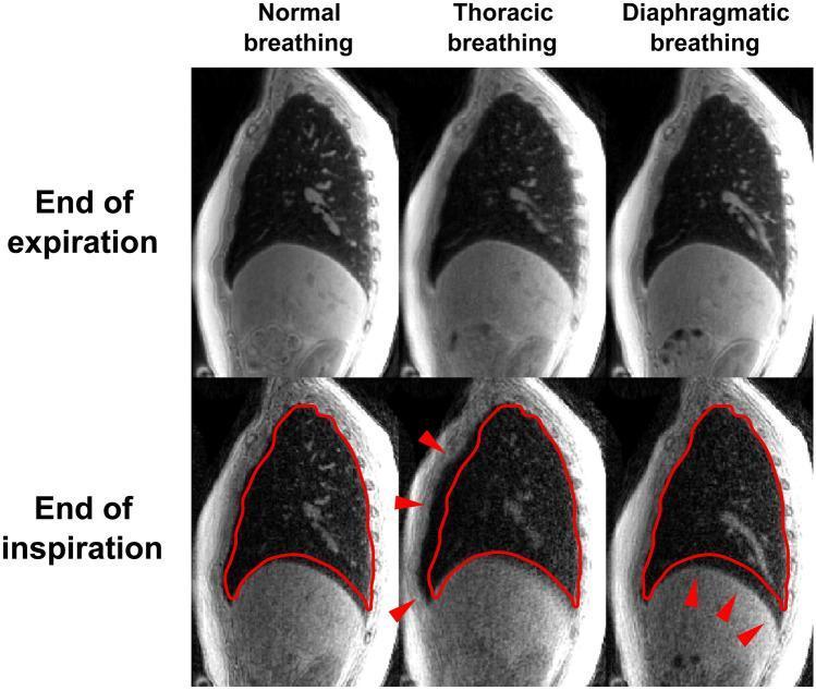
This diagnostic image provides a comparison of human respiratory dynamics using sagittal-view gated CT imaging across three breathing patterns: normal, thoracic, and diaphragmatic. The panel is organized into two rows representing physiological states: 'End of expiration' (top) and 'End of inspiration' (bottom). Each column highlights a specific respiratory technique. In the bottom row (inspiration), a red outline superimposes the lung border from the previous expiratory phase to visually demonstrate lung volume expansion. Red arrows indicate primary mechanical drivers for each pattern: the thoracic breathing column displays anterior movement of the chest wall, while the diaphragmatic breathing column emphasizes significant inferior displacement of the diaphragm and expansion of the lower thoracic cavity. This comparison illustrates the mechanical differences between costal and abdominal breathing, demonstrating how different muscle groups contribute to changes in pleural pressure and lung volume. The imagery serves as an educational tool for pulmonology and physiology, focusing on respiratory mechanics and accessory muscle recruitment.
vocal fremitus percussion dullness resonance clinical signs
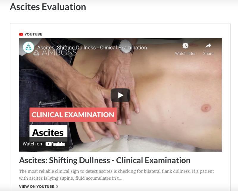
This clinical photograph is a frame from a medical educational video demonstrating an abdominal examination for ascites. The image depicts a patient in a supine position with the abdomen exposed. An examiner's hands are positioned on the patient's right flank and lateral abdominal wall, demonstrating the technique for percussion or palpation used to identify 'shifting dullness.' The patient shows signs of abdominal distension consistent with fluid accumulation. Educational overlays identify the content as a clinical examination of ascites, specifically focusing on shifting dullness as a diagnostic sign. This material is designed for medical students and clinicians to learn physical examination maneuvers for renal and gastrointestinal assessment. Key landmarks visible include the costal margin, umbilicus, and lateral flanks, which are critical for determining the transition from resonance to dullness during percussion in a patient with suspected peritoneal effusion.
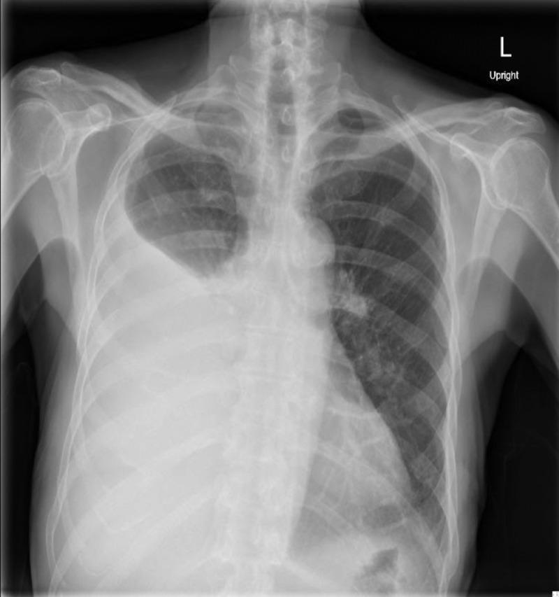
Anterior-posterior (AP) upright chest radiograph demonstrating a large, dense, homogeneous opacity occupying the lower two-thirds of the right hemithorax. The opacity exhibits a classic curvilinear superior border, or Ellis-Damoisseau line, consistent with a large pleural effusion. This finding results in complete obscuration of the right hemidiaphragm (positive silhouette sign) and the right costophrenic angle. There is a perceptible mass effect, evidenced by a slight contralateral shift of the mediastinum towards the left. In contrast, the left lung field appears clear with normal bronchovascular markings and a well-defined diaphragmatic contour. The skeletal structures and visible soft tissues are unremarkable. This diagnostic image is used to educate on the radiographic presentation of pleural fluid accumulation and its associated clinical signs, such as decreased breath sounds and dullness to percussion in the context of metastatic disease.
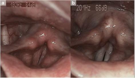
This diagnostic comparison contains two videostroboscopy frames illustrating the laryngeal manifestations of laryngopharyngeal reflux (LPR) before and after treatment. Panel (a) represents the baseline state, showing significant clinical signs including diffuse laryngeal erythema, inter-arytenoid redness, and posterior commissure hypertrophy. The vocal folds exhibit visible irritation and marginal irregularity, accompanied by pharyngolaryngeal edema. Panel (b) demonstrates the post-treatment state following a course of proton pump inhibitor therapy. Notable improvements include a marked reduction in mucosal inflammation and edema. The inter-arytenoid region appears less congested, and the vocal folds show restored smoothness and a healthier pearly-white coloration. This comparison is an educational resource for identifying Reflux Finding Score (RFS) criteria and monitoring therapeutic response in otolaryngology. The images demonstrate the reversal of tissue changes associated with chronic acid and pepsin exposure in the upper aerodigestive tract.
clubbing finger digital cyanosis peripheral signs respiratory
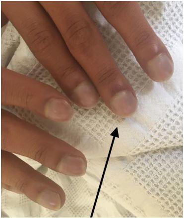
This clinical photograph shows a close-up of a patient's fingers demonstrating classic signs of digital clubbing and peripheral cyanosis. The distal phalanges exhibit a bulbous, 'drumstick' appearance with a noticeable loss of the normal Lovibond angle (the angle between the nail plate and the proximal nail fold). The nail beds appear convex and rounded. A distinct bluish-purple discoloration (cyanosis) is visible across the nail beds and distal fingertips, indicating poor blood oxygenation or circulation. A black arrow points to the most prominent area of clubbing on the index finger. These findings are clinically significant as they often indicate chronic hypoxemia secondary to congenital heart disease (such as Ebstein anomaly), pulmonary disease, or chronic inflammatory conditions. This image serves as a teaching tool for physical examination findings in cardiovascular and respiratory medicine.
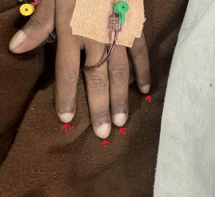
This clinical photograph displays a patient's hand demonstrating grade 3 digital clubbing, a common physical sign of chronic hypoxemia. The fingertips (distal phalanges) appear bulbous and enlarged, described as 'drumstick' morphology. The nails exhibit increased longitudinal curvature and a loss of the normal Lovibond angle at the nail base. Clinical features suggest cyanotic heart disease, supported by the visible medical equipment including a pulse oximetry sensor on the index finger and an intravenous (IV) catheter secured to the dorsal surface of the hand with adhesive dressing. There is evidence of peripheral cyanosis or hyperpigmentation of the distal digits. This visual is highly relevant for medical education regarding cardiovascular and respiratory physical examination findings and the assessment of chronic oxygen deprivation.
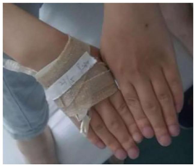
This clinical photograph displays the hands of a patient exhibiting clear signs of chronic hypoxemia and inflammatory changes. The primary visible pathology includes marked digital clubbing, characterized by the bulbous enlargement of the distal phalanges and a loss of the normal angle between the nail plate and the proximal nail fold. The fingernails and distal fingertips demonstrate a bluish-purplish discoloration consistent with peripheral cyanosis. Additionally, there is noticeable fusiform swelling of the proximal interphalangeal (PIP) joints, particularly prominent on the middle fingers, suggesting an associated inflammatory or rheumatic process. One hand is partially obscured by a tan elastic bandage and adhesive tape around the wrist and dorsum, with a visible intravenous (IV) catheter line, indicating acute medical management. The combination of digital clubbing and cyanosis is clinically significant for underlying chronic respiratory or cardiovascular disease, such as the pulmonary interstitial fibrosis mentioned in the associated clinical context.
Clinical Examination of the Respiratory System
Overview
1. GENERAL / PRELIMINARY INSPECTION
Appearance and Distress
- Does the patient look comfortable or distressed?
- Use of accessory muscles (sternocleidomastoid, scalene, intercostals) indicates increased work of breathing
- Pursed-lip breathing suggests COPD (creates auto-PEEP to prevent early airway collapse)
- Position: patients with severe dyspnea often sit upright or lean forward (tripod position)
Vital Signs and Hands
| Finding | Significance |
|---|---|
| Tachypnea (RR >20/min) | Pneumonia, PE, heart failure, metabolic acidosis |
| Bradypnea (RR <10/min) | CNS depression, opioids |
| Cyanosis (peripheral) | Reduced peripheral perfusion |
| Cyanosis (central) | SpO₂ <85%, check tongue and lips |
| Clubbing | Chronic hypoxemia (lung cancer, bronchiectasis, ILD, cyanotic heart disease, lung abscess) |
| Tremor/asterixis | CO₂ retention (type II respiratory failure) |
| Tobacco stains on fingers, teeth | Smoking history indicator |
Clubbing - Grading (Schamroth's test)
- Grade 1: Loss of nail fold angle
- Grade 2: Increased curvature of nail
- Grade 3: Drumstick appearance of the finger
- Grade 4: Hypertrophic pulmonary osteoarthropathy (HPOA) with periosteal new bone formation
Causes of Clubbing (categorized)
- Lung cancer (most common cause overall)
- Bronchiectasis, lung abscess, cystic fibrosis, empyema
- Idiopathic pulmonary fibrosis, asbestosis
- Benign and malignant pleural tumors
- Cyanotic congenital heart disease
- Subacute bacterial endocarditis
- Pulmonary arteriovenous fistula
- Inflammatory bowel disease (Crohn's, ulcerative colitis)
- Hepatic cirrhosis, hepatoma, hepatopulmonary syndrome
2. INSPECTION OF THE CHEST
"Inspection is performed with the patient sitting, after sufficient clothing has been removed to permit observation of the entire thorax." - Murray & Nadel
Chest Shape and Deformities
| Deformity | Description | Associated Condition |
|---|---|---|
| Barrel chest | AP diameter ≈ transverse diameter; hyperinflation | COPD, emphysema |
| Kyphoscoliosis | Combined kyphosis + scoliosis | Restrictive ventilatory defect, cor pulmonale |
| Pectus excavatum (funnel chest) | Sternum depressed inward | Usually cosmetic; can compress heart in severe cases |
| Pectus carinatum (pigeon chest) | Sternum protrudes anteriorly | Childhood asthma, rickets |
| Harrison's sulcus | Horizontal groove along lower ribs at diaphragm attachment | Severe childhood asthma, rickets |
| Ankylosing spondylitis | Rigid thorax, loss of chest expansion | Restrictive defect |
| Flail chest | Paradoxical movement of chest segment | Multiple rib fractures |
Breathing Patterns
| Pattern | Description | Cause |
|---|---|---|
| Cheyne-Stokes | Crescendo-decrescendo cycles with apnea | Heart failure, CNS disease |
| Kussmaul breathing | Deep, regular, rapid | Metabolic acidosis (DKA) |
| Biot's breathing | Irregular, with random apnea | Brainstem lesions |
| Ataxic breathing | Completely irregular rate and depth | Medullary lesions (ominous) |
| Apneustic breathing | Prolonged inspiratory pause | Pontine lesions |
Symmetry and Movement
- Observe both sides simultaneously: look for lag in chest wall movement on one side
- Unilateral reduced movement = consolidation, effusion, pneumothorax, collapse on that side
Olfactory Inspection
- Anaerobic lung abscess: foul, fetid odor
- Pseudomonas skin/soft tissue infection: sweet smell
- Ketones on breath: diabetic crisis
- Ethanol: alcoholism
- Tobacco leaf stains on teeth, fingers, clothing
3. PALPATION
Tracheal Position
- Palpate the trachea at the suprasternal notch with the index finger
- Central trachea = normal
- Trachea deviated toward the side of lesion: collapse/atelectasis (lung pulls trachea toward it)
- Trachea deviated away from the side of lesion: massive effusion, tension pneumothorax (pushes trachea away)
Chest Expansion
- Place both hands flat on the back (lower chest), thumbs meeting in the midline
- Ask patient to take a deep breath
- Normally thumbs move symmetrically outward 3-5 cm
- Reduced unilateral expansion: consolidation, effusion, pneumothorax, collapse
- Reduced bilateral expansion: COPD, fibrosis, ankylosing spondylitis
Vocal (Tactile) Fremitus
- Use the ulnar side of the hand (or palms) and move systematically from apex to base, comparing both sides
- Sound transmission increases through solid material (consolidation) and decreases when fluid/air intervenes
| Fremitus | Condition | Mechanism |
|---|---|---|
| Increased | Consolidation (pneumonia), lung collapse with patent airway | Solid lung transmits sound better than air |
| Decreased / absent | Pleural effusion, pneumothorax, pleural thickening | Fluid/air between lung and chest wall impedes transmission |
| Decreased | Emphysema, airway obstruction | Decreased air movement and lung density |
Other Palpatory Findings
- Tenderness: rib fractures, costochondritis (Tietze's syndrome), musculoskeletal causes of chest pain
- Subcutaneous emphysema: crepitus (crackling sensation) on palpation - air in soft tissues (pneumothorax, post-trauma)
- Empyema necessitans: fluctuant area on chest wall (empyema tracking through intercostal space)
- Cervical rib: bony abnormality palpated in supraclavicular fossa
- Subcutaneous calcinosis: seen in systemic sclerosis
- Lymphadenopathy: cervical/supraclavicular nodes - clue to thoracic malignancy or mycobacterial infection
- Palpable pleural friction rub: occasionally felt as a "to and fro" sensation
4. PERCUSSION
"Percussion as part of the physical examination follows Auenbrugger's sounding of beer barrels to determine their fluid levels." - Fishman's Pulmonary Diseases
Technique
- Middle finger of the non-dominant hand (pleximeter) is placed flat on the chest
- The tip of the middle finger of the dominant hand (plexor) strikes the pleximeter with a quick, free wrist-flick
- The response is predominantly felt (vibration) as well as heard
Percussion Notes and Their Meanings
| Note | Quality | Cause |
|---|---|---|
| Resonant | Clear, hollow | Normal air-filled lung |
| Hyperresonant | Louder, lower-pitched than normal | Pneumothorax, large bulla, emphysema |
| Tympanitic | Drum-like, very hollow | Tension pneumothorax (extreme hyperresonance) |
| Dull | Short duration, high pitch, low intensity | Consolidation (pneumonia), atelectasis, pleural mass |
| Stony dull / Flat | Absolute dullness, like percussing the thigh | Pleural effusion (large), solid tissue |
Percussion in Pleural Effusion
- Resonant zone - above the fluid (normal lung)
- Dull zone - at the fluid level
- Flat zone - at the base, completely below the fluid
Diaphragmatic Excursion
- Ask patient to breathe in and hold: percuss downward to find lower limit of resonance
- Ask patient to breathe out and hold: percuss upward to find upper limit
- Normal diaphragmatic excursion = 3-5 cm
- Reduced in emphysema, diaphragmatic palsy, large pleural effusion
5. AUSCULTATION
"Attention is focused on the intensity and quality of sounds, as well as on the presence of abnormal (adventitious) lung sounds." - Murray & Nadel
Technique
- Auscultate at both apices, over both upper and lower lobes, anteriorly and posteriorly
- Listen during both inspiration and expiration
- Compare corresponding zones on both sides
- Minimum auscultation areas: 6 posterior, 4 anterior, 2 lateral per lung
Normal Breath Sounds
| Sound | Location | Character |
|---|---|---|
| Vesicular (normal) | Peripheral lung fields | Soft, low-pitched; inspiration > expiration; inspiration:expiration ratio ~3:1 |
| Bronchovesicular | 1st and 2nd interspaces anteriorly, between scapulae | Intermediate quality; equal inspiration and expiration |
| Bronchial (tracheal) | Over trachea/manubrium | Loud, high-pitched, hollow; expiration > inspiration with gap between |
Abnormal (Adventitious) Breath Sounds
| Type | Character | Timing | Mechanism | Causes |
|---|---|---|---|---|
| Fine crackles | High-pitched, brief, non-musical | Late inspiration | Reopening of collapsed alveoli and small airways | Heart failure, ILD, early pneumonia |
| Coarse crackles | Low-pitched, bubbling, longer | Inspiration & early expiration; cleared by coughing | Secretions in large airways | Bronchitis, COPD, bronchiectasis |
- High-pitched, musical, continuous sounds (>250 ms)
- Predominantly expiratory (sometimes inspiratory too)
- Mechanism: rapid airflow through narrowed airways causing oscillations
- Causes: asthma (most common), COPD, bronchospasm, foreign body
- Monophonic wheeze: one pitch, single obstructed airway (tumor, foreign body)
- Polyphonic wheeze: multiple pitches, diffuse airway narrowing (asthma)
- Silent chest in asthma = very severe bronchospasm with insufficient airflow to generate wheeze (ominous sign)
- Low-pitched, musical, sonorous sounds
- Heard predominantly in expiration, may change or clear with coughing
- Mechanism: secretions in large airways
- Causes: bronchitis, COPD, mucus plugging
- Leathery, creaking, grating sound
- Heard in both inspiration and expiration (differentiates from crackles which are only inspiratory)
- Does NOT change with coughing
- Does change with respiration (differentiates from pericardial rub, which persists when breathing is held)
- Causes: pleuritis (pneumonia with pleuritis, TB pleuritis, malignancy, PE with infarction, connective tissue diseases)
- May be palpable as well
- Occasionally heard even in large effusions (rub generated by shape change of thickened pleura, not direct contact)
- High-pitched, loud, predominantly inspiratory continuous sound
- Heard louder over the upper airway/neck than chest
- Mechanism: turbulent flow through narrowed extrathoracic airway
- Causes: croup, epiglottitis, foreign body inhalation, anaphylaxis, laryngeal tumor, vocal cord dysfunction, post-extubation edema
- Life-threatening - requires immediate attention
- Key differentiation: stridor is inspiratory, loud over neck; wheeze is expiratory, loud over chest
- Crunching, crackling sounds synchronous with cardiac contraction
- Heard even when breathing is stopped (differentiates from pleural rub)
- Indicates pneumomediastinum
6. VOICE SOUNDS (Vocal Resonance)
| Sign | Technique | Finding | Cause |
|---|---|---|---|
| Bronchophony | Patient says "ninety-nine" | Words louder and clearer than normal | Consolidation |
| Whispering pectoriloquy | Patient whispers "one, two, three" | Whispered sounds transmitted clearly | Consolidation |
| Aegophony (E to A change) | Patient says "eee" | Heard as "aaa" through stethoscope | Upper border of pleural effusion (bronchophony in compressed lung above fluid) |
7. CLINICAL CORRELATION TABLE - Disease Patterns
| Condition | Inspection | Palpation (Expansion / Fremitus) | Percussion | Auscultation | Vocal Resonance |
|---|---|---|---|---|---|
| Consolidation (Pneumonia) | Lagging movement | Reduced expansion; Increased fremitus | Dull | Bronchial breathing; fine crackles | Increased (bronchophony, whisp. pectoriloquy) |
| Pleural Effusion | Reduced movement | Reduced expansion; Reduced/absent fremitus | Stony dull | Reduced/absent breath sounds; bronchial breathing above fluid (aegophony) | Reduced below; aegophony at upper border |
| Pneumothorax | Reduced movement | Reduced expansion; Absent fremitus | Hyperresonant/tympanitic | Absent breath sounds | Absent |
| Emphysema / COPD | Barrel chest; pursed-lip breathing; accessory muscles | Reduced expansion bilaterally; Reduced fremitus | Hyperresonant bilaterally | Reduced breath sounds; prolonged expiration; wheeze; coarse crackles | Reduced |
| Collapse/Atelectasis | Reduced movement; ipsilateral rib indrawing | Reduced expansion; trachea pulled toward; Reduced fremitus | Dull | Absent or bronchial | Reduced |
| Fibrosis (ILD) | Reduced movement | Reduced expansion; tremitus may increase | Dull | Fine (velcro) crackles at bases | Variable |
| Asthma (acute) | Use of accessory muscles; hyperinflation | Reduced bilateral expansion | Hyperresonant (air trapping) | Polyphonic wheeze (or silent chest in severe) | Normal |
| Bronchitis | Normal or mild | Normal | Resonant | Coarse crackles, rhonchi | Normal |
8. ASSOCIATED CONCEPTS
Mediastinal Shift
- Toward lesion: collapse/atelectasis (lung volume loss), fibrosis
- Away from lesion: large effusion (>1000 mL typically), tension pneumothorax, large mass
Diaphragm Paralysis
- Paradoxical inward movement of the abdomen on inspiration (abdominal wall draws in instead of pushing out)
- Reduced breath sounds at base
- Reduced diaphragmatic excursion on percussion
Cor Pulmonale (Pulmonary Heart Disease)
- Elevated JVP, loud P2, right parasternal heave
- Peripheral edema
- Tricuspid/pulmonary regurgitation murmur
- Results from chronic lung disease causing pulmonary hypertension
Superior Vena Cava (SVC) Syndrome
- Venous engorgement of head and neck
- Fixed elevated JVP (non-pulsatile)
- Facial/arm swelling, especially on waking
- Caused by tumor compressing SVC (most commonly lung cancer or lymphoma)
Auscultation Areas Reference
- Upper zones (apices): above spine of scapula
- Middle zones: between spine of scapula and inferior angle
- Lower zones: below inferior angle of scapula
- Apices: above clavicles
- Upper lobes: above 4th rib
- Lower lobes: 4th rib downward
9. CLINICAL SKILLS PEARLS
- Always compare both sides simultaneously - asymmetry is the key to diagnosis
- Absent breath sounds is more significant than reduced - suspect pneumothorax, large effusion, or complete obstruction
- Crackles that clear with coughing = secretions (coarse crackles of bronchitis); those that do not clear = structural (fine crackles of heart failure/fibrosis)
- Wheeze that disappears after bronchodilator = reversible airway disease (asthma)
- Stony dull percussion = effusion (dull without modifier = consolidation or collapse)
- Trachea is a key landmark: shifted in large effusion, tension pneumothorax, and collapse
- Aegophony at the upper border of a pleural effusion is a classic and highly specific sign
- Silent chest in an asthmatic = emergency - insufficient airflow to generate wheeze
- Pleural rub vs. pericardial rub: ask patient to hold breath - pericardial rub persists, pleural rub disappears
According to Indian scenario andvtextbooks examination
Indian MBBS respiratory system clinical examination Macleod's Das Das textbook inspection palpation percussion auscultation
respiratory system examination chest expansion trachea palpation clinical
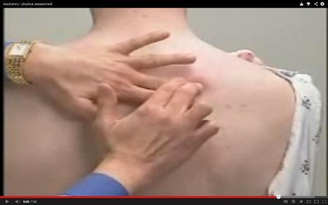
This clinical photograph demonstrates a physical examination technique, specifically palpation of the posterior thorax. The image shows a clinician's hands positioned over the upper back/scapular region of a patient who is seated and wearing a hospital gown. The clinician is using their fingers to assess for tactile fremitus, respiratory expansion, or underlying musculoskeletal irregularities. The patient has fair skin with a few scattered, benign-appearing pigmented macules (moles) on the upper back. The clinician is wearing a gold watch on their left wrist and a blue sleeve is visible. This visual serves as an educational example of a respiratory system physical assessment, emphasizing correct hand placement for evaluating pulmonary or thoracic wall characteristics in a clinical setting.
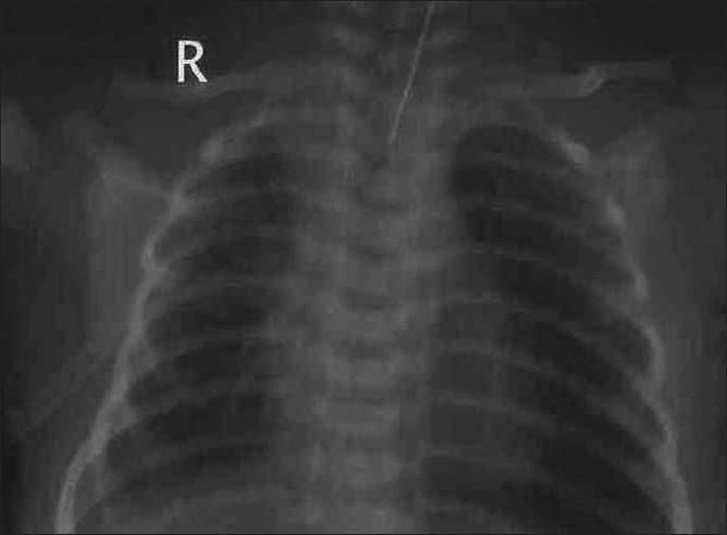
This diagnostic image is a neonatal anteroposterior (AP) chest radiograph illustrating the status post-drainage of a pleural effusion. The image shows full expansion of both lung fields with symmetric aeration and visible bronchovascular markings. The trachea is centrally located and midline, and the mediastinal shift previously caused by the effusion has resolved. A radiopaque intercostal chest tube is visible, originating from the right hemithorax and positioned within the pleural space to facilitate drainage. The costophrenic angles and diaphragmatic contours are visible, indicating successful re-expansion. This radiograph serves as a clinical example of post-procedural monitoring in neonatal respiratory distress management, specifically demonstrating the effective resolution of a large unilateral pleural effusion and the resulting restoration of normal pulmonary volume and thoracic symmetry.
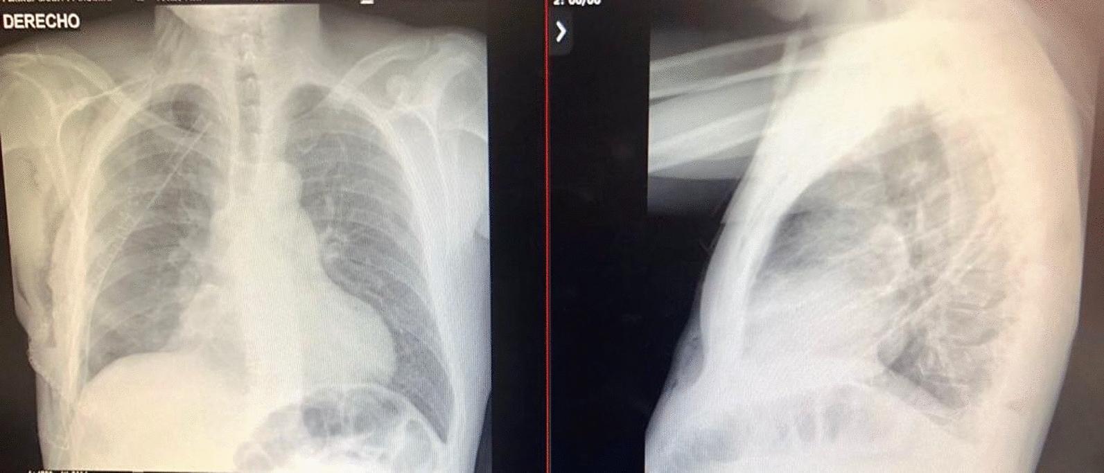
Postoperative chest radiography in anteroposterior (AP) and lateral projections following the resection of a large pleural mass. The AP view (left) demonstrates bilateral lung re-expansion with clear lung parenchyma and no residual radiopaque masses. The mediastinal contour and heart size are within normal limits, and the trachea is midline. Both costophrenic and cardiophrenic angles are sharp and well-defined, indicating the absence of pleural effusion. The lateral view (right) confirms pulmonary expansion with clear retrosternal and retrocardiac spaces. The diaphragmatic domes are smooth and distinct. Bony structures, including the ribs, sternum, and vertebrae, appear intact. Surgical sutures or clips from the previous thoracotomy are visible in the right hemithorax. This diagnostic imaging serves as a clinical baseline to confirm successful surgical intervention and recovery of respiratory function in a patient previously diagnosed with a large solitary fibrous tumor of the pleura.
pleural effusion consolidation pneumothorax chest X-ray clinical findings comparison
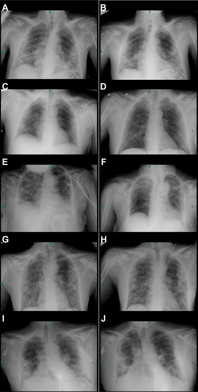
This composite educational graphic presents ten posterior-anterior (PA) or anteroposterior (AP) chest X-ray panels (A-J), organized as longitudinal comparison pairs demonstrating the progression or regression of COVID-19 pneumonia across five different patients. The images illustrate a spectrum of diagnostic findings including bilateral patchy reticular opacifications, ground-glass opacities, and perihilar and peripheral consolidations with lower zone predominance. Key pathological features visible across various stages include subsegmental atelectasis, reticulonodular patterns, and pleural effusions. For instance, panels (A) and (B) show minor regression of patchy opacifications following treatment, while panels (I) and (J) demonstrate disease progression, specifically highlighted by increased bilateral consolidation, the development of a right-sided pleural effusion, and a small right apical pneumothorax. The series serves as a clinical reference for monitoring viral pneumonia, secondary bacterial co-infections (e.g., Staphylococcus aureus, Klebsiella pneumoniae), and the radiological impact of advanced therapies like blood purification and cytokine control in critical care settings.
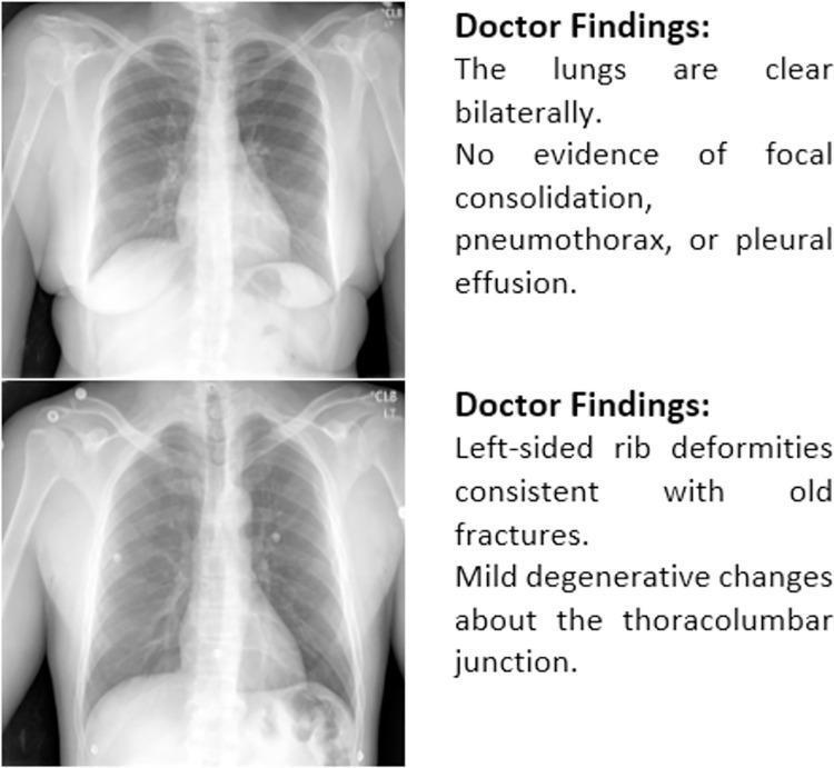
This diagnostic image provides a comparison of two posterior-anterior (PA) chest radiographs accompanied by their respective clinical findings. The upper panel displays a normal chest X-ray illustrating clear lung fields bilaterally. There is a notable absence of focal consolidation, pneumothorax, or pleural effusion, representing a standard baseline for pulmonary health. The lower panel demonstrates a chest radiograph with skeletal abnormalities, specifically identifying left-sided rib deformities suggestive of chronic or healed fractures. Additionally, it highlights mild degenerative changes located at the thoracolumbar junction of the spine. The visual comparison serves as an educational tool for differentiating between normal thoracic anatomy and common musculoskeletal findings such as old rib trauma and spinal degeneration in the context of diagnostic radiology. Key educational concepts include the identification of clear lung parenchyma versus the recognition of extrapulmonary skeletal variations.
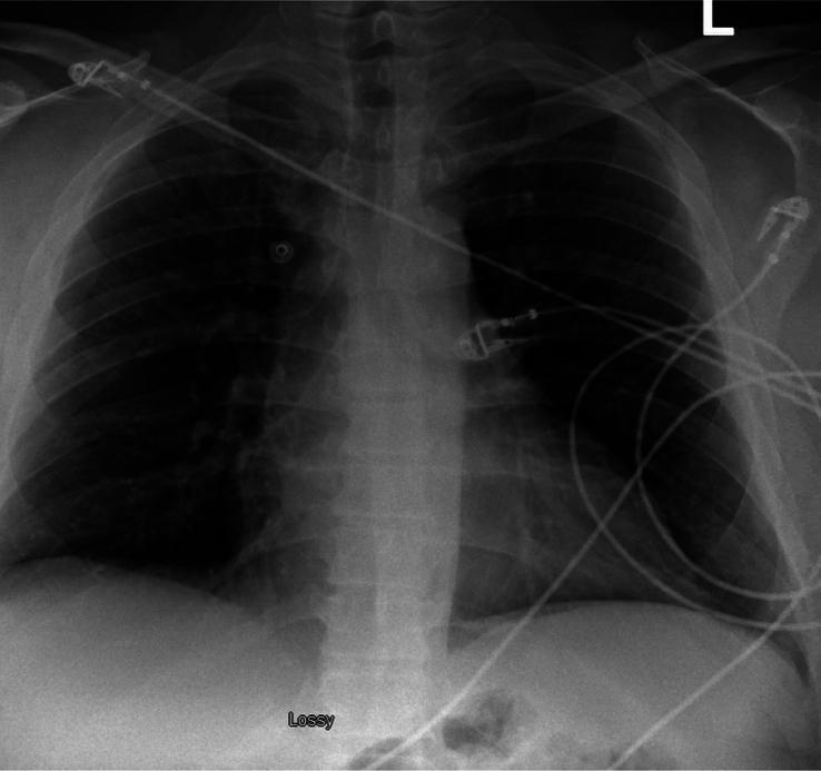
This diagnostic image is a posterior-anterior (PA) view chest X-ray of an adult patient. The radiograph demonstrates normal pulmonary and pleural findings. Both lung fields are clear, with no evidence of focal airspace consolidation, interstitial infiltrates, or pulmonary masses. The costophrenic and cardiophrenic angles are sharp and well-defined, indicating the absence of pleural effusion. No pneumothorax is visible. The cardiomediastinal silhouette is within normal limits for size and contour, and the trachea is midline. External artifacts, specifically several electrocardiogram (ECG) leads and wires, are superimposed over the thoracic cavity and upper abdominal region. The osseous structures of the rib cage, clavicles, and proximal humeri appear intact. This image serves as an educational example of a normal chest radiograph used in clinical triage to rule out acute cardiopulmonary pathology in patients presenting with symptoms such as chest pain or hypertension.
Schamroth window test clubbing finger nail
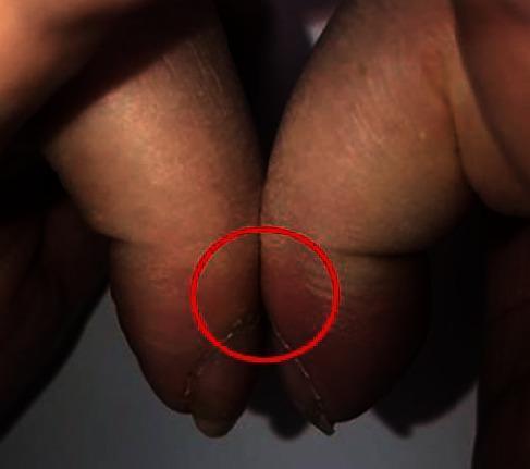
This clinical photograph displays a positive Schamroth sign, a classic physical examination finding indicative of digital clubbing (acropachy). The image shows the dorsal surfaces of the terminal phalanges of two opposite fingers placed in close apposition. In a normal presentation, a small diamond-shaped window (Schamroth's window) is visible between the nail beds; however, in this case, the window is absent. The nail beds are in direct contact, highlighted by a red circle, demonstrating the obliteration of the normal angle. The skin at the base of the nails appears thickened and shiny, consistent with the bulbous swelling seen in hypertrophic osteoarthropathy. This sign is clinically significant as an indicator of underlying chronic conditions, such as pulmonary or cardiovascular diseases, or in this context, paraneoplastic syndromes associated with metastatic malignancy.
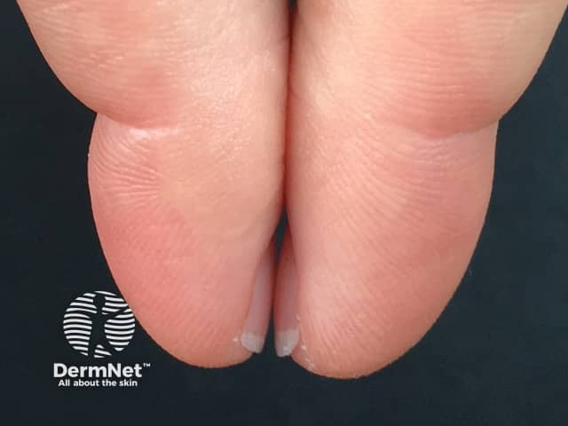
Imaging modality: Clinical photography (dermatology/nail assessment) using a macro-level optic lens. Technique: dorsal view of the fingertips with the index and neighboring digits pressed together to evaluate the Schamroth window sign. The image emphasizes the nail plates, nail beds, and distal phalanges, with attention to the diamond-shaped gap that should appear between opposed nails in healthy individuals. In this photograph the fingertips are oriented to allow direct appraisal of the Schamroth window, facilitating rapid assessment for digital clubbing. Key visual features include nail bed rounding, convex terminal phalanges, and altered relative positioning of the nail folds. Pathology-oriented interpretation centers on clubbing physiology: when the diamond window is reduced or abolished, suggesting acquired or hereditary digital clubbing; otherwise a preserved window is reassuring. Diagnostic significance lies in correlating visible nail changes with systemic disease risk, particularly chronic cardiopulmonary disorders (interstitial lung disease, COPD, cystic fibrosis, lung neoplasms) and certain hepatic conditions. Differential considerations include a normal variant of nail morphology, hypertrophic osteoarthropathy, and secondary clubbing due to chronic inflammation. Clinical correlation should trigger evaluation for breathlessness, hypoxia, and chest or abdominal pathology, with follow-up imaging (chest radiography/CT) and laboratory workup as indicated. Uses: educational demonstrations, bedside assessment, and longitudinal monitoring of clubbing progression or resolution.
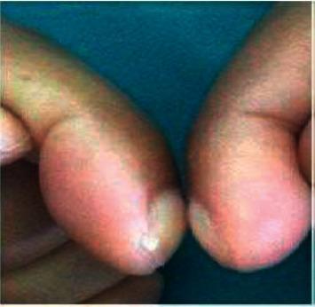
This clinical photograph displays a close-up view of two fingers positioned dorsal surface to dorsal surface to assess for digital clubbing. The image demonstrates a classic presentation of clubbing, characterized by the bulbous, drumstick-like enlargement of the distal phalanges. There is a visible increase in the longitudinal and transverse curvature of the nails, along with the softening of the nail bed. Crucially, the image illustrates a positive Schamroth's sign, identified by the complete absence of the normal diamond-shaped window (Schamroth's window) between the opposed nail beds. This physical finding is a hallmark of hypertrophic osteoarthropathy (HOA) and can be associated with chronic underlying conditions such as pulmonary malignancy, cystic fibrosis, or cyanotic heart disease. The photograph serves as an educational reference for medical trainees to recognize morphological fingertip changes and the clinical examination technique for thoracic and systemic disease markers.
Clinical Examination of the Respiratory System
As Per Indian Medical Curriculum (Harrison's, Macleod's Framework)
"A systematic physical examination is critical for accurate diagnosis. Even in young adults, persistent respiratory symptoms are associated with a greater likelihood of developing chronic lung disease." - Harrison's Principles of Internal Medicine, 22E
SEQUENCE OF EXAMINATION
- General examination (from foot of bed)
- Inspection of chest
- Palpation
- Percussion
- Auscultation
- Voice sounds
PART 1: GENERAL EXAMINATION
A. Examination from the End of the Bed ("End of Bed Look")
- Breathlessness at rest - can the patient complete a sentence?
- Respiratory rate (normal: 12-20/min)
- Respiratory pattern - labored, paradoxical, use of accessory muscles
- Position - tripod position (sitting, hands on knees), orthopnea (heart failure), tachypnea
- Consciousness - confusion = hypoxia or hypercapnia
B. Hands
| Sign | Description | Significance |
|---|---|---|
| Clubbing | Bulbous enlargement of distal phalanges, loss of nail-fold angle, increased sponginess | Lung cancer, bronchiectasis, ILD, cyanotic CHD, lung abscess, CF |
| Peripheral cyanosis | Bluish discoloration of fingertips + nail beds | Reduced peripheral perfusion |
| Peripheral tremor / asterixis | Flapping tremor (CO₂ narcosis) | Type II respiratory failure (hypercapnia) |
| Nicotine staining | Yellow-brown staining of index and middle fingers | Chronic smoking |
| Warm, bounding pulse | Vasodilation | CO₂ retention |
Schamroth's Window Test for Clubbing
Grading of Clubbing (Indian Textbook Standard - Schamroth Grades)
| Grade | Features |
|---|---|
| Grade 1 | Increased sponginess / fluctuation of nail bed; loss of normal angle at nail base |
| Grade 2 | Obliteration of Lovibond angle (normal nail-bed-to-finger angle = 160°; clubbing = ≥180°) |
| Grade 3 | Drumstick appearance - bulbous enlargement of all distal phalanges |
| Grade 4 | Hypertrophic Pulmonary Osteoarthropathy (HPOA) - periosteal new bone formation, painful swollen joints |
Causes of Clubbing - Mnemonic: CLUBBING
- Cardiac: cyanotic congenital heart disease, infective endocarditis
- Lung cancer (most common acquired cause)
- Ulcerative colitis, Crohn's disease
- Bronchiectasis, lung abscess, cystic fibrosis, TB
- Biliary cirrhosis, hepatic cirrhosis
- Idiopathic / hereditary (no disease)
- Nothing (unilateral clubbing = local vascular disease)
- Gastrointestinal: inflammatory bowel disease
Harrison's 22E notes: "Clubbing may be associated with hypertrophic osteoarthropathy - subperiosteal formation of new bone in the distal diaphyses of the long bones, causing pain and symmetric arthritis-like changes in shoulders, knees, ankles, wrists, and elbows."
C. Central Cyanosis
- Check tongue and mucous membranes (not just lips - lips can appear cyanotic in cold weather)
- Central cyanosis = visible when deoxyhemoglobin >5 g/dL in capillaries; SpO₂ typically <85%
| Feature | Central | Peripheral |
|---|---|---|
| Site | Tongue, lips, mucous membranes | Fingers, toes, earlobes |
| Temperature | Warm | Cold |
| Cause | Cardiorespiratory disease, Hb abnormality | Cold, poor cardiac output, venous obstruction |
| Warming test | Persists after warming | Disappears after warming |
| Clubbing | May be present | Absent |
- Respiratory: massive pneumonia, massive effusion, ARDS, severe COPD, status asthmaticus
- Cardiac: cyanotic CHD (Fallot's tetralogy, Eisenmenger's), pulmonary AV fistula
- Hb abnormalities: metHb, sulfHb (need spectroscopy)
D. Face and Neck
- JVP elevation - cor pulmonale (right heart failure from lung disease); fixed raised JVP = SVC obstruction
- Pursed-lip breathing - COPD (prolongs expiration, provides auto-PEEP)
- Supraclavicular lymph nodes - Virchow's node (left side), mycobacterial infection (TB), lung cancer
- Horner's syndrome (miosis, ptosis, anhidrosis) - Pancoast tumor (apex lung cancer)
- Wasting - malignancy, TB
- Plethoric face, dilated neck veins - SVC syndrome
PART 2: INSPECTION OF THE CHEST
A. Chest Shape and Deformities
| Deformity | What You See | Cause | Note |
|---|---|---|---|
| Barrel chest | AP diameter ≈ transverse; horizontal ribs | Emphysema, COPD | Normal in infants; abnormal in adults |
| Pectus excavatum (funnel chest) | Sternum depressed inward | Usually congenital | Rarely causes dyspnea |
| Pectus carinatum (pigeon chest) | Sternum protruding forward | Chronic childhood asthma, rickets | |
| Harrison's sulcus | Horizontal groove at costal margin along diaphragm attachment | Severe childhood asthma, rickets | Classic Indian exam question |
| Kyphosis | Forward curvature of spine | TB spine, osteoporosis | |
| Scoliosis | Lateral curvature of spine | Idiopathic, neuromuscular | Restrictive lung disease |
| Kyphoscoliosis | Combined | Marfan syndrome, childhood polio | Severe restrictive defect, cor pulmonale |
B. Chest Wall - Additional Inspection
- Surgical scars: lobectomy (posterolateral thoracotomy scar), median sternotomy
- Chest drain sites / tube thoracostomy scars
- Dilated superficial veins: SVC obstruction (flow downward), IVC obstruction (flow upward)
- Subcutaneous swellings: empyema necessitans (TB empyema tracking out through chest wall) - classic Indian finding
- Skin lesions: spider nevi (liver disease), erythema nodosum (sarcoidosis, TB), lupus pernio
C. Respiratory Pattern and Rate
| Pattern | Description | Cause |
|---|---|---|
| Normal (eupnea) | 12-20/min, regular | Normal |
| Tachypnea | >20/min | Pneumonia, PE, fever, anxiety, metabolic acidosis |
| Bradypnea | <12/min | Opioids, sedatives, hypothyroidism |
| Cheyne-Stokes | Crescendo-decrescendo with central apnea | Heart failure, uraemia, CNS disease |
| Kussmaul | Deep, rapid, sighing | Metabolic acidosis (DKA, uraemia) - classic Indian exam |
| Biot's | Irregular with random apnoeic pauses | Raised ICP, meningitis, brainstem lesion |
| Apneustic | Prolonged inspiratory hold | Pontine lesion |
D. Movement of Chest Wall
- Observe both sides from front, back, and side simultaneously
- Bilateral reduced movement: COPD, fibrosis, bilateral pleural effusion
- Unilateral reduced movement (lagging): pleural effusion, pneumothorax, collapse/consolidation on that side
E. Use of Accessory Muscles
- Sternocleidomastoid, scalenes, intercostals contracting visibly = increased work of breathing
- Intercostal recession (indrawing) = inspiratory obstruction or stiff lungs
- Subcostal recession = infants with respiratory distress
- Tracheal tug (Lemoyne's sign) = severe obstruction (SVC syndrome, tension pneumothorax)
PART 3: PALPATION
A. Tracheal Position
| Trachea | Meaning |
|---|---|
| Central | Normal, or conditions affecting both sides equally |
| Shifted toward lesion | Collapse/atelectasis (lung volume loss pulls), upper lobe fibrosis |
| Shifted away from lesion | Massive pleural effusion (>1 liter), tension pneumothorax, large mediastinal mass |
Important Indian exam pearl: In simple consolidation (pneumonia), trachea is central (no volume loss). In collapse, it shifts toward the side. In effusion, shifts away.
B. Apex Beat
- Normally in the 5th left intercostal space, mid-clavicular line
- Shifted laterally/inferiorly = cardiomegaly (cor pulmonale, dilated cardiomyopathy)
- Impalpable = emphysema (hyperinflation), large effusion
C. Chest Expansion
- Normal: thumbs move equally outward; excursion of 3-5 cm
- Reduced unilaterally: consolidation, effusion, pneumothorax, collapse on that side
- Reduced bilaterally: emphysema, bilateral fibrosis, ankylosing spondylitis
D. Vocal Fremitus (Tactile Fremitus)
| Fremitus | Clinical Condition | Mechanism |
|---|---|---|
| Increased | Consolidation (lobar pneumonia), lung collapse with patent airway | Solid medium (consolidated lung) transmits vibrations better |
| Decreased / absent | Pleural effusion | Fluid between lung and chest wall dampens vibrations |
| Decreased / absent | Pneumothorax | Air between lung and chest wall dissipates vibrations |
| Decreased | Emphysema, bronchial obstruction | Reduced air movement and lung tissue density |
| Pleural friction fremitus | Pleuritis | Palpable grating vibration during respiration |
E. Other Palpation Findings
- Point tenderness on ribs: rib fracture, bone metastases
- Costochondral tenderness: Tietze's syndrome (costochondritis) - common cause of atypical chest pain in young adults
- Crepitus (subcutaneous emphysema): crackling sensation on palpation; air in soft tissues (pneumothorax, post-intubation, post-trauma)
- Cervical rib: bony prominence in supraclavicular fossa - may cause TOS
- Empyema necessitans: fluctuant swelling on chest wall (common in India with TB)
- Spinal tenderness: TB spine (Pott's disease), an important Indian condition
PART 4: PERCUSSION
"Percussion of the thorax over normal air-containing lung produces a resonant note. The response to percussion is impaired whenever something other than air-filled lung lies beneath the chest wall." - Fishman's Pulmonary Diseases
Technique
- Pleximeter: middle finger of non-dominant hand placed firmly against the chest wall (only the middle phalanx touching)
- Plexor: tip of middle finger of dominant hand strikes a brisk, sharp blow (wrist movement only, not arm)
- The sound is both heard (pitch) and felt (vibration)
- Percuss systematically: apex → base, comparing both sides
- Minimum: 3 areas posteriorly (upper, mid, lower), 2 anteriorly (upper, lower)
- Avoid percussing over bone (scapula, ribs)
Percussion Notes
| Note | Sound Quality | Cause |
|---|---|---|
| Resonant | Clear, hollow, low-pitched | Normal air-filled lung |
| Hyperresonant | Louder, lower-pitched than normal | Pneumothorax, emphysema, large bulla |
| Tympanitic | Drum-like, very hollow | Tension pneumothorax (extreme hyperresonance) |
| Dull | Short, high-pitched, low intensity | Consolidation, atelectasis, pleural thickening |
| Stony dull | Absolute dullness, like percussing the thigh | Pleural effusion - pathognomonic sign |
Diaphragmatic Excursion (Important Indian Exam Topic)
- Technique: With patient holding a deep breath - percuss downward from resonant to dull = lower edge of lung at full inspiration
- With patient holding after full expiration - percuss upward from dull to resonant = position at expiration
- Normal excursion = 3-5 cm (approximately 2 intercostal spaces)
- Reduced: emphysema (flat diaphragm), pleural effusion, diaphragm palsy, diaphragm elevation
- Absent movement: diaphragm palsy
Important Percussion Findings in Indian Context
- Stony dull percussion at the base
- Upper border of dullness = Ellis-Damoisseau curve (highest in axilla, lower medially and laterally) - an examiner favorite question
- Above the effusion: may get Skodaic resonance (paradoxical hyperresonance above large effusion due to relaxed/compressed lung)
- On the opposite side of a large effusion: Grocco's triangle (triangle of dullness paravertebral on opposite side due to mediastinal shift)
PART 5: AUSCULTATION
"Ever since the time of Laennec, physicians have applied a stethoscope to the chest in search of sounds of disease." - Murray & Nadel
Technique
- Use the diaphragm of the stethoscope for breath sounds (high-pitched sounds)
- Use the bell for low-pitched sounds (rhonchi)
- Auscultate anteriorly (2 areas/side) + laterally (1 area/side) + posteriorly (3 areas/side) = minimum 12 areas
- Ask patient to breathe deeply through the open mouth
- Compare same zones bilaterally before moving down
- Listen during both inspiration and expiration
- Do not auscultate within 3 cm of the midline anteriorly or posteriorly (may pick up tracheal sounds directly)
Normal Breath Sounds
| Sound | Location | Character | Ratio (I:E) |
|---|---|---|---|
| Vesicular | Peripheral lung (most of lung fields) | Soft, low-pitched, gentle rustling; inspiration longer than expiration; no gap between I and E | 3:1 |
| Bronchovesicular | 1st-2nd ICS anteriorly, between scapulae | Intermediate pitch and intensity; equal I and E | 1:1 |
| Bronchial (tubular) | Over trachea, manubrium | Loud, high-pitched, hollow; gap between I and E; expiration longer than inspiration | 1:2 |
Key Indian exam point: If bronchial breathing is heard over the lung field (not trachea), it is always abnormal and indicates consolidation, compression, or cavitation.
Abnormal (Adventitious) Sounds
1. Crackles (Old term: Rales - no longer used)
| Type | Character | Timing | Cleared by cough? | Common Causes in India |
|---|---|---|---|---|
| Fine crackles | High-pitched, brief, non-musical, like crackling cellophane | Late inspiration | No | Heart failure, ILD (pulmonary fibrosis), early pneumonia |
| Coarse crackles | Low-pitched, bubbling, "wet" sounds | Inspiration and early expiration | Yes | Bronchitis, COPD, bronchiectasis, pulmonary oedema |
Pearl: Fine crackles at the bases that do not clear with coughing and have a "Velcro quality" = interstitial pulmonary fibrosis. Important differentiating sign.
2. Wheeze
- High-pitched, musical, continuous sound (>250 ms), predominantly expiratory
- Mechanism: rapid airflow through narrowed airways → wall oscillation
| Type | Description | Cause |
|---|---|---|
| Polyphonic wheeze | Multiple pitches simultaneously | Diffuse airway narrowing (asthma, COPD) |
| Monophonic wheeze | Single pitch, same throughout | Single obstructed airway (endobronchial tumor, foreign body) |
| Fixed monophonic | Unchanging, same site | Tumour, fixed obstruction |
| Silent chest | No wheeze despite obvious respiratory distress | Very severe asthma (life-threatening - not enough airflow to generate wheeze) |
3. Rhonchi
- Low-pitched, snoring/sonorous, musical sounds, mainly expiratory
- Cleared by coughing (generated by secretions in large airways)
- Common in: bronchitis, COPD, bronchiectasis
4. Pleural Friction Rub
- Leathery, creaking, grating sound
- Heard in BOTH inspiration AND expiration
- Does NOT change with coughing
- Does change with respiration (disappears when patient holds breath) - differentiates from pericardial rub
- Causes: pleuritis (pneumonia + pleuritis, TB pleuritis, malignant pleural infiltration, PE with infarction, connective tissue diseases - SLE, RA)
- Palpable rub also felt on chest wall in some patients
- Occasionally heard even in large effusions
5. Stridor
- High-pitched, harsh, predominantly inspiratory continuous sound
- Loudest over the neck/larynx (NOT chest - differentiates from wheeze)
- Causes: croup (children - common in India), epiglottitis, foreign body (children), bilateral vocal cord palsy, laryngeal/tracheal tumor, post-extubation edema, anaphylaxis
- Emergency sign - requires immediate assessment
| Stridor | Wheeze | |
|---|---|---|
| Phase | Predominantly inspiratory | Predominantly expiratory |
| Loudest site | Neck/upper airway | Chest |
| Origin | Extrathoracic large airway | Intrathoracic small airways |
6. Bronchial Breath Sounds at the Periphery (Pathological Bronchial Breathing)
- Consolidation (air replaced by fluid/exudate, but airway patent) - most common cause = lobar pneumonia
- Collapse with patent bronchus
- Cavity (resonance effect)
- Top of large pleural effusion (compressed lung above fluid conducts bronchial sounds)
PART 6: VOICE SOUNDS (Vocal Resonance)
| Sign | Method | Abnormal Finding | Meaning |
|---|---|---|---|
| Vocal resonance | Ask patient to say "ninety-nine" | Louder/clearer than normal | Consolidation |
| Bronchophony | Ask patient to say "ninety-nine" | Words sound louder and clearer ("ninety-nine" distinctly heard) | Consolidation |
| Whispering pectoriloquy | Ask patient to whisper "one, two, three" | Whispered words clearly transmitted | Consolidation (highly specific sign) |
| Aegophony (E to A change) | Ask patient to say "eee" slowly | Heard as "aaa" bleating/nasal quality | Upper border of pleural effusion (bronchophony in compressed/relaxed lung just above fluid) |
Key point: All three (bronchophony, whispering pectoriloquy, aegophony at upper border) = increased transmission through solid medium = consolidation or compressed lung above effusion.Over pleural effusion: voice sounds are reduced or absent (fluid blocks transmission).
PART 7: COMPLETE CLINICAL SYNDROME TABLE
| Condition | Inspection | Trachea | Expansion | Fremitus | Percussion | Breath Sounds | Added Sounds | Voice Sounds |
|---|---|---|---|---|---|---|---|---|
| Normal | Normal | Central | Equal | Normal | Resonant | Vesicular | None | Normal |
| Consolidation (Lobar pneumonia) | Reduced movement on affected side | Central | Reduced on affected side | Increased | Dull | Bronchial | Fine crackles | Increased (bronchophony, whispering pectoriloquy) |
| Pleural Effusion | Fullness on affected side | Away from affected side (large) | Absent/greatly reduced | Absent | Stony dull | Absent (reduced to absent) | Pleural rub (occasionally); aegophony at upper border | Absent below; aegophony at upper border |
| Pneumothorax | Reduced movement on affected side | Away from affected side (tension) | Reduced/absent | Absent | Hyperresonant/tympanitic | Absent | None | Absent |
| Collapse/Atelectasis | Reduced movement; rib indrawing | Toward affected side | Reduced | Reduced | Dull | Absent or bronchial (if bronchus patent) | None | Reduced |
| Emphysema/COPD | Barrel chest; accessory muscles; pursed lips | Central | Reduced bilaterally | Reduced | Hyperresonant bilaterally | Reduced (vesicular); prolonged expiration | Wheeze; coarse crackles | Reduced |
| Pulmonary Fibrosis (ILD) | Reduced movement bilateral | Central or deviated to worse side | Reduced bilateral | Variable | Dull | Reduced vesicular | Fine "Velcro" crackles at bases | Variable/reduced |
| Asthma (Acute) | Hyperinflation; accessory muscles | Central | Reduced bilateral | Reduced | Hyperresonant (air trapping) | Prolonged expiration; reduced | Polyphonic wheeze (or silent chest in severe) | Normal |
| Bronchiectasis | May have clubbing; purulent sputum | Central | Reduced | Variable | Normal/dull | Bronchial (if consolidated) | Coarse crackles (not cleared easily); rhonchi | Increased if consolidation |
| Cavity (TB/Lung abscess) | Reduced movement | Central | Reduced on affected side | Increased | Amphoric resonance (hollow, resonant) | Amphoric/cavernous breathing | Crackles; amphoric rub | Whispering pectoriloquy; amphoric voice |
PART 8: ASSOCIATED CONCEPTS (Indian Exam Favourites)
1. Mediastinal Shift Rules
- Trachea pulled toward = volume loss: collapse, fibrosis, post-surgical lung removal
- Trachea pushed away = space occupation: large effusion (>1000 mL), tension pneumothorax, large mass
2. Massive Pleural Effusion (Common in India - TB, malignancy)
3. Empyema Necessitans (India-specific - TB)
- Empyema (pus in pleural space) that bursts through chest wall
- Fluctuant swelling palpable on chest wall
- Associated signs of TB: wasting, evening fever, night sweats, hilar lymphadenopathy on CXR
4. Pott's Disease (TB Spine) with Respiratory Signs
- Gibbus deformity (angular kyphosis)
- Spinal tenderness on palpation
- Restrictive lung disease due to kyphoscoliosis
- Associated psoas abscess (flank swelling)
5. Cor Pulmonale (Right Heart Failure from Lung Disease)
- Elevated JVP (can be pulsatile if tricuspid regurgitation)
- Right parasternal heave (RV hypertrophy)
- Loud P2 (pulmonary hypertension)
- Ankle/sacral oedema
- Hepatomegaly, ascites
- Causes: COPD (most common in India), pulmonary fibrosis, recurrent PE, kyphoscoliosis
6. Superior Vena Cava (SVC) Syndrome
- Fixed, raised, non-pulsatile JVP
- Dilated neck and chest wall veins (flow downward toward abdomen)
- Oedema of face, neck, arms (worse on waking)
- Plethoric, cyanotic facies
- Causes: Lung cancer (right upper lobe - most common), lymphoma, mediastinal fibrosis (TB), thymoma
7. Pancoast Tumor (Apex Lung Cancer)
- Horner's syndrome (sympathetic chain involvement): unilateral ptosis, miosis, anhidrosis, enophthalmos
- Brachial plexus involvement: pain/weakness/wasting of hand and arm (T1, C8)
- Rib destruction (1st/2nd ribs)
8. Tracheal Deviation - Summary
| Condition | Trachea |
|---|---|
| Large pleural effusion | Away |
| Tension pneumothorax | Away |
| Collapse/atelectasis | Toward |
| Fibrosis | Toward |
| Large mediastinal mass | Away (if one side) |
| Pneumonia (simple) | Central |
| Small effusion | Central |
CLINICAL SKILLS PEARLS FOR INDIAN EXAMS
- "Stony dull" = pleural effusion - this specific modifier differentiates from simple "dull" (consolidation)
- Aegophony at the upper border of an effusion is one of the most specific clinical signs in medicine
- Trachea in pneumonia is central - a common trap in viva questions
- Silent chest in asthma = life-threatening - do not be falsely reassured by absence of wheeze
- Empyema necessitans is a classic Indian scenario (TB empyema) - palpable fluctuant chest wall mass
- Whispering pectoriloquy is more specific for consolidation than bronchophony
- Clubbing + TB - think bronchiectasis, lung abscess, empyema, not TB itself (TB alone rarely causes clubbing)
- Pleural rub vs pericardial rub: ask patient to hold breath - rub disappearing = pleural; persisting = pericardial
- Kussmaul breathing (deep, sighing) = metabolic acidosis; common in DKA, renal failure, sepsis
- Fine crackles not clearing with cough = always structural (ILD/heart failure), not secretions