
Taut meaning
Taut means stretched tight, with no slack or looseness.
Core meanings:
- Physical - pulled or drawn tight: "The rope was taut." / "Her muscles were taut."
- Tense/stressed - showing strain or anxiety: "His face was taut with worry."
- Well-organized/efficient (of writing, a story, a ship) - concise, trim, with no wasted elements: "A taut thriller." / "A taut narrative."
Pronunciation: /tɔːt/ (rhymes with caught, taught)
Do not confuse with:
- Taught - past tense of teach (same pronunciation, different spelling/meaning)
- Taut vs slack - opposites
Quick examples:
- "The sailor kept the rigging taut."
- "She felt taut with nervous energy."
- "Critics praised the film's taut screenplay."
Flexion of knee pictures
Knee flexion is the movement that decreases the angle between the thigh and lower leg - essentially bending the knee. Let me pull up relevant images from the medical library.
Searching sources...
Reading File
Finding Sources
Searching the Web
knee flexion anatomy diagram muscles range of motion
Reading File
Loading Image
Loading Image
Loading Image
Loading Image
Now let me also grab the textbook diagram showing flexion/extension axis from the same section:
Reading File
Loading Image
Here is a visual overview of knee flexion with images from the THIEME Atlas of Anatomy:
Knee Flexion
Flexion = bending the knee, decreasing the angle between the thigh and the lower leg (bringing the heel toward the buttock).
Range of Motion

- Flexion: 120-150° (active); up to 160° passively
- Extension (hyperextension): 5-10° beyond neutral (0°)
Axis of Flexion / Extension and Cruciate Ligaments
From the THIEME Atlas of Anatomy (General Anatomy & Musculoskeletal System):
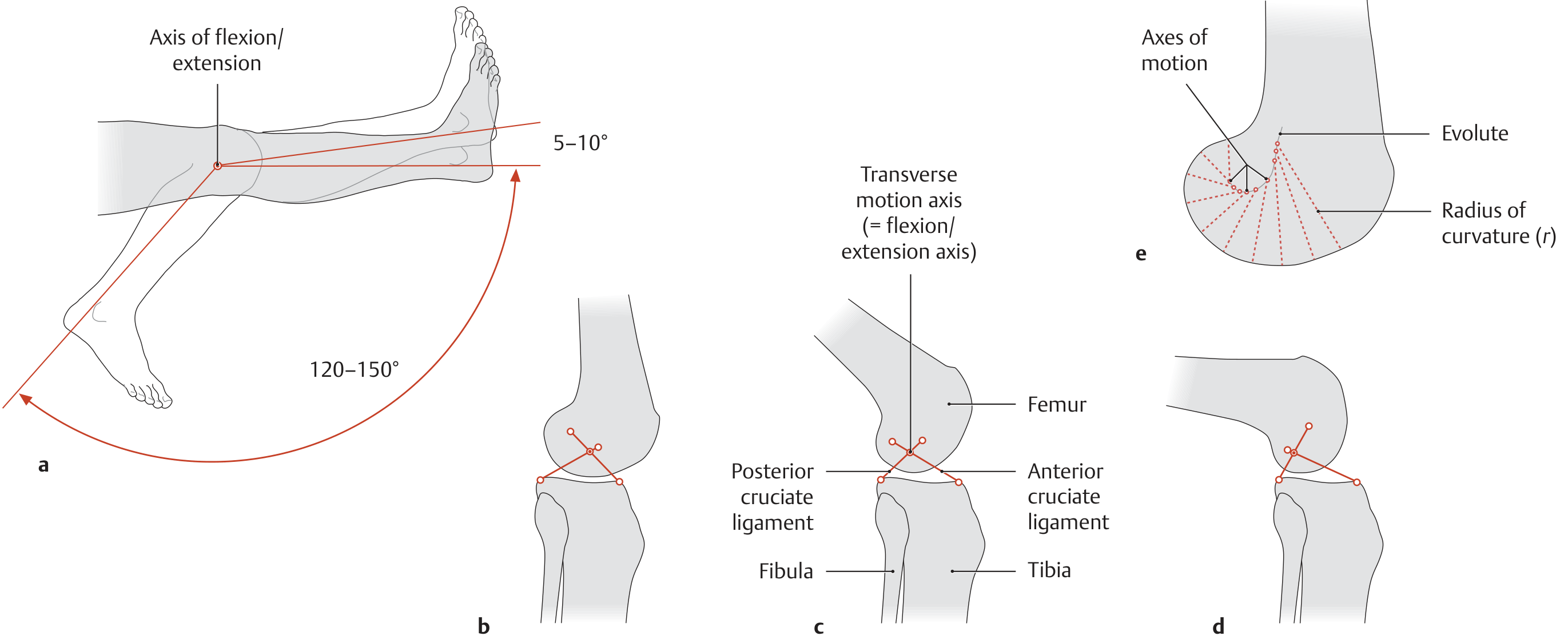
Key points from this diagram:
- Flexion/extension takes place around a transverse axis that passes through the dynamic center of rotation
- That center is where the collateral and cruciate ligaments intersect
- During flexion, the axis moves upward and backward along a curved path (the evolute), due to the changing radius of curvature of the femoral condyle
- This is why the knee is not a pure hinge - it also involves a rolling and gliding motion
Tibial Rotation with the Knee Flexed (the "Screw-Home" Mechanism)
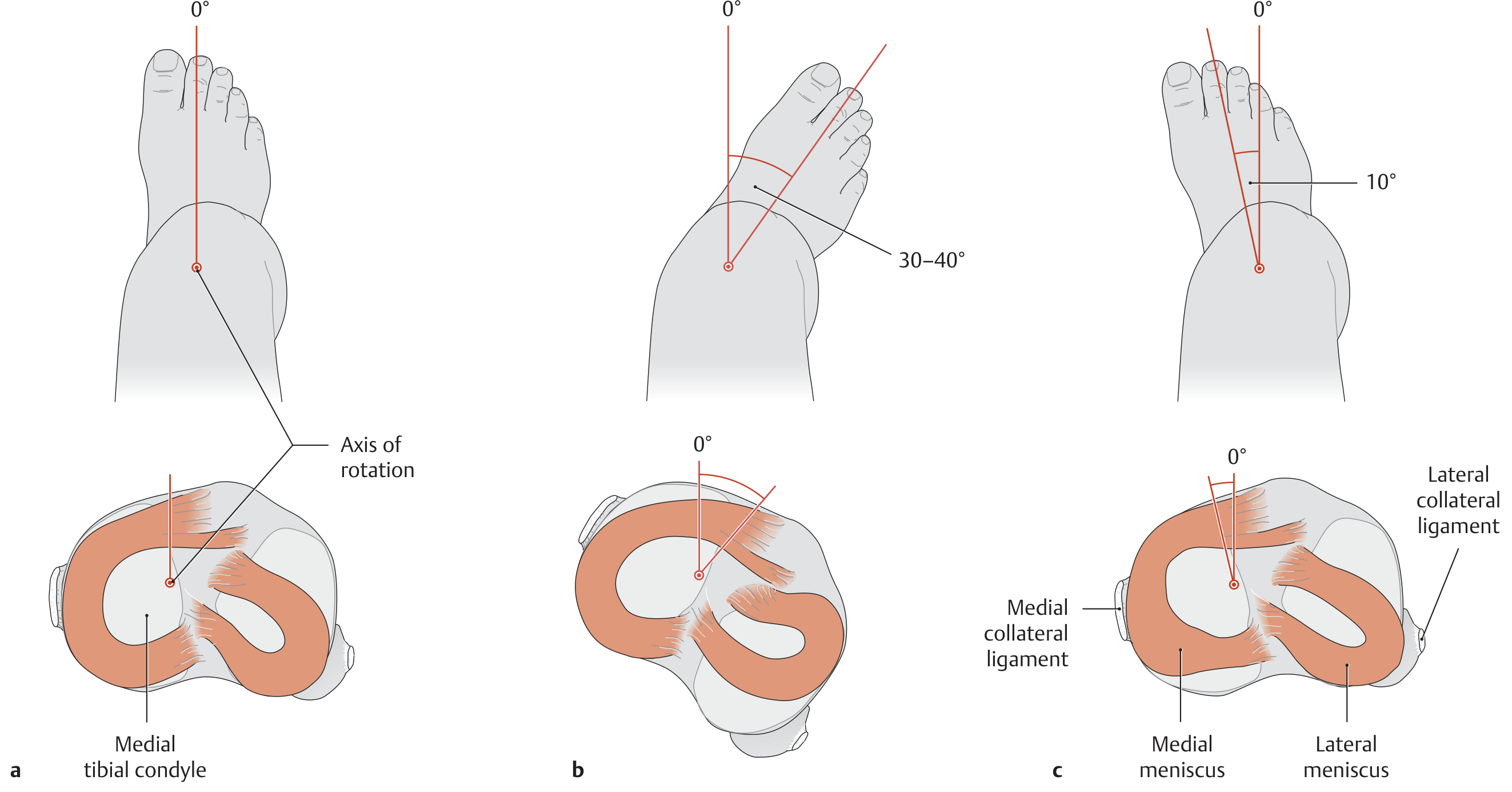
With the knee flexed to 90°:
- External rotation: 30-40°
- Internal rotation: ~10° only (cruciate ligaments wrap around each other, limiting it)
- The rotation axis runs through the medial tibial condyle
Muscles Producing Knee Flexion
| Muscle | Notes |
|---|---|
| Biceps femoris (long + short heads) | Primary flexor |
| Semitendinosus | Medial hamstring |
| Semimembranosus | Medial hamstring |
| Sartorius | Also flexes hip |
| Gracilis | Also adducts hip |
| Popliteus | Initiates flexion from full extension ("unlocks" the knee) |
| Gastrocnemius | Accessory flexor |
Knee extensors (opposing flexion): The quadriceps group (rectus femoris, vastus lateralis, vastus medialis, vastus intermedius) via the patellar tendon.
Source: General Anatomy and Musculoskeletal System, THIEME Atlas of Anatomy; TeachMeAnatomy - Knee Joint
What is EHB in dorsalis pedis artery vishram singh
EHB stands for Extensor Hallucis Brevis - a small intrinsic muscle of the foot that arises from the dorsum of the calcaneus and is closely related to the dorsalis pedis artery.
Searching sources...
Reading File
Finding Sources
Finding Sources
Searching the Web
EHB extensor hallucis brevis dorsalis pedis artery Vishram Singh anatomy
Reading File
Finding Sources
Finding Sources
Loading Image
Now let me look specifically at the Vishram Singh context - the question is about how EHB (Extensor Hallucis Brevis) relates to the dorsalis pedis artery as a surgical/anatomical landmark. Let me get more specific content:
Reading File
I now have enough information to give a thorough answer. The library doesn't have Vishram Singh's textbook directly, but the anatomy is well-covered across multiple sources.
EHB in Relation to the Dorsalis Pedis Artery (Vishram Singh context)
EHB = Extensor Hallucis Brevis
In Vishram Singh's anatomy (Lower Limb), EHB is used as an important surgical/surface landmark for locating and accessing the dorsalis pedis artery on the dorsum of the foot.
Key Anatomical Relationship
The dorsalis pedis artery (continuation of the anterior tibial artery) runs on the dorsum of the foot. Its relations are:
| Structure | Position relative to artery |
|---|---|
| Extensor Hallucis Longus (EHL) tendon | Medial |
| Extensor Digitorum Longus (EDL) to 2nd toe | Lateral |
| Extensor Hallucis Brevis (EHB) | Superficial / overlying the artery |
| Deep peroneal nerve | Immediately lateral |
| Accompanying veins | One on each side |
The dorsalis pedis artery passes deep to (under) the EHB muscle belly as it crosses the tarsal bones. This is why EHB is specifically highlighted by Vishram Singh - it is the superficial landmark you retract or pass under to expose the artery.
Why EHB Matters Clinically
-
Pulse palpation - the dorsalis pedis pulse is felt on the dorsum of the foot, between the EHL tendon and EDL tendon to the 2nd toe, with EHB lying just superficial to the artery at this point.
-
Surgical exposure - when exposing the dorsalis pedis artery (e.g., for bypass graft, flap harvest, or tarsometatarsal joint surgery), EHB must be retracted or divided to access the vessel.
-
Surface marking - EHB acts as a reliable soft-tissue marker for identifying the artery intraoperatively.
Dorsalis Pedis Artery - Full Course and Branches
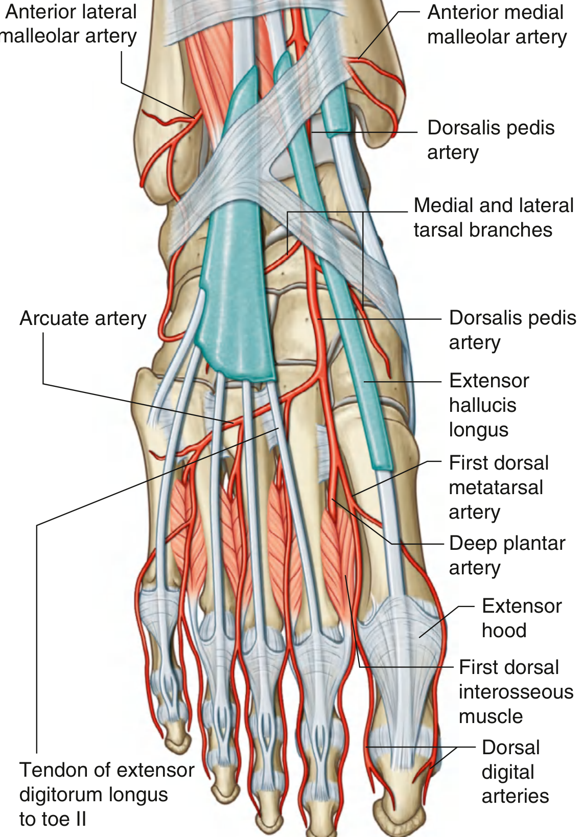
Course: Anterior tibial artery → crosses ankle joint → passes over dorsum of talus, navicular, intermediate cuneiform → dips between the two heads of 1st dorsal interosseous → becomes deep plantar artery → joins deep plantar arch
Branches:
- Medial and lateral tarsal arteries
- Arcuate artery (gives 2nd, 3rd, 4th dorsal metatarsal arteries)
- 1st dorsal metatarsal artery
- Deep plantar artery (terminal branch)
In short: In Vishram Singh's description, EHB (Extensor Hallucis Brevis) is the muscle belly that lies directly superficial to the dorsalis pedis artery on the dorsum of the foot, making it a key anatomical landmark for palpating the pulse and surgically exposing the vessel.
- Gray's Anatomy for Students, p. 765
- Campbell's Operative Orthopaedics 15th Ed, Neurovascular Anatomy section
This is a shared conversation. Sign in to Orris to start your own chat.