
teach me aortic stenosis starting from rheumatic heart disease to surgical management, mch cardiac surgery level in detailed from standard textbooks and literature, pictures lacked notes plz
AORTIC STENOSIS
MCh Cardiac Surgery Level - Comprehensive Notes
Sources: Braunwald's Heart Disease, Schwartz's Principles of Surgery, Sabiston Textbook of Surgery, Fuster & Hurst's The Heart, Textbook of Clinical Echocardiography
PART 1: RHEUMATIC HEART DISEASE - THE INFLAMMATORY ORIGIN
Acute Rheumatic Fever (ARF) - The Trigger
- Molecular mimicry is the driving pathologic process - streptococcal M-protein epitopes structurally resemble host cardiac proteins (particularly laminin, an extracellular matrix protein in cardiac valves)
- Cross-reactive T-cell and antibody responses attack the endocardium, myocardium, and pericardium 2-6 weeks after the streptococcal infection
- The immune response targets the valve leaflets, producing acute rheumatic valvulitis
Pathology of Acute Rheumatic Valvulitis
- Aschoff bodies - pathognomonic granulomatous lesions with central fibrinoid necrosis surrounded by Anitschkow cells (caterpillar cells - activated macrophages) and multinucleated Aschoff giant cells
- Verrucous endocarditis - small (1-2 mm) sterile vegetations along the line of closure of valve leaflets
- The mitral valve is most commonly affected (65-70%), followed by combined mitral + aortic (25%), isolated aortic (rare acutely)
- In a small minority, this acute response causes clinically evident inflammation severe enough to cause acute severe MR requiring surgical "peeling" of inflammatory material off the valve
Chronic Rheumatic Heart Disease - The Long Game
- Repeated bouts of inflammation → commissural fusion
- Progressive thickening, fibrosis, and calcification of valve leaflets
- Chordal fusion and shortening (primarily mitral)
- Net result: combined stenosis and regurgitation (mixed lesion) is most common
- Isolated stenosis develops over 10-40 years
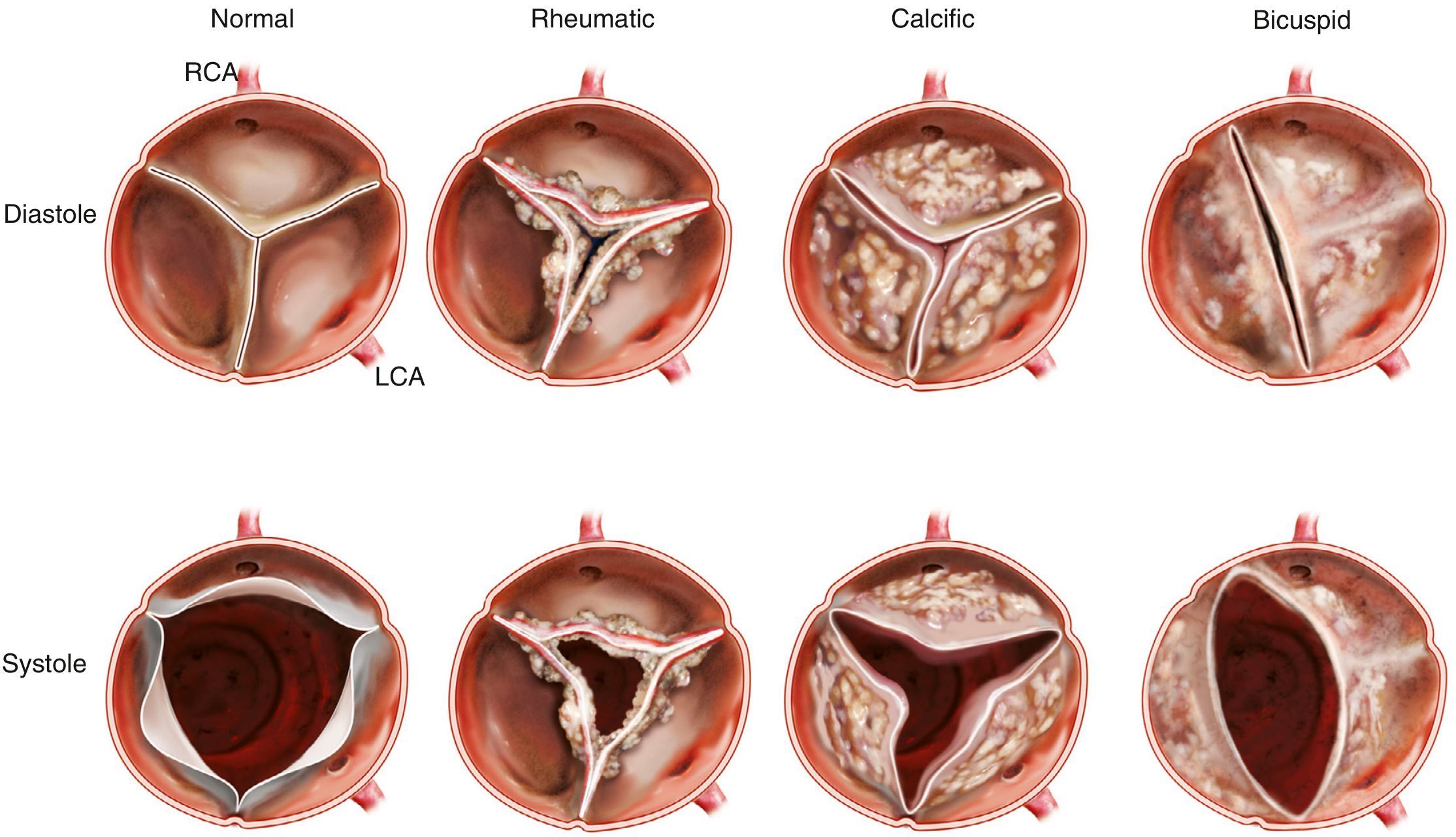
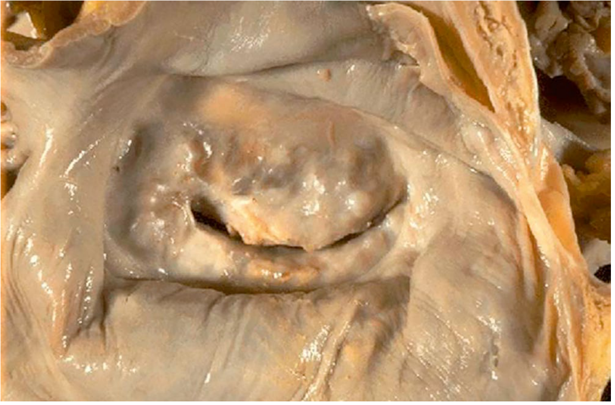
| Feature | Rheumatic AS | Calcific (Degenerative) AS |
|---|---|---|
| Age of presentation | 40s-60s | 70s-80s |
| Commissural fusion | YES - pathognomonic | No |
| Calcification pattern | Leaflet tips + commissures | Base of leaflets, aortic side |
| Echo systolic orifice shape | Triangular | Stellate/irregular |
| Mitral valve involvement | Almost always present | Incidental/separate |
| Leaflet architecture | All 3 cusps fused together | Independent leaflet stiffening |
| Regurgitation | Common (mixed lesion) | Less common |
PART 2: ETIOLOGY OF AORTIC STENOSIS - COMPLETE CLASSIFICATION
1. Calcific (Degenerative) AS - Most Common in Developed World
- Affects ~2% of adults >65 years with tricuspid valves; presents 7-8th decade
- Begins as aortic sclerosis (focal leaflet thickening without obstruction) in ~25% of adults >65 years
- Progressive fibrocalcification of the aortic side of the leaflets, predominantly at the base
- Risk factors mirror atherosclerosis: age, male sex, hypertension, hyperlipidemia, smoking, diabetes
- No commissural fusion (key distinction from rheumatic)
- Stellate orifice in systole due to independent stiffening of all three cusps
2. Bicuspid Aortic Valve (BAV) - Most Common Cause of AS in Young Adults
- Prevalence: ~1-2% of general population; male:female = 3:1
- Accounts for two-thirds of severe AS in adults <70 years and one-third in >70 years
- Due to accelerated calcification of a congenitally abnormal valve
- Right-left cusp fusion (most common, ~75%), right-noncoronary fusion, left-noncoronary fusion (rarest)
- Presents 1-2 decades earlier than calcific tricuspid AS
- Often associated with aortopathy (dilation of sinuses of Valsalva, ascending aorta) due to abnormal fibrillin/elastin in aortic wall - requires surveillance and potential concomitant aortic replacement
- Echo: elliptical orifice in systole; diastolic sagging and systolic doming of leaflets on parasternal long-axis
3. Rheumatic AS
- Commissural fusion with coexistent mitral valve disease
- Triangular orifice in systole
- Presents 4th-6th decade
- Decreasing in prevalence with improved streptococcal treatment programs
Other/Rare Causes
- Radiation-induced valvulopathy (post-mediastinal irradiation)
- Systemic lupus erythematosus (Libman-Sacks endocarditis)
- Familial hypercholesterolemia (premature calcification)
PART 3: PATHOPHYSIOLOGY - THE PRESSURE OVERLOAD STORY
Normal Aortic Valve
- Area: 3.0-4.0 cm²
- Opens fully during systole with minimal gradient (<5 mmHg)
- Symptoms emerge only when area reduced to <25% of normal (~0.75 cm²)
Hemodynamic Cascade of Progressive AS
Progression Rates (Schwartz, p. 851)
- Peak jet velocity increases: +0.3 m/s per year
- Mean pressure gradient increases: +7 mmHg per year
- Valve area decreases: -0.1 cm² per year
LV Response Stages
| Stage | LV Change | Hemodynamics |
|---|---|---|
| Compensation | Concentric hypertrophy, normal EF | Increased LVESP, normal CO |
| Early decompensation | Diastolic dysfunction (impaired relaxation) | Elevated LVEDP, reduced compliance |
| Late decompensation | Systolic dysfunction, EF drops | Low CO, pulmonary congestion |
| End-stage | Dilated LV, markedly reduced EF | Low-flow, low-gradient AS |
The Classic Symptom Triad - and Their Meaning
- Mechanism: Increased O₂ demand (hypertrophied muscle mass) + reduced O₂ supply (compression of subendocardial vessels by high intramural pressures + reduced diastolic filling time due to elevated LVEDP)
- Also: Reduced coronary perfusion pressure (low aortic diastolic pressure relative to LVEDP)
- CAD coexists in 50% of AS patients - always evaluate
- Survival without AVR after angina onset: ~5 years
- Mechanism: Inability to increase CO with exercise → cerebral hypoperfusion; also exertional hypotension
- Peripheral vasodilation during exercise cannot be matched by fixed CO
- Baroreceptor dysfunction and paroxysmal arrhythmia contribute
- Survival without AVR after syncope onset: ~3 years
- Initially: diastolic dysfunction → elevated LVEDP → pulmonary congestion
- Late: systolic dysfunction → low output + pulmonary edema
- Survival without AVR after HF onset: <2 years
- Historical survival rule: "5-3-2" (angina 5 years, syncope 3 years, heart failure <2 years)
PART 4: CLINICAL FEATURES
History
- Long asymptomatic period (often decades)
- Symptoms: exertional dyspnea, angina, syncope/presyncope, reduced exercise tolerance
- History of rheumatic fever (sore throat in childhood, joint pains) - important for rheumatic etiology
- History of known cardiac murmur since childhood (bicuspid)
Physical Examination
| Finding | Sign | Mechanism |
|---|---|---|
| Pulse | Pulsus parvus et tardus - slow-rising, small-volume, delayed carotid upstroke | Fixed reduced CO and delayed ejection |
| Apical impulse | Sustained, forceful, non-displaced apex beat | Concentric hypertrophy (increased wall thickness, not chamber dilation) |
| Auscultation | Harsh, crescendo-decrescendo (diamond-shaped) systolic ejection murmur at right 2nd ICS, radiating to carotids | Turbulent flow through stenotic valve |
| Murmur timing | Late-peaking murmur = more severe AS | The later the peak, the longer systole is needed to empty LV |
| S2 | Absent A2 or single/paradoxically split S2 | Reduced/absent aortic valve closure sound due to calcification and reduced leaflet mobility |
| S4 | Prominent S4 gallop | Atrial "kick" against stiff, non-compliant LV |
| Ejection click | Early systolic click (in bicuspid/mobile valves) | Abrupt halting of mobile valve at maximal opening |
| Gallavardin phenomenon | Musical/squeaky component at apex, distinct from the harsher basal murmur | High-frequency component of AS murmur radiates to apex and can mimic MR |
PART 5: INVESTIGATIONS
ECG
- LV hypertrophy (Sokolow-Lyon criteria: S in V1 + R in V5/V6 ≥35 mm; strain pattern: ST depression and T-wave inversion in lateral leads)
- Left axis deviation
- Left atrial enlargement (P-mitrale in rheumatic AS with associated MS)
- Bundle branch blocks (especially LBBB in advanced disease)
- AF (especially in rheumatic AS with MS)
Chest X-Ray
- Normal heart size (concentric hypertrophy does NOT increase cardiac silhouette initially)
- Post-stenotic dilation of ascending aorta (especially in bicuspid AS)
- Aortic valve calcification (lateral view) - best seen on fluoroscopy
- Signs of pulmonary venous hypertension in decompensated disease
- Cardiomegaly - late sign indicating systolic dysfunction
Cardiac Catheterization (Gorlin Formula)
PART 6: ECHOCARDIOGRAPHY - THE DEFINITIVE DIAGNOSTIC TOOL
Complete Echocardiographic Evaluation Must Include:
- Imaging of the valve to define the cause of stenosis
- Quantitation of stenosis severity
- Evaluation of coexisting valvular lesions
- Assessment of LV systolic function
- Response to chronic pressure overload in other upstream chambers
2D Imaging Findings by Etiology
| Type | 2D Appearance | Doppler Orifice Shape (Systole) |
|---|---|---|
| Normal | Thin, fully opening leaflets | Wide, triangular/round |
| Rheumatic | Commissural fusion, leaflet thickening (tips), mitral involvement | Triangular |
| Calcific | Bright echogenic deposits at leaflet bases, restricted opening, NO commissural fusion | Stellate/irregular |
| Bicuspid | 2 leaflets, raphe, diastolic sagging and systolic doming | Elliptical |
Quantitation of AS Severity
| Indicator | Mild | Moderate | Severe |
|---|---|---|---|
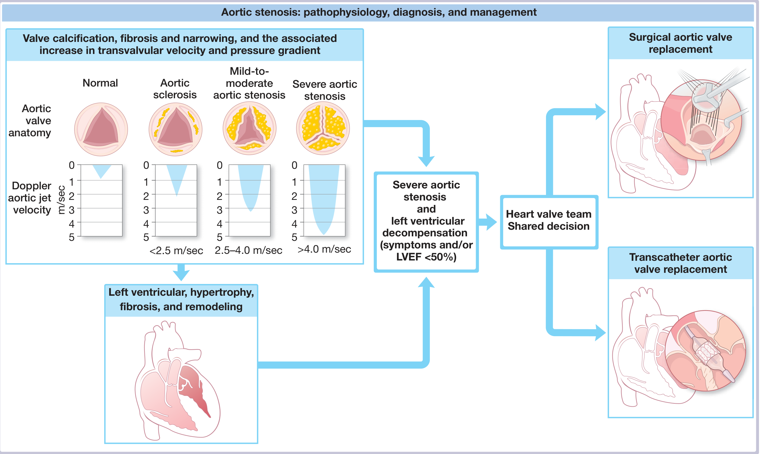
| Peak jet velocity (m/s) | <3.0 | 3.0-4.0 | >4.0 |
| Mean gradient (mmHg) | <25 | 25-40 | >40 |
| Valve area (cm²) | >1.5 | 1.0-1.5 | <1.0 |
| Indexed valve area (cm²/m²) | - | - | <0.6 |
- Measured by continuous wave (CW) Doppler from multiple windows (apical 5-chamber, right parasternal, suprasternal)
- The highest velocity obtained is used (not averaged)
- Peak velocity ≥4 m/s = severe AS
- Calculated by tracing the CW Doppler velocity envelope
- Software automatically converts using the simplified Bernoulli equation: ΔP = 4v²
- Mean gradient >40 mmHg = severe AS
- CSA_LVOT = cross-sectional area of LVOT = π(D/2)²
- D_LVOT = LVOT diameter measured in PLAX view
- VTI_LVOT = velocity-time integral of LVOT (pulsed-wave Doppler, apical 5-chamber)
- VTI_AV = velocity-time integral of aortic valve (CW Doppler)
- DVI = VTI_LVOT / VTI_AV
- DVI <0.25 = severe AS (equivalent to AVA <1.0 cm²)
- Useful when LVOT diameter measurement is unreliable (avoids squaring the diameter error)
CW Doppler Waveform Differentiation
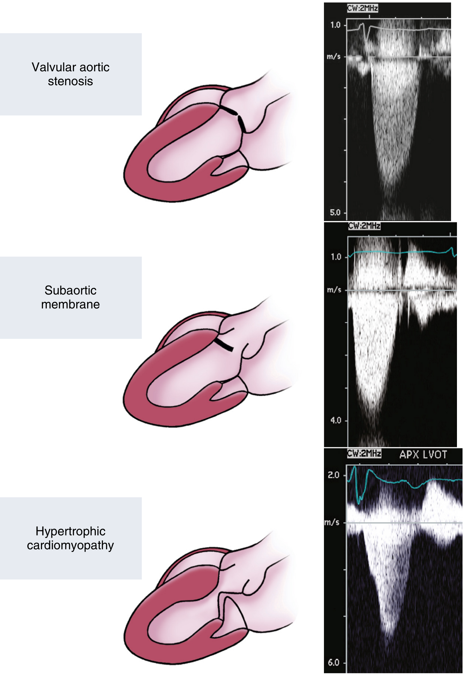
Low-Flow, Low-Gradient AS - A Clinical Challenge
40% of older patients have lower gradients/velocities despite severe AS:
- AVA <1.0 cm² BUT mean gradient <40 mmHg AND LVEF <50%
- Low gradient may reflect reduced forward flow, not necessarily truly "moderate" stenosis
- Dobutamine stress echo: Infuse dobutamine to increase flow
- True severe AS: AVA remains <1.0 cm², gradient rises
- Pseudo-severe AS: AVA increases to >1.0 cm² as improved contractility opens the valve further
- Surgery is indicated if true severe AS, but operative risk is higher with low EF
- AVA <1.0 cm², mean gradient <40 mmHg, BUT LVEF ≥50%
- Small, hypertrophied LV with reduced stroke volume index (<35 mL/m²)
- More common in women, elderly, hypertensive patients
- CT calcium scoring confirms truly severe AS (calcium score >2000 AU in men, >1200 AU in women)
- Prognosis similar to typical high-gradient AS; mortality benefit from AVR
Additional Imaging
- CT aortic valve calcium scoring - best objective measure of calcification burden
- Aortic valve anatomy for TAVI planning (annulus sizing, coronary height, access vessels)
- CT angiography of peripheral vasculature for TAVI access
- LV fibrosis quantification by late gadolinium enhancement (LGE) - predicts irreversible damage
- Important prognostic marker: >2 segments of LGE associated with worse post-AVR outcomes
- Flow measurement across the aortic valve
PART 7: STAGING OF AS (2021 ACC/AHA Valve Guidelines)
| Stage | Definition | Hemodynamics | Symptoms | LV Function |
|---|---|---|---|---|
| A | At risk | None | None | Normal |
| B | Progressive | Mild-moderate (Vmax 2-3.9 m/s; mean gradient <40 mmHg; AVA >1.0) | None | Normal/early remodeling |
| C1 | Severe asymptomatic, preserved EF | Severe (Vmax ≥4 m/s; mean gradient ≥40 mmHg; AVA ≤1.0 cm²) | None | LVEF ≥50%, possible early decompensation |
| C2 | Severe asymptomatic, reduced EF | As above | None | LVEF <50% |
| D1 | Severe symptomatic, high-gradient | Vmax ≥4 m/s; mean gradient ≥40 mmHg | Angina, syncope, HF | Usually preserved EF initially |
| D2 | Severe symptomatic, low-flow/low-gradient reduced EF | AVA <1.0 cm², mean gradient <40 mmHg | Symptoms | LVEF <50% |
| D3 | Severe symptomatic, low-gradient preserved EF | AVA <1.0 cm², mean gradient <40 mmHg, SVi <35 mL/m² | Symptoms | LVEF ≥50% |
PART 8: NATURAL HISTORY
- Long latent period - patients with severe AS remain asymptomatic for many years
- Annual risk of sudden death in asymptomatic severe AS: <1%
- Once symptomatic: 50% mortality at 2-3 years without intervention
- The symptom triad dictates prognosis:
- Angina → mean survival 5 years
- Syncope → mean survival 3 years
- Heart failure → mean survival <2 years
- Rate of progression of mild-moderate AS: highly variable, ~0.3 m/s per year increase in velocity
- In very severe AS (Vmax >5 m/s), annual risk of progression to symptoms: 50-80%
PART 9: MEDICAL MANAGEMENT
- Statins were hypothesized to slow progression (shared pathophysiology with atherosclerosis) but randomized trials (SEAS, ASTRONOMER) showed NO benefit in slowing progression
- ACE inhibitors/ARBs: caution - excessive afterload reduction with fixed outflow obstruction can cause dramatic hypotension; may be carefully used for hypertension
- Diuretics: symptomatic relief for pulmonary congestion but do not change natural history
- Beta-blockers: may be used for angina but depress contractility - use cautiously
- Antibiotic prophylaxis for GAS - critical in rheumatic AS to prevent recurrence
- Optimize hemodynamics pre-operatively
- Treat AF (rate control, anticoagulation)
- Treat heart failure (judicious diuresis, cautious vasodilator therapy)
- Risk factor modification (hypertension, diabetes, dyslipidemia)
- NOT a definitive treatment for calcific AS
- AVA improves only transiently; restenosis occurs within 6-12 months in virtually all patients
- Appropriate only as a bridge to surgery or TAVI in hemodynamically unstable patients
- Role in rheumatic AS is better (commissural fusion can be mechanically opened), analogous to percutaneous mitral commissurotomy
- Complications: acute AR, stroke, vascular access complications
PART 10: INDICATIONS FOR INTERVENTION (2021 ACC/AHA Guidelines)
Class I (Benefit >> Risk - MUST DO)
- Symptomatic severe high-gradient AS (Stage D1) - AVR is indicated (COR I, LOE B-NR)
- Severe AS (any stage) undergoing other cardiac surgery - concomitant AVR (COR I, LOE B-NR)
- Asymptomatic severe AS with LVEF <50% (Stage C2) - AVR indicated (COR I, LOE B-NR)
Class IIa (Benefit > Risk - SHOULD DO)
- Asymptomatic severe AS, normal EF, with very severe hemodynamics (Stage C1 with Vmax ≥5 m/s or mean gradient ≥60 mmHg) - AVR reasonable
- Asymptomatic severe AS undergoing non-cardiac surgery - AVR first if feasible
- Low-flow, low-gradient severe AS with reduced EF (Stage D2) - confirmed by dobutamine stress echo
Class IIb (Uncertain Benefit)
- Asymptomatic severe AS with normal EF and rapid disease progression (increase in Vmax ≥0.3 m/s/year) OR very high surgical risk and favorable anatomy for TAVI
- Moderate AS in patients undergoing cardiac surgery
PART 11: OPERATIVE APPROACH - GENERAL PRINCIPLES
Pre-operative Workup
- Doppler TTE - confirm severity, assess LV function, screen other valves
- Coronary angiography - mandatory before elective AVR (CAD in 30-50% of AS patients)
- TEE - detailed valve morphology, annulus sizing for prosthesis selection
- CT angiography (for TAVI) - annulus diameter, aortic root anatomy, coronary heights, iliofemoral access
- Carotid Doppler - assess for significant carotid stenosis
- STS/EuroSCORE II risk stratification
- Frailty assessment (for TAVI candidates) - 5-meter walk test, grip strength, serum albumin, Katz ADL score
Conduct of Open Heart Surgery - Standard Setup
- Aortic cannulation (distal ascending aorta), venous cannulation (right atrium, bicaval for combined procedures)
- Mild to moderate hypothermia (28-32°C)
- Antegrade and/or retrograde cardioplegia (cold blood cardioplegia preferred)
- Myocardial protection is paramount - the hypertrophied LV is less tolerant of ischemia
- Transverse or oblique aortotomy 1.5-2 cm above the right coronary ostium
- Extends into the non-coronary sinus for better exposure
- Traction sutures on aortotomy edges improve visualization
- Leaflets excised at their bases, commissures cut
- Complete debridement of calcium - calcium in the annulus and aortic root must be meticulously removed
- Caution: avoid injury to the left coronary ostium (just above the left-noncoronary commissure) and the right coronary ostium (above the right coronary sinus)
- Avoid annular perforation - can lead to aorto-ventricular discontinuity
- Special care at the non-coronary/right coronary commissure - AV nodal artery passes nearby
- Multiple sizers used to determine maximum prosthesis size
- Choice of prosthesis size impacts hemodynamics and risk of patient-prosthesis mismatch (PPM)
- PPM: indexed effective orifice area (EOA) <0.85 cm²/m² (moderate) or <0.65 cm²/m² (severe)
- For small aortic annuli: consider aortic root enlargement procedures (Nicks, Manougian, Konno)
PART 12: PROSTHETIC VALVES - SELECTION AND TYPES
Mechanical Valves
- Types: Bileaflet (St. Jude Medical, CarboMedics - most common), Tilting disc (Medtronic-Hall, Bjork-Shiley - largely historic), Caged ball (Starr-Edwards - largely historic)
- Material: Pyrolytic carbon (leaflets), titanium/carbon housing
- Durability: Essentially indefinite (30+ years)
- Requirement: Lifelong anticoagulation (warfarin, target INR 2.0-3.0 for aortic mechanical)
- Thromboembolic risk: ~1-2% per year stroke risk even with anticoagulation
- Hemolysis: Low with modern bileaflet valves
- Indications: Young patients (<60 years) who can tolerate anticoagulation, patients already on anticoagulation
Bioprosthetic Valves (Tissue Valves)
| Type | Examples | Advantage | Disadvantage |
|---|---|---|---|
| Porcine xenograft | Medtronic Mosaic, St. Jude Epic | No anticoagulation | Durability 10-20 years |
| Bovine pericardial | Edwards Perimount Magna, Sorin Mitroflow | Better hemodynamics, more durable | Same as above |
| Stentless | Toronto SPV, Medtronic Freestyle | Excellent hemodynamics, larger EOA | Technically demanding |
| Homograft (allograft) | Cryopreserved human aortic valve | Best hemodynamics, infection-resistant | Limited availability, complex implantation |
- Deterioration slower in older patients (70+ years) - biological calcification rate decreases with age
- Structural valve deterioration (SVD): calcification, leaflet tear, degeneration - more rapid in young patients
- Current guidelines: Tissue valve preferred over mechanical in patients >65 years (2020 ACC/AHA)
- For patients 50-65 years: shared decision-making between mechanical and bioprosthetic
Suturing Techniques
- Interrupted pledgeted sutures (mattress sutures) - most common; allows for secure annular fixation; easier to achieve good results with calcium-laden annuli
- Continuous suture technique - faster, fewer sutures
- Supra-annular placement - used for smaller annuli to obtain larger valve; supra-annular position of bioprosthetic valve improves EOA
Aortic Root Enlargement (for Small Annuli/PPM Prevention)
- Incise the non-coronary sinus down through the annulus into the anterior mitral leaflet and repair with a patch
- Allows 1-2 size increase in prosthesis
- Incision through the non-coronary/left coronary commissure into the anterior mitral leaflet
- More extensive enlargement possible
- Most extensive - incision extends through the right coronary/right ventricular outflow tract, patching the ventricular septum and aortic annulus
- Allows very large enlargements; used in children and reoperations
- Risk: VSD, complete heart block
PART 13: SURGICAL AORTIC VALVE REPLACEMENT (SAVR) - STEP-BY-STEP
Standard SAVR Operative Steps:
- Midline skin incision from sternal notch to xiphoid
- Median sternotomy with oscillating saw; sternal retractor
- Pericardial incision with suspension sutures
- Heparin administration (300-400 IU/kg; target ACT >400 seconds)
- Aortic cannulation - purse-string sutures in distal ascending aorta
- Venous cannulation - right atrial appendage (single 2-stage cannula or bicaval)
- CPB established; vent through right superior pulmonary vein or LV apex
- Aortic cross-clamp applied
- Cardioplegia delivery - antegrade via aortic root or direct coronary ostia; retrograde via coronary sinus
- Transverse aortotomy ~1.5-2 cm above right coronary ostium
- Valve excision and annular debridement
- Annular sizing
- Prosthesis implantation with interrupted or continuous sutures
- Aortotomy closure (usually two-layer running suture with felt reinforcement)
- De-airing maneuvers - vigorous (fill LV, aspirate from aortic root)
- Aortic cross-clamp release - ventricular fibrillation or spontaneous rhythm
- Weaning from CPB
- TEE assessment of prosthesis function: paravalvular leak, prosthesis gradient, LV function
- Protamine reversal, hemostasis, sternal closure
PART 14: TRANSCATHETER AORTIC VALVE REPLACEMENT (TAVI/TAVR)
Indication Evolution
- Originally: Inoperable patients (PARTNER B - 2010: 50% reduction in mortality vs. medical therapy)
- Then: High surgical risk (PARTNER A - 2011; CoreValve High-Risk - 2014)
- Then: Intermediate surgical risk (PARTNER 2 - 2016; SURTAVI - 2017)
- Now: Low surgical risk (PARTNER 3 - 2019; Evolut Low Risk - 2019)
- Current indication: Severe symptomatic AS + STS-PROM ≥3% OR low-risk patients ≥65 years (shared decision with heart team)
Valve Types
- Cobalt-chromium stent frame with bovine pericardial leaflets
- Requires balloon inflation during rapid ventricular pacing (to minimize cardiac output during deployment)
- Deployed in the native aortic annulus; more precise positioning
- Available: SAPIEN 3, SAPIEN 3 Ultra
- Nitinol frame with porcine pericardial leaflets
- Self-expands upon delivery sheath retraction; no rapid pacing required
- Supra-annular leaflet position → better hemodynamics and EOA
- Allows repositioning/retrieval before full deployment (Evolut R/PRO)
- Higher rate of pacemaker implantation (10-25%) due to AV nodal compression in LVOT
Access Routes
- Transfemoral (TF) - MOST COMMON (~90%): Retrograde crossing of native AV via femoral artery; requires iliofemoral artery ≥5.5 mm diameter; best outcomes
- Transaortic (TAo): Direct puncture of ascending aorta via upper J ministernotomy; for severe peripheral vascular disease
- Transapical (TA): Anterolateral thoracotomy, LV apex puncture, antegrade delivery; used when femoral and transaortic not feasible; higher morbidity
- Transsubclavian/Transaxillary: Alternative arterial access when femoral unsuitable; good option with arterial diameter ≥5.5 mm
TAVI Procedure Steps (Transfemoral):
- Femoral access (percutaneous or surgical cutdown)
- Temporary pacemaker placed in right ventricular apex
- Aortic valve crossing with straight/angled wire from contralateral femoral artery (or directly); confirm position with fluoroscopy
- Pre-dilatation balloon aortic valvuloplasty (balloon-expandable TAVI)
- Valve delivery system advanced over stiff wire to aortic annulus
- Precise positioning using fluoroscopic landmarks (aortic annulus at device positioning zone)
- Deployment:
- Balloon-expandable: rapid ventricular pacing (160-200 bpm) → balloon inflation → valve deploys
- Self-expanding: slow sheath retraction without rapid pacing
- Post-deployment assessment with TEE and fluoroscopy: paravalvular leak (PVL), gradient, coronary flow, pacemaker need
- Post-dilatation if significant PVL
TAVI Complications
| Complication | Frequency | Management |
|---|---|---|
| Stroke | 2-5% | Neurological support; cerebral embolic protection devices (SENTINEL) reducing risk |
| Paravalvular leak (PVL) | 5-15% (trace-mild); moderate-severe <5% | Post-dilatation; valve-in-valve if severe |
| Permanent pacemaker | Self-expanding: 15-25%; Balloon-expandable: 5-10% | Pacemaker implantation |
| Vascular access complications | 5-10% | Surgical repair; covered stent |
| Annular rupture | <1% | Emergent surgery; often fatal |
| Coronary occlusion | <1% | Emergency PCI or CABG; prevented by coronary protection wire |
| Aortic root injury | <1% | Surgery |
| AV conduction block (new LBBB) | 20-30% self-expanding | Observation; pacemaker if complete block |
TAVI vs. SAVR - Key Trial Data
| Trial | Population | Key Finding |
|---|---|---|
| PARTNER B (2010) | Inoperable | TAVI vs. medical: 50% RRR in all-cause mortality at 1 year |
| PARTNER A (2011) | High risk | TAVI non-inferior to SAVR in mortality |
| PARTNER 2 (2016) | Intermediate risk | TAVI non-inferior to SAVR; better with transfemoral |
| PARTNER 3 (2019) | Low risk | TAVI superior to SAVR at 1 year (composite endpoint) |
| Evolut Low Risk (2019) | Low risk | TAVI non-inferior to SAVR; self-expanding shows excellent hemodynamics |
- For patients <65 years: SAVR preferred (better long-term data; TAVI durability uncertain)
- For patients 65-80 years: shared decision-making (TAVI or SAVR)
- For patients >80 years or prohibitive surgical risk: TAVI preferred
PART 15: SPECIAL SURGICAL SITUATIONS
Concomitant Coronary Artery Disease
- CAD present in 30-50% of AS patients requiring AVR
- CABG should be performed concomitantly with AVR when stenosis >70% in major coronary arteries
- Does not significantly increase operative mortality in most cases
- Off-pump CABG + TAVI is possible in selected centers
Concomitant Mitral Valve Disease (Mixed Valve RHD)
- Significant MV disease (moderate-severe MS or MR) should be addressed simultaneously
- Double valve replacement (DVR) carries higher risk than single-valve surgery
- MV repair preferable to replacement when feasible
- Careful pre-operative TEE and catheterization required
Aortic Root Replacement (Bentall/Modified Bentall Procedure)
- Composite graft with mechanical valve + Dacron tube graft
- Coronary reimplantation (buttons) into the graft
- Alternatively: tissue valve conduit (BioValsalva graft)
- Reimplantation of native aortic valve into Dacron tube graft
- Preserves native valve (no anticoagulation)
- Suitable when valve morphology is good (no calcification)
Reoperative AVR (Redo Surgery)
- Previous CABG, prior valve surgery
- Significantly higher risk: adhesions, injury to patent grafts, RIMA
- Valve-in-Valve TAVI - excellent option: TAVI inside a degenerated bioprosthesis
- Avoids redo sternotomy
- VIVID registry: good outcomes; risk of coronary obstruction (if prior TAVI/SAVR with tall leaflets)
Ross Procedure (Pulmonary Autograft)
- Replaces the diseased AV with the patient's own pulmonary valve (autograft), then uses a homograft to replace the pulmonary valve
- Advantages: Living autograft grows with the patient, no anticoagulation, best hemodynamics, resistant to endocarditis
- Best suited for: Children, young adults, women of childbearing age, athletes
- Disadvantages: Two valves at risk instead of one; autograft dilatation over time; complex operation
- 10-year survival 90%; freedom from autograft reoperation 75-80% at 15-20 years
- (Schwartz, p. 856)
PART 16: POST-OPERATIVE MANAGEMENT
Immediate Post-op ICU Care
- Hemodynamic monitoring: arterial line, PA catheter or CVP, urine output, cardiac output
- Vasopressors (norepinephrine) for vasodilatory shock (rewarming post-CPB)
- Inotropes (dobutamine/milrinone) for low-output syndrome
- Avoid tachycardia and hypotension - hypertrophied LV requires adequate preload and afterload
- Pacing threshold testing; temporary pacing via epicardial wires
- Chest tube management
Early Complications
- Low cardiac output syndrome (hypertrophied LV requires adequate filling pressure)
- Perioperative MI - usually from inadequate myocardial protection or coronary ostia injury
- Complete AV block (especially after aortic root enlargement or septal debridement)
- Prosthesis-patient mismatch - residual high gradient; consider reoperation if severe (EOA index <0.65 cm²/m²)
- Paravalvular leak - requires early reoperation if significant
- Bleeding - coagulopathy, inadequate hemostasis
- Respiratory failure - prolonged ventilation
Anticoagulation Protocol
- Mechanical valve: Warfarin (INR 2.0-3.0 for isolated aortic mechanical); start heparin bridge post-op day 1-2
- Bioprosthetic SAVR: Aspirin 81mg indefinitely; some advocate warfarin (INR 2-2.5) for first 3-6 months (2020 ACC/AHA: IIb indication), then aspirin alone
- TAVI: Dual antiplatelet therapy (DAPT) for 3-6 months, then aspirin monotherapy; anticoagulation if AF
Valve Durability and Follow-up
- Annual echocardiography for TAVI recipients
- Every 3-5 years for stable bioprosthetic SAVR
- Serial monitoring for SVD: increasing gradients, worsening regurgitation
- Transcatheter heart valve thrombosis: TAVI valves susceptible to subclinical leaflet thrombosis (HALT/HAVEC on CT) - managed with anticoagulation
PART 17: OUTCOMES AND PROGNOSIS
SAVR Outcomes
- Isolated AVR operative mortality: 1-3% (experienced centers)
- Combined AVR+CABG: 3-5%
- Redo sternotomy AVR: 5-10%
- 5-year survival after AVR: ~80%
- 10-year survival: ~60-65%
- Functional improvement: dramatic - most patients return to NYHA Class I-II
TAVI Outcomes (Modern Era)
- In-hospital mortality: <2% (transfemoral, low-risk)
- 1-year mortality comparable to SAVR in most risk categories
- Stroke at 30 days: 2-3%
- Vascular complications: 5-10%
- Pacemaker implantation: 10-25% (self-expanding)
- Valve durability: excellent at 5 years; data emerging for 10 years
Factors Predicting Worse Outcomes
- Pre-op LVEF <40%
- Severe LV fibrosis on MRI
- Pulmonary hypertension
- Severe comorbidities (renal failure, COPD, frailty)
- Emergency surgery
- Concomitant procedures
- Small aortic annulus with PPM
SUMMARY FLOWCHART
Group A Strep Pharyngitis
↓
Molecular Mimicry
↓
Acute Rheumatic Fever → Acute Rheumatic Carditis → Rheumatic Valvulitis
↓
Repeated Exposures + Ongoing Damage
↓
Commissural Fusion + Leaflet Thickening
↓
RHEUMATIC AORTIC STENOSIS
(+ Congenital Bicuspid, + Calcific Degenerative)
↓
Progressive Obstruction to LV Outflow
↓
LV Pressure Overload → Concentric Hypertrophy (Laplace, Frank-Starling)
↓
LONG ASYMPTOMATIC PERIOD (Stage B/C)
↓
SYMPTOM ONSET (Stage D): Angina - Syncope - Heart Failure
↓
50% Mortality at 2-3 years without intervention
↓
HEART VALVE TEAM DECISION:
↓ ↓
SAVR (Preferred <65y TAVI (Preferred >80y,
or young/complex) high/intermediate risk,
or low-risk ≥65y)
↓
Prosthesis Selection:
Mechanical (lifelong warfarin, durable)
Bioprosthetic (no anticoagulation, 10-20yr durability)
Homograft / Ross (young, special cases)
↓
DRAMATIC SYMPTOM IMPROVEMENT
80% 5-year survival post-AVR
- Braunwald's Heart Disease, 10th Ed. - Chapter 72 (AS), Chapter on Aging and CV Disease
- Schwartz's Principles of Surgery, 11th Ed. - Chapter 21 (Cardiac Surgery: Aortic Valve Operative Techniques)
- Sabiston Textbook of Surgery, 21st Ed. - Chapter 112 (Cardiac Surgery: Valve Disease)
- Fuster and Hurst's The Heart, 15th Ed. - Chapter 28 (Aortic Stenosis)
- Textbook of Clinical Echocardiography (Otto) - Chapter 11 (Stenotic Valves)
- Goldman-Cecil Medicine, International Edition (Valve Disease)These are your comprehensive MCh Cardiac Surgery level notes on Aortic Stenosis, built directly from six standard textbooks. Here's a quick orientation to what was covered:
What These Notes Cover (17 Parts)
| Part | Topic |
|---|---|
| 1 | Rheumatic Heart Disease - molecular mimicry, Aschoff bodies, Jones criteria, chronic RHD pathology |
| 2 | Full etiology classification - Calcific, Bicuspid, Rheumatic, rare causes |
| 3 | Pathophysiology - Laplace law, Frank-Starling, concentric hypertrophy, progression rates |
| 4 | Clinical features - pulsus parvus et tardus, Gallavardin phenomenon, symptom triad |
| 5 | Investigations - ECG, CXR, Gorlin formula |
| 6 | Echocardiography - 2D imaging, continuity equation, DVI, low-flow low-gradient AS |
| 7 | 2021 ACC/AHA Staging (Stage A through D3) |
| 8 | Natural history - "5-3-2" survival rule |
| 9 | Medical management - why no drugs work, BAV as bridge |
| 10 | Indications for intervention (Class I, IIa, IIb) |
| 11 | Operative approach - CPB setup, aortotomy, annular debridement |
| 12 | Prosthetic valves - mechanical vs. tissue, suturing techniques, root enlargement (Nicks/Manougian/Konno) |
| 13 | SAVR step-by-step |
| 14 | TAVI - valve types, access routes, steps, complications, key trials (PARTNER, Evolut) |
| 15 | Special situations - redo surgery, Bentall, David, Ross procedure |
| 16 | Post-op management - ICU care, anticoagulation, follow-up |
| 17 | Outcomes and prognostic factors |
Generate a PowerPoint presentation on aortic stenosis for MCh cardiology teaching rounds
ls /home/daytona/workspace/ 2>/dev/null && mkdir -p /home/daytona/workspace/aortic-stenosis-ppt
node /home/daytona/skills/shared/scripts/fetch_images.js "https://cdn.orris.care/cdss_images/e3476113f8ca1b8c3587f3768a9ec0d115a289ba8d5a8ad55c7e1e90cf221438.png" "https://cdn.orris.care/cdss_images/210bcc14d6feb9ac6b12973ea2114c1196e7959d82d1f7abe2da295c61b20bed.png" "https://cdn.orris.care/cdss_images/e0dde8d70e5315322961d837763d57b676c7d3df92e896906b5ae49eb88f7302.png" "https://cdn.orris.care/cdss_images/b066f763c551005c2db9e9831280415a205a914074c4321c8f35b78c09f77000.png" 2>/dev/null | node -e "const d=require('fs');let x='';process.stdin.on('data',c=>x+=c);process.stdin.on('end',()=>{const arr=JSON.parse(x);arr.forEach((item,i)=>{d.writeFileSync('/home/daytona/workspace/aortic-stenosis-ppt/img'+i+'.txt',item.error?'ERROR:'+item.error:item.base64)});console.log('Done, errors:',arr.filter(a=>a.error).length)})"
~/aortic-stenosis-ppt/build.js
const pptxgen = require("pptxgenjs");
const fs = require("fs");
// Load pre-fetched images
const img0 = fs.readFileSync("/home/daytona/workspace/aortic-stenosis-ppt/img0.txt", "utf8"); // valve types
const img1 = fs.readFileSync("/home/daytona/workspace/aortic-stenosis-ppt/img1.txt", "utf8"); // natural history flowchart
const img2 = fs.readFileSync("/home/daytona/workspace/aortic-stenosis-ppt/img2.txt", "utf8"); // CW Doppler
const img3 = fs.readFileSync("/home/daytona/workspace/aortic-stenosis-ppt/img3.txt", "utf8"); // gross pathology
let pres = new pptxgen();
pres.layout = "LAYOUT_16x9";
pres.author = "MCh Cardiac Surgery Teaching";
pres.title = "Aortic Stenosis – MCh Cardiology Teaching Rounds";
// ─── COLOUR PALETTE ───────────────────────────────────────────────
// Deep navy dominant, crimson accent, light slate content areas
const C = {
navy: "0A1628", // dominant dark bg
navyMid: "122040", // card/panel bg
navyLt: "1A3060", // subtle divider / sub-panel
crimson: "C0392B", // accent / highlight
crimsonLt:"E74C3C", // lighter accent
gold: "F39C12", // 2nd accent / warning
teal: "1ABC9C", // 3rd accent / positive
white: "FFFFFF",
offWhite: "ECF0F1",
gray: "95A5A6",
grayLt: "BDC3C7",
};
// ─── HELPER: Slide Header Bar ─────────────────────────────────────
function addHeader(slide, title, subtitle) {
// Full-width top bar
slide.addShape(pres.shapes.RECTANGLE, {
x: 0, y: 0, w: 10, h: 0.82,
fill: { color: C.navyMid },
line: { color: C.navyMid }
});
// Crimson accent left strip
slide.addShape(pres.shapes.RECTANGLE, {
x: 0, y: 0, w: 0.06, h: 0.82,
fill: { color: C.crimson },
line: { color: C.crimson }
});
// Title text
slide.addText(title.toUpperCase(), {
x: 0.18, y: 0, w: subtitle ? 7.5 : 9.7, h: 0.82,
fontSize: 20, bold: true, color: C.white,
fontFace: "Calibri", valign: "middle", margin: 0, charSpacing: 1
});
if (subtitle) {
slide.addText(subtitle, {
x: 7.7, y: 0, w: 2.2, h: 0.82,
fontSize: 9, color: C.grayLt,
fontFace: "Calibri", valign: "middle", align: "right", margin: 0
});
}
}
// ─── HELPER: Content Card ─────────────────────────────────────────
function addCard(slide, x, y, w, h, opts = {}) {
slide.addShape(pres.shapes.RECTANGLE, {
x, y, w, h,
fill: { color: opts.fill || C.navyMid },
line: { color: opts.border || C.navyLt, pt: opts.borderPt || 1 },
shadow: opts.shadow ? { type: "outer", color: "000000", blur: 8, offset: 2, angle: 135, opacity: 0.25 } : undefined
});
if (opts.accent) {
slide.addShape(pres.shapes.RECTANGLE, {
x, y, w: 0.05, h,
fill: { color: opts.accent },
line: { color: opts.accent }
});
}
}
// ─── HELPER: Section Label ────────────────────────────────────────
function addLabel(slide, text, x, y, color) {
slide.addShape(pres.shapes.RECTANGLE, {
x, y, w: text.length * 0.095 + 0.25, h: 0.25,
fill: { color: color || C.crimson },
line: { color: color || C.crimson }
});
slide.addText(text, {
x, y, w: text.length * 0.095 + 0.25, h: 0.25,
fontSize: 8, bold: true, color: C.white,
fontFace: "Calibri", align: "center", valign: "middle", margin: 0
});
}
// ═══════════════════════════════════════════════════════════════════
// SLIDE 1 – TITLE SLIDE
// ═══════════════════════════════════════════════════════════════════
{
let s = pres.addSlide();
s.background = { color: C.navy };
// Large crimson left accent bar
s.addShape(pres.shapes.RECTANGLE, {
x: 0, y: 0, w: 0.18, h: 5.625,
fill: { color: C.crimson }, line: { color: C.crimson }
});
// Horizontal mid divider
s.addShape(pres.shapes.RECTANGLE, {
x: 0.18, y: 2.55, w: 9.82, h: 0.04,
fill: { color: C.navyLt }, line: { color: C.navyLt }
});
// Main title
s.addText("AORTIC STENOSIS", {
x: 0.4, y: 0.9, w: 9.3, h: 1.2,
fontSize: 52, bold: true, color: C.white,
fontFace: "Calibri", charSpacing: 2
});
// Subtitle
s.addText("A Comprehensive Review for MCh Cardiology Teaching Rounds", {
x: 0.4, y: 2.1, w: 9.3, h: 0.6,
fontSize: 18, color: C.grayLt, fontFace: "Calibri", italic: true
});
// Topic tags
const tags = ["Rheumatic Heart Disease", "Pathophysiology", "Echocardiography", "Surgical Management", "TAVI"];
tags.forEach((tag, i) => {
s.addShape(pres.shapes.RECTANGLE, {
x: 0.4 + i * 1.92, y: 2.82, w: 1.78, h: 0.3,
fill: { color: C.navyMid }, line: { color: C.navyLt, pt: 1 }
});
s.addText(tag, {
x: 0.4 + i * 1.92, y: 2.82, w: 1.78, h: 0.3,
fontSize: 8, color: C.grayLt, fontFace: "Calibri",
align: "center", valign: "middle", margin: 0
});
});
// Key stat box
addCard(s, 0.4, 3.3, 4.5, 1.9, { fill: C.navyMid, border: C.navyLt, accent: C.crimson, shadow: true });
s.addText("KEY EPIDEMIOLOGY", {
x: 0.6, y: 3.3, w: 4.2, h: 0.35,
fontSize: 9, bold: true, color: C.crimson, fontFace: "Calibri",
valign: "middle", margin: 0, charSpacing: 1
});
s.addText([
{ text: "• 2-5% of adults >65 years have significant AS\n", options: {} },
{ text: "• Most common valvular heart disease in developed world\n", options: {} },
{ text: "• 50% mortality at 2-3 years once symptomatic\n", options: {} },
{ text: "• 100,000+ AVR procedures performed annually in USA", options: {} },
], {
x: 0.55, y: 3.65, w: 4.25, h: 1.4,
fontSize: 11, color: C.offWhite, fontFace: "Calibri", valign: "top"
});
// Source box
addCard(s, 5.1, 3.3, 4.7, 1.9, { fill: C.navyMid, border: C.navyLt, accent: C.gold, shadow: true });
s.addText("SOURCES", {
x: 5.3, y: 3.3, w: 4.4, h: 0.35,
fontSize: 9, bold: true, color: C.gold, fontFace: "Calibri",
valign: "middle", margin: 0, charSpacing: 1
});
s.addText([
{ text: "• Braunwald's Heart Disease, 10th Ed.\n", options: {} },
{ text: "• Schwartz's Principles of Surgery, 11th Ed.\n", options: {} },
{ text: "• Sabiston Textbook of Surgery, 21st Ed.\n", options: {} },
{ text: "• Fuster & Hurst's The Heart, 15th Ed.\n", options: {} },
{ text: "• Otto: Textbook of Clinical Echocardiography", options: {} },
], {
x: 5.3, y: 3.65, w: 4.4, h: 1.4,
fontSize: 10, color: C.offWhite, fontFace: "Calibri", valign: "top"
});
// Bottom bar
s.addShape(pres.shapes.RECTANGLE, {
x: 0, y: 5.37, w: 10, h: 0.255,
fill: { color: C.navyMid }, line: { color: C.navyMid }
});
s.addText("MCh Cardiology Teaching Rounds | Aortic Stenosis", {
x: 0.2, y: 5.37, w: 9.6, h: 0.255,
fontSize: 8, color: C.gray, fontFace: "Calibri", valign: "middle"
});
}
// ═══════════════════════════════════════════════════════════════════
// SLIDE 2 – OUTLINE / TABLE OF CONTENTS
// ═══════════════════════════════════════════════════════════════════
{
let s = pres.addSlide();
s.background = { color: C.navy };
addHeader(s, "Lecture Outline", "MCh Cardiology Teaching Rounds");
const sections = [
{ num: "01", title: "Rheumatic Heart Disease", sub: "ARF → Molecular mimicry → Chronic RHD → Valve pathology", color: C.crimson },
{ num: "02", title: "Etiology & Classification", sub: "Calcific · Bicuspid · Rheumatic – comparative features", color: C.crimsonLt },
{ num: "03", title: "Pathophysiology", sub: "Pressure overload · LV hypertrophy · Laplace law · Symptom triad", color: C.gold },
{ num: "04", title: "Clinical Features & Investigations", sub: "Examination findings · ECG · CXR · Cardiac catheterization", color: C.teal },
{ num: "05", title: "Echocardiography", sub: "Quantitation · Continuity equation · Low-flow low-gradient AS", color: "#3498DB" },
{ num: "06", title: "Staging & Indications for Intervention", sub: "2021 ACC/AHA Guidelines · Stage A–D3", color: "#9B59B6" },
{ num: "07", title: "Surgical Management", sub: "SAVR · Prosthetic valves · Root enlargement · TAVI · Ross", color: "#27AE60" },
{ num: "08", title: "Outcomes & Follow-up", sub: "Mortality data · Anticoagulation · SVD · Key trials", color: "#E67E22" },
];
sections.forEach((sec, i) => {
const col = i < 4 ? 0 : 1;
const row = i % 4;
const x = 0.18 + col * 4.95;
const y = 0.95 + row * 1.15;
addCard(s, x, y, 4.7, 1.0, { fill: C.navyMid, border: C.navyLt, accent: sec.color, shadow: true });
s.addText(sec.num, {
x: x + 0.15, y: y, w: 0.55, h: 1.0,
fontSize: 26, bold: true, color: sec.color,
fontFace: "Calibri", valign: "middle", align: "center", margin: 0
});
s.addShape(pres.shapes.RECTANGLE, {
x: x + 0.72, y: y + 0.22, w: 0.02, h: 0.56,
fill: { color: C.navyLt }, line: { color: C.navyLt }
});
s.addText(sec.title, {
x: x + 0.82, y: y + 0.08, w: 3.76, h: 0.38,
fontSize: 12, bold: true, color: C.white,
fontFace: "Calibri", valign: "middle", margin: 0
});
s.addText(sec.sub, {
x: x + 0.82, y: y + 0.48, w: 3.76, h: 0.42,
fontSize: 9, color: C.gray,
fontFace: "Calibri", valign: "top", margin: 0
});
});
}
// ═══════════════════════════════════════════════════════════════════
// SLIDE 3 – RHEUMATIC HEART DISEASE
// ═══════════════════════════════════════════════════════════════════
{
let s = pres.addSlide();
s.background = { color: C.navy };
addHeader(s, "Rheumatic Heart Disease", "Sabiston Surgery 21e · Braunwald's 10e");
// Left panel – cascade
addCard(s, 0.18, 0.95, 4.6, 4.5, { fill: C.navyMid, border: C.navyLt, accent: C.crimson, shadow: true });
s.addText("THE RHEUMATIC CASCADE", {
x: 0.35, y: 0.98, w: 4.3, h: 0.3,
fontSize: 9, bold: true, color: C.crimson, fontFace: "Calibri",
valign: "middle", margin: 0, charSpacing: 1
});
const cascade = [
{ step: "01", text: "Group A β-hemolytic Streptococcal pharyngitis (GAS)", color: C.crimson },
{ step: "02", text: "Genetically susceptible host (HLA-DR2, DR4)", color: C.crimsonLt },
{ step: "03", text: "Molecular mimicry — M-protein vs. cardiac laminin", color: C.gold },
{ step: "04", text: "Cross-reactive T-cells & antibodies attack endocardium", color: C.gold },
{ step: "05", text: "Acute Rheumatic Valvulitis (2-6 wks post-infection)", color: C.teal },
{ step: "06", text: "Repeated strep exposures → cumulative valve damage", color: C.teal },
{ step: "07", text: "Commissural fusion + fibrosis → CHRONIC RHD", color: "#3498DB" },
];
cascade.forEach((c, i) => {
const y = 1.4 + i * 0.55;
s.addShape(pres.shapes.RECTANGLE, {
x: 0.3, y, w: 0.42, h: 0.37,
fill: { color: c.color }, line: { color: c.color }
});
s.addText(c.step, {
x: 0.3, y, w: 0.42, h: 0.37,
fontSize: 9, bold: true, color: C.navy,
fontFace: "Calibri", align: "center", valign: "middle", margin: 0
});
s.addText(c.text, {
x: 0.82, y, w: 3.85, h: 0.37,
fontSize: 9.5, color: C.offWhite, fontFace: "Calibri", valign: "middle", margin: 0
});
if (i < cascade.length - 1) {
s.addShape(pres.shapes.RECTANGLE, {
x: 0.48, y: y + 0.37, w: 0.05, h: 0.18,
fill: { color: C.gray }, line: { color: C.gray }
});
}
});
// Right panel – pathology details
addCard(s, 4.95, 0.95, 4.87, 2.1, { fill: C.navyMid, border: C.navyLt, accent: C.gold, shadow: true });
s.addText("ASCHOFF BODY — PATHOGNOMONIC LESION", {
x: 5.12, y: 0.98, w: 4.6, h: 0.3,
fontSize: 9, bold: true, color: C.gold, fontFace: "Calibri",
valign: "middle", margin: 0, charSpacing: 1
});
s.addText([
{ text: "• Central fibrinoid necrosis\n", options: {} },
{ text: "• Anitschkow (caterpillar) cells — activated macrophages\n", options: {} },
{ text: "• Aschoff giant cells (multinucleated)\n", options: {} },
{ text: "• Verrucous endocarditis — 1-2 mm sterile vegetations at valve closure line\n", options: {} },
{ text: "• Pancarditis: endocardium + myocardium + pericardium", options: {} },
], {
x: 5.12, y: 1.32, w: 4.6, h: 1.65,
fontSize: 10, color: C.offWhite, fontFace: "Calibri", valign: "top"
});
// Gross pathology image
addCard(s, 4.95, 3.18, 2.3, 2.2, { fill: C.navyMid, border: C.navyLt, shadow: true });
s.addImage({ data: img3, x: 5.0, y: 3.22, w: 2.2, h: 1.85 });
s.addText("Rheumatic MV — 'fish-mouth' orifice", {
x: 4.95, y: 5.05, w: 2.3, h: 0.25,
fontSize: 7.5, color: C.gray, fontFace: "Calibri", align: "center", italic: true, margin: 0
});
// Valve features comparison
addCard(s, 7.35, 3.18, 2.47, 2.27, { fill: C.navyMid, border: C.navyLt, accent: C.teal, shadow: true });
s.addText("RHEUMATIC AV — KEY FEATURES", {
x: 7.5, y: 3.21, w: 2.2, h: 0.3,
fontSize: 8, bold: true, color: C.teal, fontFace: "Calibri", valign: "middle", margin: 0
});
s.addText([
{ text: "✦ Commissural fusion\n", options: { color: C.teal } },
{ text: "✦ Triangular orifice in systole\n", options: { color: C.teal } },
{ text: "✦ Mitral valve always involved\n", options: { color: C.offWhite } },
{ text: "✦ Mixed lesion: stenosis + regurgitation\n", options: { color: C.offWhite } },
{ text: "✦ Presents 4th–6th decade", options: { color: C.offWhite } },
], {
x: 7.5, y: 3.55, w: 2.2, h: 1.85,
fontSize: 9.5, fontFace: "Calibri", valign: "top"
});
}
// ═══════════════════════════════════════════════════════════════════
// SLIDE 4 – ETIOLOGY & MORPHOLOGY (with image)
// ═══════════════════════════════════════════════════════════════════
{
let s = pres.addSlide();
s.background = { color: C.navy };
addHeader(s, "Etiology & Valve Morphology", "Textbook of Clinical Echocardiography · Braunwald's");
// Main image
addCard(s, 0.18, 0.95, 5.5, 4.35, { fill: C.navyMid, border: C.navyLt, shadow: true });
s.addImage({ data: img0, x: 0.25, y: 1.0, w: 5.36, h: 4.0 });
s.addText("Fig 11.5 — Textbook of Clinical Echocardiography: Aortic valve viewed from above. Top row: diastole. Bottom row: systole.", {
x: 0.18, y: 5.05, w: 5.5, h: 0.3,
fontSize: 7.5, color: C.gray, fontFace: "Calibri", italic: true, align: "center"
});
// Three etiology cards on right
const etiologies = [
{
title: "CALCIFIC / DEGENERATIVE", color: C.gold, age: "70s–80s",
items: [
"Most common in developed world",
"Fibrocalcification at leaflet bases, aortic side",
"NO commissural fusion (key!)",
"Stellate-shaped orifice in systole",
"Shared risk factors with atherosclerosis",
"25% adults >65y have aortic sclerosis",
]
},
{
title: "BICUSPID AORTIC VALVE", color: C.teal, age: "50s–60s",
items: [
"2/3 of severe AS in adults <70 y",
"Prevalence ~1-2%, M:F = 3:1",
"Accelerated calcification of congenital valve",
"Elliptical orifice; diastolic sagging/systolic doming",
"50% have associated aortopathy",
"Right-left cusp fusion most common (~75%)",
]
},
{
title: "RHEUMATIC", color: C.crimson, age: "40s–60s",
items: [
"Commissural fusion — pathognomonic",
"Triangular orifice in systole",
"Mitral valve ALWAYS involved",
"Common in developing world",
"Decreasing incidence with GAS treatment",
"Mixed stenosis + regurgitation typical",
]
},
];
etiologies.forEach((et, i) => {
const y = 0.95 + i * 1.55;
addCard(s, 5.85, y, 3.97, 1.42, { fill: C.navyMid, border: C.navyLt, accent: et.color, shadow: true });
s.addText(et.title, {
x: 6.05, y: y + 0.05, w: 3.4, h: 0.3,
fontSize: 9, bold: true, color: et.color, fontFace: "Calibri",
valign: "middle", margin: 0, charSpacing: 0.5
});
s.addText(`Peak age: ${et.age}`, {
x: 6.05, y: y + 0.05, w: 3.65, h: 0.3,
fontSize: 8.5, color: C.gray, fontFace: "Calibri",
align: "right", valign: "middle", margin: 0
});
s.addText(et.items.map(x => `• ${x}`).join("\n"), {
x: 6.05, y: y + 0.36, w: 3.65, h: 1.0,
fontSize: 9, color: C.offWhite, fontFace: "Calibri", valign: "top", margin: 0
});
});
}
// ═══════════════════════════════════════════════════════════════════
// SLIDE 5 – PATHOPHYSIOLOGY
// ═══════════════════════════════════════════════════════════════════
{
let s = pres.addSlide();
s.background = { color: C.navy };
addHeader(s, "Pathophysiology", "Sabiston · Schwartz Principles of Surgery");
// Top: Core mechanism
addCard(s, 0.18, 0.95, 9.65, 0.82, { fill: C.navyMid, border: C.navyLt, accent: C.crimson, shadow: true });
s.addText("CORE MECHANISM: Fixed outflow obstruction → ↑LV systolic pressure → Concentric LV Hypertrophy (Laplace Law: Wall Stress ∝ P × r / 2h)", {
x: 0.3, y: 0.95, w: 9.4, h: 0.82,
fontSize: 11.5, bold: true, color: C.white, fontFace: "Calibri", valign: "middle", margin: 0
});
// LV response stages
addCard(s, 0.18, 1.88, 6.1, 1.32, { fill: C.navyMid, border: C.navyLt, accent: C.teal, shadow: true });
s.addText("LV RESPONSE STAGES", {
x: 0.35, y: 1.91, w: 5.8, h: 0.28,
fontSize: 9, bold: true, color: C.teal, fontFace: "Calibri", valign: "middle", margin: 0, charSpacing: 1
});
const stages = [
{ stage: "Compensation", lv: "Concentric hypertrophy, EF normal", hemo: "↑LVESP, normal CO", col: C.teal },
{ stage: "Early decompensation", lv: "Diastolic dysfunction", hemo: "↑LVEDP, ↓compliance", col: C.gold },
{ stage: "Late decompensation", lv: "Systolic dysfunction", hemo: "Low CO, pulm. congestion", col: C.crimsonLt },
{ stage: "End-stage", lv: "Dilated LV, ↓EF", hemo: "Low-flow, low-gradient", col: C.crimson },
];
const stageHeaders = ["Stage", "LV Change", "Hemodynamics"];
const stageData = [
stageHeaders.map(h => ({ text: h, options: { bold: true, color: C.navy, fill: { color: C.navyLt }, fontSize: 8 } })),
...stages.map(st => [
{ text: st.stage, options: { bold: true, color: st.col, fontSize: 8.5 } },
{ text: st.lv, options: { color: C.offWhite, fontSize: 8.5 } },
{ text: st.hemo, options: { color: C.offWhite, fontSize: 8.5 } }
])
];
s.addTable(stageData, {
x: 0.25, y: 2.22, w: 5.9, h: 0.9,
colW: [1.6, 2.3, 2.0],
border: { pt: 0.5, color: C.navyLt },
fill: { color: C.navyMid }
});
// Symptom triad
addCard(s, 6.45, 1.88, 3.38, 1.32, { fill: C.navyMid, border: C.navyLt, accent: C.crimson, shadow: true });
s.addText("CLASSIC SYMPTOM TRIAD", {
x: 6.62, y: 1.91, w: 3.1, h: 0.28,
fontSize: 9, bold: true, color: C.crimson, fontFace: "Calibri", valign: "middle", margin: 0, charSpacing: 1
});
const triad = [
{ s: "Angina", y: "5-yr survival", col: C.gold },
{ s: "Syncope", y: "3-yr survival", col: C.crimsonLt },
{ s: "Heart Failure", y: "<2-yr survival", col: C.crimson },
];
triad.forEach((t, i) => {
s.addShape(pres.shapes.RECTANGLE, {
x: 6.55 + i * 1.1, y: 2.28, w: 1.0, h: 0.82,
fill: { color: C.navyLt }, line: { color: t.col, pt: 1.5 }
});
s.addText(t.s, {
x: 6.55 + i * 1.1, y: 2.32, w: 1.0, h: 0.35,
fontSize: 9, bold: true, color: t.col,
fontFace: "Calibri", align: "center", margin: 0
});
s.addText(t.y, {
x: 6.55 + i * 1.1, y: 2.68, w: 1.0, h: 0.35,
fontSize: 7.5, color: C.offWhite,
fontFace: "Calibri", align: "center", margin: 0
});
});
// Progression rates
addCard(s, 0.18, 3.28, 4.6, 1.0, { fill: C.navyMid, border: C.navyLt, accent: C.gold, shadow: true });
s.addText("AVERAGE ANNUAL PROGRESSION (Schwartz)", {
x: 0.35, y: 3.31, w: 4.3, h: 0.28,
fontSize: 9, bold: true, color: C.gold, fontFace: "Calibri", valign: "middle", margin: 0, charSpacing: 1
});
const prog = [
{ label: "Peak Jet Velocity", val: "+0.3 m/s / yr" },
{ label: "Mean Gradient", val: "+7 mmHg / yr" },
{ label: "Valve Area", val: "−0.1 cm² / yr" },
];
prog.forEach((p, i) => {
s.addText(`${p.label}:`, {
x: 0.32 + i * 1.52, y: 3.65, w: 1.1, h: 0.55,
fontSize: 8.5, color: C.gray, fontFace: "Calibri", valign: "top", margin: 0
});
s.addText(p.val, {
x: 0.32 + i * 1.52, y: 3.98, w: 1.45, h: 0.25,
fontSize: 10, bold: true, color: C.gold, fontFace: "Calibri", valign: "middle", margin: 0
});
});
// Natural history flowchart image (bottom half)
addCard(s, 4.92, 3.28, 5.0, 2.1, { fill: C.navyMid, border: C.navyLt, shadow: true });
s.addImage({ data: img1, x: 4.97, y: 3.32, w: 4.9, h: 1.9 });
s.addText("Natural History & Management Overview (Fuster & Hurst, Ch. 28)", {
x: 4.92, y: 5.25, w: 5.0, h: 0.2,
fontSize: 7, color: C.gray, fontFace: "Calibri", italic: true, align: "center"
});
// Medical Rx note
addCard(s, 0.18, 4.32, 4.6, 1.0, { fill: C.navyMid, border: C.navyLt, accent: C.crimson, shadow: true });
s.addText("MEDICAL MANAGEMENT — KEY POINT", {
x: 0.35, y: 4.35, w: 4.3, h: 0.28,
fontSize: 9, bold: true, color: C.crimson, fontFace: "Calibri", valign: "middle", margin: 0, charSpacing: 1
});
s.addText("NO proven medical therapy modifies the natural history or slows progression of AS. Statins (SEAS/ASTRONOMER trials) showed no benefit. Medical Rx = bridge to surgery only.", {
x: 0.32, y: 4.66, w: 4.35, h: 0.6,
fontSize: 9, color: C.offWhite, fontFace: "Calibri", valign: "top", margin: 0
});
}
// ═══════════════════════════════════════════════════════════════════
// SLIDE 6 – CLINICAL FEATURES & EXAMINATION
// ═══════════════════════════════════════════════════════════════════
{
let s = pres.addSlide();
s.background = { color: C.navy };
addHeader(s, "Clinical Features & Physical Examination", "Braunwald's Heart Disease 10e");
// Examination findings table
addCard(s, 0.18, 0.95, 9.65, 0.35, { fill: C.navyLt, border: C.navyLt });
s.addText("PHYSICAL EXAMINATION FINDINGS", {
x: 0.25, y: 0.95, w: 9.5, h: 0.35,
fontSize: 9, bold: true, color: C.white, fontFace: "Calibri",
valign: "middle", margin: 0, charSpacing: 1
});
const examData = [
[
{ text: "Finding", options: { bold: true, color: C.navy, fill: { color: C.navyLt } } },
{ text: "Sign", options: { bold: true, color: C.navy, fill: { color: C.navyLt } } },
{ text: "Mechanism", options: { bold: true, color: C.navy, fill: { color: C.navyLt } } }
],
[
{ text: "Pulse", options: { bold: true, color: C.gold } },
{ text: "Pulsus parvus et tardus", options: { bold: true, color: C.offWhite } },
{ text: "Fixed reduced CO and delayed ejection", options: { color: C.grayLt } }
],
[
{ text: "Apex Beat", options: { bold: true, color: C.gold } },
{ text: "Sustained, forceful, non-displaced", options: { bold: true, color: C.offWhite } },
{ text: "Concentric hypertrophy (thick walls, no dilation)", options: { color: C.grayLt } }
],
[
{ text: "Murmur", options: { bold: true, color: C.crimson } },
{ text: "Harsh crescendo-decrescendo SEM, 2nd RICS → carotids", options: { bold: true, color: C.offWhite } },
{ text: "Turbulent flow through stenotic valve; late-peaking = more severe", options: { color: C.grayLt } }
],
[
{ text: "S2", options: { bold: true, color: C.gold } },
{ text: "Absent A2 or single / paradoxically split S2", options: { bold: true, color: C.offWhite } },
{ text: "Calcified immobile leaflets; reduced aortic valve closure sound", options: { color: C.grayLt } }
],
[
{ text: "S4 Gallop", options: { bold: true, color: C.gold } },
{ text: "Prominent S4", options: { bold: true, color: C.offWhite } },
{ text: "Forceful atrial kick into stiff non-compliant LV", options: { color: C.grayLt } }
],
[
{ text: "Ejection Click", options: { bold: true, color: C.teal } },
{ text: "Early systolic click", options: { bold: true, color: C.offWhite } },
{ text: "Abrupt halt of mobile valve at max opening (bicuspid/early calcific)", options: { color: C.grayLt } }
],
[
{ text: "Gallavardin Phenomenon", options: { bold: true, color: C.teal } },
{ text: "Musical component at apex (mimics MR)", options: { bold: true, color: C.offWhite } },
{ text: "High-frequency component of AS murmur radiates to apex", options: { color: C.grayLt } }
],
];
s.addTable(examData, {
x: 0.18, y: 1.33, w: 9.65, h: 3.2,
colW: [1.8, 2.95, 4.9],
border: { pt: 0.5, color: C.navyLt },
fill: { color: C.navyMid }
});
// Pitfall box
addCard(s, 0.18, 4.6, 9.65, 0.78, { fill: C.navyMid, border: C.crimson, accent: C.crimson, shadow: true });
s.addText("⚠ CLINICAL PITFALL:", {
x: 0.32, y: 4.63, w: 1.5, h: 0.72,
fontSize: 10, bold: true, color: C.crimson, fontFace: "Calibri", valign: "middle", margin: 0
});
s.addText("Pulsus parvus et tardus may be ABSENT in elderly patients due to large artery stiffening — a common false-reassurance trap. Always confirm with Doppler echocardiography. The murmur intensity does NOT correlate with stenosis severity. (Braunwald's Heart Disease, Ch. 83)", {
x: 1.85, y: 4.63, w: 7.9, h: 0.72,
fontSize: 9.5, color: C.offWhite, fontFace: "Calibri", valign: "middle", margin: 0
});
}
// ═══════════════════════════════════════════════════════════════════
// SLIDE 7 – ECHOCARDIOGRAPHY
// ═══════════════════════════════════════════════════════════════════
{
let s = pres.addSlide();
s.background = { color: C.navy };
addHeader(s, "Echocardiographic Assessment", "Textbook of Clinical Echocardiography (Otto)");
// Left: Severity grading table
addCard(s, 0.18, 0.95, 4.5, 3.0, { fill: C.navyMid, border: C.navyLt, accent: C.teal, shadow: true });
s.addText("ACC / AHA SEVERITY GRADING", {
x: 0.35, y: 0.98, w: 4.2, h: 0.28,
fontSize: 9, bold: true, color: C.teal, fontFace: "Calibri",
valign: "middle", margin: 0, charSpacing: 1
});
const gradeData = [
[
{ text: "Parameter", options: { bold: true, color: C.navy, fill: { color: C.navyLt }, fontSize: 8.5 } },
{ text: "Mild", options: { bold: true, color: C.navy, fill: { color: C.navyLt }, fontSize: 8.5 } },
{ text: "Moderate", options: { bold: true, color: C.navy, fill: { color: C.navyLt }, fontSize: 8.5 } },
{ text: "Severe", options: { bold: true, color: C.navy, fill: { color: C.navyLt }, fontSize: 8.5 } }
],
[
{ text: "Peak Jet Velocity (m/s)", options: { color: C.offWhite, fontSize: 9 } },
{ text: "< 3.0", options: { color: C.teal, fontSize: 9 } },
{ text: "3.0–4.0", options: { color: C.gold, fontSize: 9 } },
{ text: "> 4.0", options: { bold: true, color: C.crimson, fontSize: 10 } }
],
[
{ text: "Mean Gradient (mmHg)", options: { color: C.offWhite, fontSize: 9 } },
{ text: "< 25", options: { color: C.teal, fontSize: 9 } },
{ text: "25–40", options: { color: C.gold, fontSize: 9 } },
{ text: "> 40", options: { bold: true, color: C.crimson, fontSize: 10 } }
],
[
{ text: "Valve Area (cm²)", options: { color: C.offWhite, fontSize: 9 } },
{ text: "> 1.5", options: { color: C.teal, fontSize: 9 } },
{ text: "1.0–1.5", options: { color: C.gold, fontSize: 9 } },
{ text: "< 1.0", options: { bold: true, color: C.crimson, fontSize: 10 } }
],
[
{ text: "AVA Index (cm²/m²)", options: { color: C.offWhite, fontSize: 9 } },
{ text: "—", options: { color: C.gray, fontSize: 9 } },
{ text: "—", options: { color: C.gray, fontSize: 9 } },
{ text: "< 0.6", options: { bold: true, color: C.crimson, fontSize: 10 } }
],
[
{ text: "DVI (velocity ratio)", options: { color: C.offWhite, fontSize: 9 } },
{ text: "> 0.50", options: { color: C.teal, fontSize: 9 } },
{ text: "0.25–0.50", options: { color: C.gold, fontSize: 9 } },
{ text: "< 0.25", options: { bold: true, color: C.crimson, fontSize: 10 } }
],
];
s.addTable(gradeData, {
x: 0.25, y: 1.3, w: 4.35, h: 2.55,
colW: [1.75, 0.7, 0.85, 1.05],
border: { pt: 0.5, color: C.navyLt },
fill: { color: C.navyMid }
});
// Continuity equation
addCard(s, 0.18, 4.02, 4.5, 1.37, { fill: C.navyMid, border: C.navyLt, accent: C.gold, shadow: true });
s.addText("CONTINUITY EQUATION (Most Accurate)", {
x: 0.35, y: 4.05, w: 4.2, h: 0.28,
fontSize: 9, bold: true, color: C.gold, fontFace: "Calibri",
valign: "middle", margin: 0, charSpacing: 1
});
s.addText("AVA = (CSA_LVOT × VTI_LVOT) / VTI_AV\n= 0.785 × D_LVOT² × VTI_LVOT / VTI_AV", {
x: 0.32, y: 4.35, w: 4.25, h: 0.55,
fontSize: 11, bold: true, color: C.white, fontFace: "Courier New", valign: "top", margin: 0
});
s.addText("Based on conservation of mass: flow through LVOT = flow through stenotic valve", {
x: 0.32, y: 4.92, w: 4.25, h: 0.4,
fontSize: 8.5, color: C.gray, fontFace: "Calibri", valign: "top", margin: 0
});
// Right: CW Doppler image
addCard(s, 4.85, 0.95, 2.35, 4.44, { fill: C.navyMid, border: C.navyLt, shadow: true });
s.addImage({ data: img2, x: 4.9, y: 1.0, w: 2.25, h: 3.85 });
s.addText("CW Doppler: valvular AS vs. subaortic membrane vs. HCM (Otto)", {
x: 4.85, y: 4.88, w: 2.35, h: 0.45,
fontSize: 7, color: C.gray, fontFace: "Calibri", italic: true, align: "center"
});
// Low-flow low-gradient
addCard(s, 7.32, 0.95, 2.52, 4.44, { fill: C.navyMid, border: C.navyLt, accent: C.crimson, shadow: true });
s.addText("LOW-FLOW, LOW-GRADIENT AS", {
x: 7.48, y: 0.98, w: 2.25, h: 0.3,
fontSize: 8.5, bold: true, color: C.crimson, fontFace: "Calibri",
valign: "middle", margin: 0, charSpacing: 0.5
});
s.addText("TYPE 1 — Reduced EF (<50%)", {
x: 7.48, y: 1.32, w: 2.25, h: 0.25,
fontSize: 8.5, bold: true, color: C.gold, fontFace: "Calibri", valign: "middle", margin: 0
});
s.addText([
{ text: "• AVA <1.0 cm², mean grad <40 mmHg, EF <50%\n", options: {} },
{ text: "• 'Pseudo-severe' vs true severe AS\n", options: {} },
{ text: "• Dobutamine stress echo: true severe → AVA stays <1.0, gradient rises\n", options: {} },
{ text: "• False severe → AVA opens >1.0 cm²", options: {} },
], {
x: 7.48, y: 1.6, w: 2.25, h: 1.2,
fontSize: 8.5, color: C.offWhite, fontFace: "Calibri", valign: "top", margin: 0
});
s.addShape(pres.shapes.RECTANGLE, {
x: 7.4, y: 2.85, w: 2.4, h: 0.02,
fill: { color: C.navyLt }, line: { color: C.navyLt }
});
s.addText("TYPE 2 — Preserved EF ≥50% (Paradoxical)", {
x: 7.48, y: 2.9, w: 2.25, h: 0.3,
fontSize: 8.5, bold: true, color: C.teal, fontFace: "Calibri", valign: "middle", margin: 0
});
s.addText([
{ text: "• AVA <1.0, grad <40, EF ≥50%, SVi <35 mL/m²\n", options: {} },
{ text: "• Small hypertrophied LV with ↓SV\n", options: {} },
{ text: "• More common in women/elderly/hypertensive\n", options: {} },
{ text: "• CT Ca score confirms: >2000 AU (men), >1200 AU (women)\n", options: {} },
{ text: "• Prognosis same as high-gradient AS; AVR beneficial", options: {} },
], {
x: 7.48, y: 3.22, w: 2.25, h: 2.1,
fontSize: 8.5, color: C.offWhite, fontFace: "Calibri", valign: "top", margin: 0
});
}
// ═══════════════════════════════════════════════════════════════════
// SLIDE 8 – STAGING & INDICATIONS
// ═══════════════════════════════════════════════════════════════════
{
let s = pres.addSlide();
s.background = { color: C.navy };
addHeader(s, "Staging & Indications for Intervention", "2021 ACC/AHA Valvular Heart Disease Guidelines");
// Staging table
addCard(s, 0.18, 0.95, 9.65, 0.28, { fill: C.navyLt, border: C.navyLt });
s.addText("2021 ACC/AHA STAGING OF AORTIC STENOSIS", {
x: 0.25, y: 0.95, w: 9.5, h: 0.28,
fontSize: 8.5, bold: true, color: C.white, fontFace: "Calibri",
valign: "middle", margin: 0, charSpacing: 1
});
const stageData2 = [
["Stage", "Definition", "Hemodynamics", "Symptoms", "LV Function"].map(h => ({
text: h, options: { bold: true, color: C.navy, fill: { color: C.navyLt }, fontSize: 8 }
})),
[
{ text: "A — At Risk", options: { bold: true, color: C.teal, fontSize: 8.5 } },
{ text: "Risk factors for AS", options: { color: C.offWhite, fontSize: 8.5 } },
{ text: "None", options: { color: C.gray, fontSize: 8.5 } },
{ text: "None", options: { color: C.gray, fontSize: 8.5 } },
{ text: "Normal", options: { color: C.teal, fontSize: 8.5 } }
],
[
{ text: "B — Progressive", options: { bold: true, color: C.teal, fontSize: 8.5 } },
{ text: "Mild–moderate AS", options: { color: C.offWhite, fontSize: 8.5 } },
{ text: "Vmax 2–3.9 m/s; mean grad <40; AVA >1.0", options: { color: C.offWhite, fontSize: 8.5 } },
{ text: "None", options: { color: C.gray, fontSize: 8.5 } },
{ text: "Normal or early remodeling", options: { color: C.teal, fontSize: 8.5 } }
],
[
{ text: "C1 — Severe Asymptomatic EF≥50%", options: { bold: true, color: C.gold, fontSize: 8.5 } },
{ text: "Severe AS", options: { color: C.offWhite, fontSize: 8.5 } },
{ text: "Vmax ≥4 m/s; mean grad ≥40; AVA ≤1.0 cm²", options: { color: C.offWhite, fontSize: 8.5 } },
{ text: "None", options: { color: C.gray, fontSize: 8.5 } },
{ text: "LVEF ≥50%", options: { color: C.gold, fontSize: 8.5 } }
],
[
{ text: "C2 — Severe Asymptomatic EF<50%", options: { bold: true, color: C.gold, fontSize: 8.5 } },
{ text: "Severe AS", options: { color: C.offWhite, fontSize: 8.5 } },
{ text: "As above", options: { color: C.offWhite, fontSize: 8.5 } },
{ text: "None", options: { color: C.gray, fontSize: 8.5 } },
{ text: "LVEF <50%", options: { bold: true, color: C.crimson, fontSize: 8.5 } }
],
[
{ text: "D1 — Severe Symptomatic High-Gradient", options: { bold: true, color: C.crimson, fontSize: 8.5 } },
{ text: "Severe AS", options: { color: C.offWhite, fontSize: 8.5 } },
{ text: "Vmax ≥4 m/s; mean grad ≥40", options: { color: C.offWhite, fontSize: 8.5 } },
{ text: "Angina / Syncope / HF", options: { bold: true, color: C.crimson, fontSize: 8.5 } },
{ text: "Usually preserved EF", options: { color: C.offWhite, fontSize: 8.5 } }
],
[
{ text: "D2 — Low-Flow, Low-Gradient, Low EF", options: { bold: true, color: C.crimson, fontSize: 8.5 } },
{ text: "Severe AS", options: { color: C.offWhite, fontSize: 8.5 } },
{ text: "AVA <1.0, grad <40", options: { color: C.offWhite, fontSize: 8.5 } },
{ text: "Symptoms present", options: { bold: true, color: C.crimson, fontSize: 8.5 } },
{ text: "LVEF <50%", options: { bold: true, color: C.crimson, fontSize: 8.5 } }
],
[
{ text: "D3 — Paradoxical Low-Flow, Low-Gradient, EF≥50%", options: { bold: true, color: C.crimsonLt, fontSize: 8.5 } },
{ text: "Severe AS", options: { color: C.offWhite, fontSize: 8.5 } },
{ text: "AVA <1.0, grad <40, SVi <35 mL/m²", options: { color: C.offWhite, fontSize: 8.5 } },
{ text: "Symptoms present", options: { bold: true, color: C.crimson, fontSize: 8.5 } },
{ text: "LVEF ≥50%", options: { color: C.gold, fontSize: 8.5 } }
],
];
s.addTable(stageData2, {
x: 0.18, y: 1.26, w: 9.65, h: 2.55,
colW: [2.3, 1.0, 2.4, 1.55, 2.4],
border: { pt: 0.5, color: C.navyLt },
fill: { color: C.navyMid }
});
// Indications
const indBoxes = [
{
label: "CLASS I (MUST DO)", color: C.teal, items: [
"Symptomatic severe AS (Stage D1) → AVR",
"Severe AS (any stage) undergoing other cardiac surgery",
"Asymptomatic severe AS with LVEF <50% (Stage C2)",
]
},
{
label: "CLASS IIa (SHOULD DO)", color: C.gold, items: [
"Asymptomatic very severe AS: Vmax ≥5 m/s or mean grad ≥60 mmHg (Stage C1)",
"Asymptomatic severe AS undergoing non-cardiac surgery",
"Low-flow, low-gradient AS with reduced EF confirmed by dobutamine stress echo (D2)",
]
},
{
label: "CLASS IIb (CONSIDER)", color: C.crimsonLt, items: [
"Asymptomatic severe AS with rapid progression (+0.3 m/s/yr) or high institutional TAVI expertise",
"Moderate AS undergoing cardiac surgery for other indications",
]
},
];
indBoxes.forEach((box, i) => {
const x = 0.18 + i * 3.27;
addCard(s, x, 3.88, 3.15, 1.5, { fill: C.navyMid, border: box.color, accent: box.color, shadow: true });
s.addText(box.label, {
x: x + 0.15, y: 3.91, w: 2.95, h: 0.28,
fontSize: 8.5, bold: true, color: box.color, fontFace: "Calibri",
valign: "middle", margin: 0, charSpacing: 0.5
});
s.addText(box.items.map(it => `• ${it}`).join("\n"), {
x: x + 0.15, y: 4.22, w: 2.95, h: 1.1,
fontSize: 8.5, color: C.offWhite, fontFace: "Calibri", valign: "top", margin: 0
});
});
}
// ═══════════════════════════════════════════════════════════════════
// SLIDE 9 – SURGICAL MANAGEMENT: SAVR
// ═══════════════════════════════════════════════════════════════════
{
let s = pres.addSlide();
s.background = { color: C.navy };
addHeader(s, "Surgical Aortic Valve Replacement (SAVR)", "Schwartz's Principles of Surgery 11e · Sabiston 21e");
// Left: Step-by-step
addCard(s, 0.18, 0.95, 4.6, 4.5, { fill: C.navyMid, border: C.navyLt, accent: C.teal, shadow: true });
s.addText("OPERATIVE STEPS", {
x: 0.35, y: 0.98, w: 4.3, h: 0.28,
fontSize: 9, bold: true, color: C.teal, fontFace: "Calibri",
valign: "middle", margin: 0, charSpacing: 1
});
const steps = [
"Median sternotomy — pericardial suspension",
"Heparin 300–400 IU/kg (ACT >400 s)",
"Aortic cannulation (distal ascending aorta)",
"Venous cannulation (RA single stage or bicaval)",
"CPB established — mild hypothermia 28–32°C",
"Aortic cross-clamp applied",
"Antegrade ± retrograde cold blood cardioplegia",
"Transverse/oblique aortotomy 1.5-2 cm above RCA ostium",
"Valve excision + meticulous annular decalcification",
"Annular sizing — avoid patient-prosthesis mismatch",
"Prosthesis implanted (interrupted pledgeted sutures)",
"Aortotomy closed (two-layer felt-reinforced suture)",
"De-airing maneuvers",
"Aortic cross-clamp release; wean from CPB",
"TEE assessment: gradient, paravalvular leak, LV function",
"Protamine reversal; hemostasis; sternal closure",
];
steps.forEach((step, i) => {
const y = 1.33 + i * 0.26;
s.addText(`${i + 1}.`, {
x: 0.28, y, w: 0.28, h: 0.25,
fontSize: 8.5, bold: true, color: C.teal,
fontFace: "Calibri", valign: "middle", margin: 0
});
s.addText(step, {
x: 0.55, y, w: 4.08, h: 0.25,
fontSize: 8.5, color: C.offWhite,
fontFace: "Calibri", valign: "middle", margin: 0
});
});
// Top right: Prosthesis types
addCard(s, 4.95, 0.95, 4.87, 2.25, { fill: C.navyMid, border: C.navyLt, accent: C.gold, shadow: true });
s.addText("PROSTHETIC VALVE SELECTION", {
x: 5.12, y: 0.98, w: 4.6, h: 0.28,
fontSize: 9, bold: true, color: C.gold, fontFace: "Calibri",
valign: "middle", margin: 0, charSpacing: 1
});
const prosthData = [
["", "Mechanical", "Bioprosthetic"].map(h => ({
text: h, options: { bold: true, color: C.navy, fill: { color: C.navyLt }, fontSize: 8.5 }
})),
[
{ text: "Durability", options: { color: C.offWhite, fontSize: 8.5 } },
{ text: "30+ years (indefinite)", options: { color: C.teal, fontSize: 8.5, bold: true } },
{ text: "10–20 years (SVD risk)", options: { color: C.gold, fontSize: 8.5 } }
],
[
{ text: "Anticoagulation", options: { color: C.offWhite, fontSize: 8.5 } },
{ text: "LIFELONG warfarin (INR 2.0–3.0)", options: { color: C.crimson, fontSize: 8.5 } },
{ text: "Aspirin only after 3–6 months", options: { color: C.teal, fontSize: 8.5, bold: true } }
],
[
{ text: "Thromboembolism", options: { color: C.offWhite, fontSize: 8.5 } },
{ text: "~1–2%/year (with anticoag)", options: { color: C.gold, fontSize: 8.5 } },
{ text: "Lower", options: { color: C.teal, fontSize: 8.5 } }
],
[
{ text: "Best for", options: { color: C.offWhite, fontSize: 8.5 } },
{ text: "Age <60 y, can tolerate anticoag", options: { color: C.offWhite, fontSize: 8.5 } },
{ text: "Age >65 y; sedentary; anticoag risk", options: { color: C.offWhite, fontSize: 8.5 } }
],
[
{ text: "Types", options: { color: C.offWhite, fontSize: 8.5 } },
{ text: "Bileaflet (St Jude, CarboMedics)", options: { color: C.grayLt, fontSize: 8.5 } },
{ text: "Porcine, Bovine pericardial, Homograft", options: { color: C.grayLt, fontSize: 8.5 } }
],
];
s.addTable(prosthData, {
x: 5.05, y: 1.3, w: 4.7, h: 1.85,
colW: [1.35, 1.7, 1.65],
border: { pt: 0.5, color: C.navyLt },
fill: { color: C.navyMid }
});
// Mid right: Root enlargement
addCard(s, 4.95, 3.28, 4.87, 1.3, { fill: C.navyMid, border: C.navyLt, accent: C.crimsonLt, shadow: true });
s.addText("AORTIC ROOT ENLARGEMENT (for small annuli / PPM prevention)", {
x: 5.12, y: 3.31, w: 4.6, h: 0.28,
fontSize: 9, bold: true, color: C.crimsonLt, fontFace: "Calibri",
valign: "middle", margin: 0, charSpacing: 0.5
});
const rootPros = [
{ name: "Nicks", desc: "Incise non-coronary sinus into ant. MV leaflet → +1-2 sizes" },
{ name: "Manougian", desc: "NC/LC commissure through ant. MV leaflet → larger enlargement" },
{ name: "Konno-Rastan", desc: "Through RCA/RVOT into septum → maximal enlargement; risk: VSD, CHB" },
];
rootPros.forEach((p, i) => {
s.addText(`${p.name}:`, { x: 5.08, y: 3.62 + i * 0.3, w: 1.0, h: 0.28, fontSize: 9, bold: true, color: C.crimsonLt, fontFace: "Calibri", valign: "middle", margin: 0 });
s.addText(p.desc, { x: 6.1, y: 3.62 + i * 0.3, w: 3.65, h: 0.28, fontSize: 9, color: C.offWhite, fontFace: "Calibri", valign: "middle", margin: 0 });
});
// PPM definition
addCard(s, 4.95, 4.63, 4.87, 0.78, { fill: C.navyMid, border: C.navyLt, accent: C.gold, shadow: true });
s.addText("PATIENT-PROSTHESIS MISMATCH (PPM):", {
x: 5.12, y: 4.65, w: 4.6, h: 0.28,
fontSize: 9, bold: true, color: C.gold, fontFace: "Calibri", valign: "middle", margin: 0
});
s.addText("Moderate PPM: indexed EOA <0.85 cm²/m² | Severe PPM: <0.65 cm²/m². Prevent with oversizing or root enlargement. Surgical mortality 1–3% (isolated AVR at experienced centres).", {
x: 5.12, y: 4.95, w: 4.6, h: 0.43,
fontSize: 9, color: C.offWhite, fontFace: "Calibri", valign: "top", margin: 0
});
}
// ═══════════════════════════════════════════════════════════════════
// SLIDE 10 – TAVI
// ═══════════════════════════════════════════════════════════════════
{
let s = pres.addSlide();
s.background = { color: C.navy };
addHeader(s, "Transcatheter Aortic Valve Implantation (TAVI/TAVR)", "Schwartz 11e · Braunwald's 10e · PARTNER / Evolut Trials");
// Valve types side by side
addCard(s, 0.18, 0.95, 4.6, 1.65, { fill: C.navyMid, border: C.navyLt, accent: C.teal, shadow: true });
s.addText("TRANSCATHETER VALVE TYPES", {
x: 0.35, y: 0.98, w: 4.3, h: 0.28,
fontSize: 9, bold: true, color: C.teal, fontFace: "Calibri",
valign: "middle", margin: 0, charSpacing: 1
});
// Balloon-expandable
s.addShape(pres.shapes.RECTANGLE, { x: 0.25, y: 1.3, w: 2.15, h: 1.22, fill: { color: C.navyLt }, line: { color: C.navyLt } });
s.addText("BALLOON-EXPANDABLE\n(Edwards SAPIEN 3/Ultra)", {
x: 0.28, y: 1.32, w: 2.1, h: 0.38,
fontSize: 8.5, bold: true, color: "#3498DB", fontFace: "Calibri", align: "center", margin: 0
});
s.addText("• Co-Cr stent + bovine pericardial leaflets\n• Rapid ventricular pacing during deployment\n• Precise annular positioning\n• PPM: 5–10%", {
x: 0.28, y: 1.72, w: 2.1, h: 0.78,
fontSize: 8.5, color: C.offWhite, fontFace: "Calibri", valign: "top", margin: 0
});
// Self-expanding
s.addShape(pres.shapes.RECTANGLE, { x: 2.55, y: 1.3, w: 2.12, h: 1.22, fill: { color: C.navyLt }, line: { color: C.navyLt } });
s.addText("SELF-EXPANDING\n(Medtronic CoreValve / Evolut)", {
x: 2.58, y: 1.32, w: 2.09, h: 0.38,
fontSize: 8.5, bold: true, color: C.teal, fontFace: "Calibri", align: "center", margin: 0
});
s.addText("• Nitinol frame + porcine pericardial\n• No rapid pacing; repositionable (Evolut R)\n• Supra-annular leaflets → better hemodynamics\n• PPM: 15–25% (AV node compression)", {
x: 2.58, y: 1.72, w: 2.09, h: 0.78,
fontSize: 8.5, color: C.offWhite, fontFace: "Calibri", valign: "top", margin: 0
});
// Access routes
addCard(s, 0.18, 2.68, 4.6, 1.35, { fill: C.navyMid, border: C.navyLt, accent: C.gold, shadow: true });
s.addText("ACCESS ROUTES", {
x: 0.35, y: 2.71, w: 4.3, h: 0.28,
fontSize: 9, bold: true, color: C.gold, fontFace: "Calibri",
valign: "middle", margin: 0, charSpacing: 1
});
const routes = [
{ r: "Transfemoral (TF)", note: "~90% of cases — gold standard; ≥5.5 mm artery required", col: C.teal },
{ r: "Transaortic (TAo)", note: "Upper J ministernotomy; for severe PVD", col: C.gold },
{ r: "Transapical (TA)", note: "Anterolateral thoracotomy; LV apex antegrade", col: C.crimsonLt },
{ r: "Transaxillary/Subclavian", note: "Alternative when femoral unsuitable; ≥5.5 mm artery", col: "#9B59B6" },
];
routes.forEach((r, i) => {
s.addText(`${r.r}:`, { x: 0.32, y: 3.02 + i * 0.24, w: 1.75, h: 0.22, fontSize: 8.5, bold: true, color: r.col, fontFace: "Calibri", valign: "middle", margin: 0 });
s.addText(r.note, { x: 2.1, y: 3.02 + i * 0.24, w: 2.55, h: 0.22, fontSize: 8.5, color: C.offWhite, fontFace: "Calibri", valign: "middle", margin: 0 });
});
// Key Trials table
addCard(s, 0.18, 4.1, 4.6, 1.3, { fill: C.navyMid, border: C.navyLt, accent: C.crimson, shadow: true });
s.addText("KEY LANDMARK TRIALS", {
x: 0.35, y: 4.13, w: 4.3, h: 0.28,
fontSize: 9, bold: true, color: C.crimson, fontFace: "Calibri",
valign: "middle", margin: 0, charSpacing: 1
});
const trialsData = [
["Trial (Year)", "Population", "Key Finding"].map(h => ({
text: h, options: { bold: true, color: C.navy, fill: { color: C.navyLt }, fontSize: 7.5 }
})),
[
{ text: "PARTNER B (2010)", options: { color: C.teal, fontSize: 8 } },
{ text: "Inoperable", options: { color: C.offWhite, fontSize: 8 } },
{ text: "TAVI: 50% RRR all-cause mortality vs. medical Rx", options: { color: C.offWhite, fontSize: 8 } }
],
[
{ text: "PARTNER A (2011)", options: { color: C.teal, fontSize: 8 } },
{ text: "High risk", options: { color: C.offWhite, fontSize: 8 } },
{ text: "TAVI non-inferior to SAVR at 1 year", options: { color: C.offWhite, fontSize: 8 } }
],
[
{ text: "PARTNER 2 (2016)", options: { color: C.gold, fontSize: 8 } },
{ text: "Intermediate risk", options: { color: C.offWhite, fontSize: 8 } },
{ text: "TAVI non-inferior; TF subgroup superior", options: { color: C.offWhite, fontSize: 8 } }
],
[
{ text: "PARTNER 3 (2019)", options: { bold: true, color: C.crimson, fontSize: 8 } },
{ text: "Low risk", options: { color: C.offWhite, fontSize: 8 } },
{ text: "TAVI SUPERIOR to SAVR (composite endpoint at 1 yr)", options: { bold: true, color: C.crimson, fontSize: 8 } }
],
];
s.addTable(trialsData, {
x: 0.25, y: 4.44, w: 4.45, h: 0.9,
colW: [1.3, 0.95, 2.2],
border: { pt: 0.5, color: C.navyLt },
fill: { color: C.navyMid }
});
// Right: Complications + SAVR vs TAVI guidance
addCard(s, 4.95, 0.95, 4.87, 2.38, { fill: C.navyMid, border: C.navyLt, accent: C.crimson, shadow: true });
s.addText("TAVI COMPLICATIONS", {
x: 5.12, y: 0.98, w: 4.6, h: 0.28,
fontSize: 9, bold: true, color: C.crimson, fontFace: "Calibri",
valign: "middle", margin: 0, charSpacing: 1
});
const compsData = [
["Complication", "Rate", "Management"].map(h => ({
text: h, options: { bold: true, color: C.navy, fill: { color: C.navyLt }, fontSize: 8 }
})),
[{ text: "Stroke", options: { color: C.offWhite, fontSize: 8 } }, { text: "2–5%", options: { color: C.crimson, fontSize: 8, bold: true } }, { text: "Cerebral embolic protection (Sentinel)", options: { color: C.grayLt, fontSize: 8 } }],
[{ text: "Paravalvular leak", options: { color: C.offWhite, fontSize: 8 } }, { text: "5–15% trace-mild", options: { color: C.gold, fontSize: 8 } }, { text: "Post-dilatation; valve-in-valve if severe", options: { color: C.grayLt, fontSize: 8 } }],
[{ text: "Permanent pacemaker", options: { color: C.offWhite, fontSize: 8 } }, { text: "SE 15–25%; BE 5–10%", options: { color: C.gold, fontSize: 8 } }, { text: "Permanent pacemaker implant", options: { color: C.grayLt, fontSize: 8 } }],
[{ text: "Vascular access complications", options: { color: C.offWhite, fontSize: 8 } }, { text: "5–10%", options: { color: C.gold, fontSize: 8 } }, { text: "Surgical repair / covered stent", options: { color: C.grayLt, fontSize: 8 } }],
[{ text: "Annular rupture", options: { color: C.offWhite, fontSize: 8 } }, { text: "<1%", options: { color: C.crimson, fontSize: 8, bold: true } }, { text: "Emergency surgery; often fatal", options: { color: C.crimson, fontSize: 8 } }],
[{ text: "Coronary occlusion", options: { color: C.offWhite, fontSize: 8 } }, { text: "<1%", options: { color: C.crimson, fontSize: 8, bold: true } }, { text: "Emergency PCI/CABG; coronary protection wire", options: { color: C.grayLt, fontSize: 8 } }],
];
s.addTable(compsData, {
x: 5.05, y: 1.3, w: 4.7, h: 1.96,
colW: [1.7, 1.2, 1.8],
border: { pt: 0.5, color: C.navyLt },
fill: { color: C.navyMid }
});
// SAVR vs TAVI decision
addCard(s, 4.95, 3.42, 4.87, 1.98, { fill: C.navyMid, border: C.navyLt, accent: C.gold, shadow: true });
s.addText("TAVI vs. SAVR — 2021 ACC/AHA DECISION GUIDE", {
x: 5.12, y: 3.45, w: 4.6, h: 0.28,
fontSize: 9, bold: true, color: C.gold, fontFace: "Calibri",
valign: "middle", margin: 0, charSpacing: 0.5
});
const decData = [
["Age / Risk", "Preferred Approach"].map(h => ({
text: h, options: { bold: true, color: C.navy, fill: { color: C.navyLt }, fontSize: 8.5 }
})),
[{ text: "< 65 years", options: { bold: true, color: C.teal, fontSize: 9 } }, { text: "SAVR preferred (better long-term data; TAVI durability uncertain)", options: { color: C.offWhite, fontSize: 8.5 } }],
[{ text: "65–80 years", options: { bold: true, color: C.gold, fontSize: 9 } }, { text: "Shared decision — TAVI or SAVR (heart valve team)", options: { color: C.offWhite, fontSize: 8.5 } }],
[{ text: "> 80 years / Prohibitive risk", options: { bold: true, color: C.crimson, fontSize: 9 } }, { text: "TAVI preferred", options: { bold: true, color: C.crimson, fontSize: 9 } }],
[{ text: "Redo surgery / degenerated bioprosthesis", options: { color: C.offWhite, fontSize: 8.5 } }, { text: "Valve-in-Valve TAVI (avoids redo sternotomy)", options: { color: C.teal, fontSize: 8.5 } }],
];
s.addTable(decData, {
x: 5.05, y: 3.77, w: 4.7, h: 1.56,
colW: [1.75, 2.95],
border: { pt: 0.5, color: C.navyLt },
fill: { color: C.navyMid }
});
}
// ═══════════════════════════════════════════════════════════════════
// SLIDE 11 – SPECIAL PROCEDURES & OUTCOMES
// ═══════════════════════════════════════════════════════════════════
{
let s = pres.addSlide();
s.background = { color: C.navy };
addHeader(s, "Special Procedures, Outcomes & Follow-up", "Schwartz 11e · Sabiston · Braunwald's");
// Ross procedure
addCard(s, 0.18, 0.95, 3.1, 2.68, { fill: C.navyMid, border: C.navyLt, accent: "#9B59B6", shadow: true });
s.addText("ROSS PROCEDURE", {
x: 0.35, y: 0.98, w: 2.85, h: 0.28,
fontSize: 9, bold: true, color: "#9B59B6", fontFace: "Calibri",
valign: "middle", margin: 0, charSpacing: 1
});
s.addText([
{ text: "Concept:\n", options: { bold: true, color: "#9B59B6" } },
{ text: "Native pulmonary valve → aortic position (autograft)\nHomograft → pulmonary position\n\n", options: { color: C.offWhite } },
{ text: "Advantages:\n", options: { bold: true, color: C.teal } },
{ text: "• Living autograft — grows with patient\n• No anticoagulation\n• Best hemodynamics\n• Endocarditis-resistant\n\n", options: { color: C.offWhite } },
{ text: "Best for:\n", options: { bold: true, color: C.gold } },
{ text: "Children, young adults, women of childbearing age, athletes\n\n", options: { color: C.offWhite } },
{ text: "Outcomes:\n", options: { bold: true, color: C.gray } },
{ text: "10-yr survival 90%; freedom from reoperation 75-80% at 15-20 yrs\nRisk: autograft dilatation; pulmonary homograft calcification", options: { color: C.offWhite } },
], {
x: 0.32, y: 1.3, w: 2.9, h: 2.28,
fontSize: 8.5, fontFace: "Calibri", valign: "top", margin: 0
});
// Bentall procedure
addCard(s, 3.42, 0.95, 3.1, 2.68, { fill: C.navyMid, border: C.navyLt, accent: C.gold, shadow: true });
s.addText("BENTALL PROCEDURE (Composite Root Replacement)", {
x: 3.58, y: 0.98, w: 2.85, h: 0.36,
fontSize: 9, bold: true, color: C.gold, fontFace: "Calibri",
valign: "middle", margin: 0
});
s.addText([
{ text: "Indication:\n", options: { bold: true, color: C.gold } },
{ text: "AS + ascending aortic aneurysm OR bicuspid AS with aortopathy (≥4.5-5.0 cm)\n\n", options: { color: C.offWhite } },
{ text: "Classic Bentall:\n", options: { bold: true, color: C.crimsonLt } },
{ text: "Mechanical valve + Dacron graft — composite conduit; coronary reimplantation as buttons\n\n", options: { color: C.offWhite } },
{ text: "Tissue Bentall:\n", options: { bold: true, color: C.crimsonLt } },
{ text: "Bioprosthetic valve + Dacron graft (BioValsalva) — avoids anticoagulation\n\n", options: { color: C.offWhite } },
{ text: "David Procedure (Valve-Sparing):\n", options: { bold: true, color: C.teal } },
{ text: "Native valve reimplanted into Dacron tube. Ideal when valve morphology is good (no calcification). Avoids prosthesis entirely.", options: { color: C.offWhite } },
], {
x: 3.58, y: 1.38, w: 2.88, h: 2.2,
fontSize: 8.5, fontFace: "Calibri", valign: "top", margin: 0
});
// Redo AVR
addCard(s, 6.66, 0.95, 3.17, 2.68, { fill: C.navyMid, border: C.navyLt, accent: C.crimson, shadow: true });
s.addText("REDO AVR — SPECIAL CONSIDERATIONS", {
x: 6.82, y: 0.98, w: 2.92, h: 0.36,
fontSize: 9, bold: true, color: C.crimson, fontFace: "Calibri",
valign: "middle", margin: 0
});
s.addText([
{ text: "Redo sternotomy risks:\n", options: { bold: true, color: C.crimson } },
{ text: "• Adhesions → injury to patent grafts (RIMA, SVG)\n• Higher morbidity and mortality (5–10%)\n• Cannulation: femoral or axillary first\n\n", options: { color: C.offWhite } },
{ text: "Valve-in-Valve TAVI:\n", options: { bold: true, color: C.teal } },
{ text: "• TAVI inside degenerated bioprosthesis\n• Avoids redo sternotomy entirely\n• VIVID registry: good outcomes\n• Risk: coronary obstruction if tall leaflets → coronary protection wire mandatory\n• Ideal for high-risk redo patients", options: { color: C.offWhite } },
], {
x: 6.82, y: 1.38, w: 2.94, h: 2.2,
fontSize: 8.5, fontFace: "Calibri", valign: "top", margin: 0
});
// Outcomes
addCard(s, 0.18, 3.72, 9.65, 0.35, { fill: C.navyLt, border: C.navyLt });
s.addText("OUTCOMES & POST-OPERATIVE MANAGEMENT", {
x: 0.25, y: 3.72, w: 9.5, h: 0.35,
fontSize: 9, bold: true, color: C.white, fontFace: "Calibri",
valign: "middle", margin: 0, charSpacing: 1
});
const outData = [
[
{ text: "Procedure", options: { bold: true, color: C.navy, fill: { color: C.navyLt }, fontSize: 8.5 } },
{ text: "30-day Mortality", options: { bold: true, color: C.navy, fill: { color: C.navyLt }, fontSize: 8.5 } },
{ text: "5-yr Survival", options: { bold: true, color: C.navy, fill: { color: C.navyLt }, fontSize: 8.5 } },
{ text: "Anticoagulation", options: { bold: true, color: C.navy, fill: { color: C.navyLt }, fontSize: 8.5 } },
{ text: "Follow-up Echo", options: { bold: true, color: C.navy, fill: { color: C.navyLt }, fontSize: 8.5 } },
],
[
{ text: "Isolated SAVR (elective)", options: { color: C.offWhite, fontSize: 8.5 } },
{ text: "1–3%", options: { color: C.teal, bold: true, fontSize: 9 } },
{ text: "~80%", options: { color: C.teal, fontSize: 8.5 } },
{ text: "Tissue: Aspirin ± warfarin 3-6 mo; Mechanical: Warfarin lifelong (INR 2.0–3.0)", options: { color: C.offWhite, fontSize: 8.5 } },
{ text: "Every 3–5 years (stable bioprosthesis)", options: { color: C.offWhite, fontSize: 8.5 } }
],
[
{ text: "SAVR + CABG", options: { color: C.offWhite, fontSize: 8.5 } },
{ text: "3–5%", options: { color: C.gold, bold: true, fontSize: 9 } },
{ text: "~70%", options: { color: C.gold, fontSize: 8.5 } },
{ text: "As per prosthesis type + DAPT post-CABG", options: { color: C.offWhite, fontSize: 8.5 } },
{ text: "Every 3–5 years", options: { color: C.offWhite, fontSize: 8.5 } }
],
[
{ text: "TAVI (transfemoral, low risk)", options: { color: C.offWhite, fontSize: 8.5 } },
{ text: "<2%", options: { color: C.teal, bold: true, fontSize: 9 } },
{ text: "~85%", options: { color: C.teal, fontSize: 8.5 } },
{ text: "DAPT 3–6 months → aspirin alone; warfarin if AF", options: { color: C.offWhite, fontSize: 8.5 } },
{ text: "Annual (assess SVD, thrombosis, PVL)", options: { color: C.offWhite, fontSize: 8.5 } }
],
[
{ text: "Redo Sternotomy AVR", options: { color: C.offWhite, fontSize: 8.5 } },
{ text: "5–10%", options: { color: C.crimsonLt, bold: true, fontSize: 9 } },
{ text: "~65%", options: { color: C.gold, fontSize: 8.5 } },
{ text: "As per new prosthesis type", options: { color: C.offWhite, fontSize: 8.5 } },
{ text: "Every 3–5 years", options: { color: C.offWhite, fontSize: 8.5 } }
],
];
s.addTable(outData, {
x: 0.18, y: 4.1, w: 9.65, h: 1.3,
colW: [1.9, 1.1, 1.0, 3.5, 2.15],
border: { pt: 0.5, color: C.navyLt },
fill: { color: C.navyMid }
});
}
// ═══════════════════════════════════════════════════════════════════
// SLIDE 12 – SUMMARY / TAKE-HOME
// ═══════════════════════════════════════════════════════════════════
{
let s = pres.addSlide();
s.background = { color: C.navy };
// Top bar
s.addShape(pres.shapes.RECTANGLE, {
x: 0, y: 0, w: 10, h: 1.1,
fill: { color: C.navyMid }, line: { color: C.navyMid }
});
s.addShape(pres.shapes.RECTANGLE, {
x: 0, y: 0, w: 0.18, h: 1.1,
fill: { color: C.crimson }, line: { color: C.crimson }
});
s.addText("TAKE-HOME MESSAGES", {
x: 0.3, y: 0, w: 9.5, h: 0.65,
fontSize: 30, bold: true, color: C.white,
fontFace: "Calibri", valign: "middle", charSpacing: 2
});
s.addText("Aortic Stenosis — MCh Cardiology Teaching Rounds", {
x: 0.3, y: 0.65, w: 9.5, h: 0.45,
fontSize: 13, color: C.grayLt, fontFace: "Calibri", italic: true
});
const takeHome = [
{ n: "01", txt: "RHD mechanism is MOLECULAR MIMICRY — streptococcal M-protein mimics cardiac laminin. Commissural fusion distinguishes rheumatic from calcific AS (stellate orifice, no fusion).", col: C.crimson },
{ n: "02", txt: "Calcific AS is most common in developed world; bicuspid AV accounts for 2/3 of AS in adults <70 yrs. Bicuspid carries 50% risk of associated aortopathy — always screen the aorta.", col: C.gold },
{ n: "03", txt: "Symptom onset is the critical inflection point. The '5-3-2 rule': angina (5-yr survival), syncope (3 yr), heart failure (<2 yr). 50% mortality without intervention once symptomatic.", col: C.teal },
{ n: "04", txt: "Echo is definitive: AVA <1.0 cm², mean gradient >40 mmHg, Vmax >4 m/s = severe. The continuity equation (conservation of mass) is the most accurate method for AVA calculation.", col: "#3498DB" },
{ n: "05", txt: "NO medical therapy slows AS progression. BAV is a bridge, not a cure (in calcific AS). AVR is the only definitive treatment with dramatic improvement in survival and quality of life.", col: "#9B59B6" },
{ n: "06", txt: "TAVI has revolutionised management. PARTNER 3 (2019) showed TAVI superior to SAVR in low-risk patients at 1 year. Age <65 → SAVR preferred; 65–80 → shared decision; >80 → TAVI preferred.", col: "#E67E22" },
{ n: "07", txt: "For small annuli — prevent PPM with root enlargement (Nicks/Manougian/Konno). Ross procedure (pulmonary autograft) is best for young patients requiring durable, anticoagulation-free valve.", col: "#27AE60" },
];
takeHome.forEach((item, i) => {
const col = i < 4 ? 0 : 1;
const row = i < 4 ? i : i - 4;
const x = col === 0 ? 0.18 : 5.12;
const w = col === 0 ? 4.82 : 4.7;
const h = col === 0 ? 1.05 : (i === 6 ? 1.4 : 1.05);
const y = col === 0 ? 1.2 + row * 1.1 : (i === 4 ? 1.2 : i === 5 ? 2.3 : 3.4);
addCard(s, x, y, w, h - 0.05, { fill: C.navyMid, border: C.navyLt, accent: item.col, shadow: true });
s.addText(item.n, {
x: x + 0.1, y, w: 0.45, h: h - 0.05,
fontSize: 20, bold: true, color: item.col,
fontFace: "Calibri", valign: "middle", align: "center", margin: 0
});
s.addText(item.txt, {
x: x + 0.65, y: y + 0.08, w: w - 0.75, h: h - 0.2,
fontSize: 9.5, color: C.offWhite,
fontFace: "Calibri", valign: "middle", margin: 0
});
});
// Bottom bar
s.addShape(pres.shapes.RECTANGLE, {
x: 0, y: 5.37, w: 10, h: 0.255,
fill: { color: C.navyMid }, line: { color: C.navyMid }
});
s.addText("Sources: Braunwald's Heart Disease 10e · Schwartz's Principles of Surgery 11e · Sabiston Textbook of Surgery 21e · Fuster & Hurst's The Heart 15e · Textbook of Clinical Echocardiography (Otto)", {
x: 0.2, y: 5.37, w: 9.6, h: 0.255,
fontSize: 7, color: C.gray, fontFace: "Calibri", valign: "middle"
});
}
// ─── WRITE FILE ────────────────────────────────────────────────────
pres.writeFile({ fileName: "/home/daytona/workspace/aortic-stenosis-ppt/Aortic_Stenosis_MCh_Teaching_Rounds.pptx" })
.then(() => console.log("✅ Presentation written successfully!"))
.catch(err => { console.error("ERROR:", err); process.exit(1); });
cd /home/daytona/workspace/aortic-stenosis-ppt && node build.js 2>&1
ls -lh /home/daytona/workspace/aortic-stenosis-ppt/Aortic_Stenosis_MCh_Teaching_Rounds.pptx
Aortic Stenosis MCh Teaching Rounds
Presentation · PPTX
What's in the presentation (12 slides):
| Slide | Content |
|---|---|
| 1 - Title | Dark navy theme with epidemiology stats and source textbooks |
| 2 - Outline | 8-section colour-coded table of contents |
| 3 - Rheumatic HD | Full cascade diagram (GAS → molecular mimicry → Aschoff bodies → chronic RHD), gross pathology image |
| 4 - Etiology & Morphology | Full textbook image (normal/rheumatic/calcific/bicuspid), 3 etiology cards with distinguishing features |
| 5 - Pathophysiology | LV response stages table, symptom triad (5-3-2 rule), progression rates, natural history flowchart image, medical Rx pitfall |
| 6 - Clinical Features | Full examination findings table: pulsus parvus et tardus, Gallavardin phenomenon, S2 changes, S4, ejection click - with mechanisms |
| 7 - Echocardiography | ACC/AHA severity grading table, continuity equation formula, CW Doppler image, low-flow low-gradient AS (Type 1 & 2) |
| 8 - Staging & Indications | Full 2021 ACC/AHA Stage A–D3 table, Class I/IIa/IIb indications |
| 9 - SAVR | 16-step operative sequence, mechanical vs. tissue prosthesis table, root enlargement procedures (Nicks/Manougian/Konno), PPM definition |
| 10 - TAVI | Valve types, 4 access routes, PARTNER/Evolut trial results, complications table, age-based TAVI vs. SAVR decision guide |
| 11 - Special Procedures & Outcomes | Ross procedure, Bentall/David, Redo AVR/Valve-in-Valve TAVI, outcomes table with anticoagulation protocols |
| 12 - Take-Home Messages | 7 high-yield summary points colour-coded |
the ppt so packed increase the slide number to 30 and make it less congested
ls /home/daytona/workspace/aortic-stenosis-ppt/ && ls -lh /home/daytona/workspace/aortic-stenosis-ppt/img0.txt
ppt plz
ls /home/daytona/workspace/aortic-stenosis-ppt/img0.txt && echo "images ready"
~/aortic-stenosis-ppt/build30.js
const pptxgen = require("pptxgenjs");
const fs = require("fs");
const img0 = fs.readFileSync("/home/daytona/workspace/aortic-stenosis-ppt/img0.txt","utf8"); // valve types
const img1 = fs.readFileSync("/home/daytona/workspace/aortic-stenosis-ppt/img1.txt","utf8"); // natural history
const img2 = fs.readFileSync("/home/daytona/workspace/aortic-stenosis-ppt/img2.txt","utf8"); // CW Doppler
const img3 = fs.readFileSync("/home/daytona/workspace/aortic-stenosis-ppt/img3.txt","utf8"); // gross path
let pres = new pptxgen();
pres.layout = "LAYOUT_16x9";
pres.title = "Aortic Stenosis — MCh Cardiology Teaching Rounds";
// ── PALETTE ──────────────────────────────────────────────────────
const C = {
navy: "0B1E3D",
mid: "132540",
panel: "1A3060",
panel2: "0E2246",
crimson: "C0392B",
red2: "E74C3C",
gold: "E8A020",
teal: "1ABC9C",
blue: "2980B9",
purple: "8E44AD",
green: "27AE60",
orange: "D35400",
white: "FFFFFF",
off: "ECF0F1",
gray: "7F8C8D",
grayLt: "BDC3C7",
slate: "2C3E50",
};
// ── SHARED HELPERS ───────────────────────────────────────────────
const W = 10, H = 5.625;
function bg(s, color){ s.background = { color: color || C.navy }; }
// Top header bar with left accent stripe
function hdr(s, title, sub){
s.addShape(pres.shapes.RECTANGLE,{ x:0,y:0,w:W,h:0.78, fill:{color:C.mid}, line:{color:C.mid} });
s.addShape(pres.shapes.RECTANGLE,{ x:0,y:0,w:0.07,h:0.78, fill:{color:C.crimson}, line:{color:C.crimson} });
s.addText(title,{ x:0.2,y:0,w:sub?7.2:9.7,h:0.78, fontSize:21, bold:true, color:C.white, fontFace:"Calibri", valign:"middle", charSpacing:0.8, margin:0 });
if(sub) s.addText(sub,{ x:7.5,y:0,w:2.4,h:0.78, fontSize:8.5, color:C.grayLt, fontFace:"Calibri", valign:"middle", align:"right", margin:0 });
// bottom rule
s.addShape(pres.shapes.RECTANGLE,{ x:0.07,y:0.78,w:9.93,h:0.03, fill:{color:C.panel}, line:{color:C.panel} });
}
// Simple card (filled rectangle with optional left accent)
function card(s,x,y,w,h,fill,accent){
s.addShape(pres.shapes.RECTANGLE,{ x,y,w,h, fill:{color:fill||C.mid}, line:{color:C.panel,pt:0.8},
shadow:{type:"outer",color:"000000",blur:6,offset:2,angle:135,opacity:0.2} });
if(accent) s.addShape(pres.shapes.RECTANGLE,{ x,y,w:0.05,h, fill:{color:accent}, line:{color:accent} });
}
// Section divider label pill
function pill(s,text,x,y,color){
const w = Math.max(1.5, text.length*0.093+0.3);
s.addShape(pres.shapes.RECTANGLE,{ x,y,w,h:0.27, fill:{color:color||C.crimson}, line:{color:color||C.crimson} });
s.addText(text,{ x,y,w,h:0.27, fontSize:8, bold:true, color:C.white, fontFace:"Calibri", align:"center", valign:"middle", margin:0 });
}
// Bullet list inside a card
function bullets(s,items,x,y,w,h,opts){
const o = opts||{};
s.addText(items.map(i=>`• ${i}`).join("\n"),{
x,y,w,h, fontSize:o.fs||11, color:o.col||C.off, fontFace:"Calibri",
valign:"top", lineSpacingMultiple:1.15, margin:0
});
}
// Horizontal rule
function rule(s,x,y,w,color){
s.addShape(pres.shapes.RECTANGLE,{ x,y,w,h:0.025, fill:{color:color||C.panel}, line:{color:color||C.panel} });
}
// Footer bar
function footer(s,txt){
s.addShape(pres.shapes.RECTANGLE,{ x:0,y:5.38,w:W,h:0.245, fill:{color:C.mid}, line:{color:C.mid} });
s.addText(txt||"Aortic Stenosis · MCh Cardiology Teaching Rounds",{
x:0.2,y:5.38,w:9.6,h:0.245, fontSize:7.5, color:C.gray, fontFace:"Calibri", valign:"middle"
});
}
// Big stat box
function statBox(s,x,y,w,h,num,label,color){
card(s,x,y,w,h,C.mid,color);
s.addText(num,{ x:x+0.1,y:y+0.08,w:w-0.2,h:h*0.55, fontSize:28, bold:true, color:color||C.gold, fontFace:"Calibri", align:"center", valign:"bottom", margin:0 });
s.addText(label,{ x:x+0.1,y:y+h*0.6,w:w-0.2,h:h*0.38, fontSize:9, color:C.grayLt, fontFace:"Calibri", align:"center", valign:"top", margin:0 });
}
// Section title slide helper
function sectionTitle(s, num, title, subtitle, color){
bg(s, C.navy);
s.addShape(pres.shapes.RECTANGLE,{ x:0,y:0,w:0.22,h:H, fill:{color:color}, line:{color:color} });
s.addShape(pres.shapes.RECTANGLE,{ x:0.22,y:0,w:W-0.22,h:H, fill:{color:C.navy}, line:{color:C.navy} });
s.addText(num,{ x:0.5,y:1.0,w:3,h:1.5, fontSize:96, bold:true, color:color, fontFace:"Calibri", align:"left", valign:"middle", margin:0, transparency:20 });
s.addText(title,{ x:0.5,y:2.3,w:9.2,h:1.1, fontSize:40, bold:true, color:C.white, fontFace:"Calibri", valign:"middle", margin:0 });
s.addText(subtitle,{ x:0.5,y:3.4,w:9.2,h:0.7, fontSize:15, color:C.grayLt, fontFace:"Calibri", italic:true, valign:"top", margin:0 });
footer(s);
}
// ════════════════════════════════════════════════════════════════
// SLIDE 1 — TITLE
// ════════════════════════════════════════════════════════════════
{
let s = pres.addSlide(); bg(s);
s.addShape(pres.shapes.RECTANGLE,{x:0,y:0,w:0.22,h:H,fill:{color:C.crimson},line:{color:C.crimson}});
s.addShape(pres.shapes.RECTANGLE,{x:0.22,y:2.5,w:W-0.22,h:0.04,fill:{color:C.panel},line:{color:C.panel}});
s.addText("AORTIC STENOSIS",{x:0.42,y:0.65,w:9.3,h:1.35,fontSize:58,bold:true,color:C.white,fontFace:"Calibri",charSpacing:2});
s.addText("A Comprehensive Review for MCh Cardiology Teaching Rounds",{x:0.42,y:2.0,w:9.3,h:0.55,fontSize:16,color:C.grayLt,fontFace:"Calibri",italic:true});
// tag row
const tags=["Rheumatic HD","Pathophysiology","Echo Assessment","Staging & Indications","SAVR","TAVI","Special Procedures"];
tags.forEach((t,i)=>{
s.addShape(pres.shapes.RECTANGLE,{x:0.42+i*1.37,y:2.7,w:1.28,h:0.28,fill:{color:C.mid},line:{color:C.panel,pt:0.8}});
s.addText(t,{x:0.42+i*1.37,y:2.7,w:1.28,h:0.28,fontSize:7.5,color:C.grayLt,fontFace:"Calibri",align:"center",valign:"middle",margin:0});
});
// stat boxes
const stats=[["2–5%","Adults >65 y with AS",C.gold],["50%","Mortality at 2–3 yr (untreated symptomatic)",C.crimson],[">100k","AVR/TAVI annually, USA",C.teal],["1–3%","Operative mortality, isolated SAVR",C.green]];
stats.forEach(([n,l,c],i)=>{ statBox(s,0.42+i*2.38,3.12,2.22,1.92,n,l,c); });
footer(s,"Sources: Braunwald's Heart Disease 10e · Schwartz's Principles of Surgery 11e · Sabiston Textbook of Surgery 21e · Fuster & Hurst's The Heart 15e · Otto: Textbook of Clinical Echocardiography");
}
// ════════════════════════════════════════════════════════════════
// SLIDE 2 — CONTENTS
// ════════════════════════════════════════════════════════════════
{
let s = pres.addSlide(); bg(s);
hdr(s,"Lecture Outline");
const secs=[
{n:"01",t:"Rheumatic Heart Disease",d:"ARF · Molecular mimicry · Aschoff body · Chronic RHD",c:C.crimson},
{n:"02",t:"Etiology & Classification",d:"Calcific · Bicuspid · Rheumatic — comparative morphology",c:C.red2},
{n:"03",t:"Pathophysiology",d:"Pressure overload · Concentric hypertrophy · Laplace law",c:C.gold},
{n:"04",t:"Natural History & Symptom Triad",d:"5-3-2 rule · Angina · Syncope · Heart failure",c:C.orange},
{n:"05",t:"Clinical Features",d:"Examination · Pulsus parvus et tardus · Gallavardin · S2/S4",c:C.teal},
{n:"06",t:"Investigations",d:"ECG · CXR · Cardiac catheterization · Gorlin formula",c:C.blue},
{n:"07",t:"Echocardiography",d:"Continuity equation · Grading · Low-flow low-gradient AS",c:C.purple},
{n:"08",t:"Staging & Indications",d:"2021 ACC/AHA Stages A–D3 · Class I / IIa / IIb",c:C.green},
{n:"09",t:"Surgical Management (SAVR)",d:"Operative steps · Prosthesis types · Root enlargement",c:C.teal},
{n:"10",t:"TAVI / Special Procedures",d:"PARTNER trials · Valve types · Ross · Bentall · Redo AVR",c:C.orange},
];
secs.forEach((sec,i)=>{
const col=i<5?0:1, row=i%5;
const x=0.18+col*4.95, y=0.9+row*0.92;
card(s,x,y,4.72,0.82,C.mid,sec.c);
s.addText(sec.n,{x:x+0.12,y,w:0.55,h:0.82,fontSize:22,bold:true,color:sec.c,fontFace:"Calibri",align:"center",valign:"middle",margin:0});
rule(s,x+0.7,y+0.2,0.02,C.panel);
s.addShape(pres.shapes.RECTANGLE,{x:x+0.72,y:y+0.18,w:0.02,h:0.46,fill:{color:C.panel},line:{color:C.panel}});
s.addText(sec.t,{x:x+0.84,y:y+0.06,w:3.76,h:0.36,fontSize:12,bold:true,color:C.white,fontFace:"Calibri",valign:"middle",margin:0});
s.addText(sec.d,{x:x+0.84,y:y+0.44,w:3.76,h:0.32,fontSize:8.5,color:C.gray,fontFace:"Calibri",valign:"top",margin:0});
});
footer(s);
}
// ════════════════════════════════════════════════════════════════
// SLIDE 3 — SECTION DIVIDER: RHEUMATIC HEART DISEASE
// ════════════════════════════════════════════════════════════════
{ sectionTitle(pres.addSlide(),"01","Rheumatic Heart Disease","From Group A Streptococcal Pharyngitis to Chronic Valve Damage",C.crimson); }
// ════════════════════════════════════════════════════════════════
// SLIDE 4 — ACUTE RHEUMATIC FEVER: MECHANISM
// ════════════════════════════════════════════════════════════════
{
let s = pres.addSlide(); bg(s);
hdr(s,"Acute Rheumatic Fever — Pathogenesis","Sabiston Textbook of Surgery 21e");
// Left flow arrows
const steps=[
{t:"Group A β-hemolytic Streptococcal Pharyngitis",c:C.crimson},
{t:"Genetically susceptible host (HLA-DR2, DR4 alleles)",c:C.red2},
{t:"MOLECULAR MIMICRY\nStreptococcal M-protein ↔ Cardiac laminin",c:C.gold},
{t:"Cross-reactive T-cells & antibodies\nattack the cardiac endocardium",c:C.orange},
{t:"Acute Rheumatic Valvulitis\n(2–6 weeks post-infection)",c:C.teal},
];
steps.forEach((st,i)=>{
card(s,0.2,0.93+i*0.86,4.55,0.72,C.mid,st.c);
s.addText(st.t,{x:0.35,y:0.93+i*0.86,w:4.3,h:0.72,fontSize:11.5,bold:i===2,color:i===2?C.gold:C.off,fontFace:"Calibri",valign:"middle",margin:0});
if(i<4){
s.addShape(pres.shapes.RECTANGLE,{x:0.6,y:0.93+i*0.86+0.72,w:0.06,h:0.14,fill:{color:C.gray},line:{color:C.gray}});
s.addText("▼",{x:0.52,y:0.93+i*0.86+0.76,w:0.22,h:0.14,fontSize:9,color:C.gray,fontFace:"Calibri",align:"center",valign:"middle",margin:0});
}
});
// Right: Jones Criteria
card(s,4.93,0.93,4.9,2.15,C.mid,C.blue);
pill(s,"REVISED JONES CRITERIA (2015)",4.93,0.93,C.blue);
s.addText("MAJOR CRITERIA",{x:5.08,y:1.24,w:4.6,h:0.28,fontSize:10,bold:true,color:C.blue,fontFace:"Calibri",valign:"middle",margin:0});
bullets(s,["Carditis (clinical or subclinical on echo)","Polyarthritis / Monoarthritis","Chorea (Sydenham's)","Erythema marginatum","Subcutaneous nodules"],5.08,1.54,4.6,1.25,{fs:10.5});
card(s,4.93,3.15,4.9,1.6,C.mid,C.gold);
pill(s,"MINOR CRITERIA",4.93,3.15,C.gold);
s.addText([
{text:"• Fever (≥38.5°C)\n",options:{}},{text:"• Elevated ESR ≥60 mm/hr OR CRP ≥3 mg/dL\n",options:{}},
{text:"• Prolonged PR interval on ECG\n",options:{}},{text:"• Evidence of preceding GAS infection\n (positive throat culture / ASOT ≥200 IU/mL)",options:{}},
],{x:5.08,y:3.45,w:4.6,h:1.25,fontSize:10.5,color:C.off,fontFace:"Calibri",valign:"top",margin:0});
// ARF note
card(s,0.2,5.1,9.63,0.32,C.panel2,C.crimson);
s.addText("⚠ DIAGNOSIS: 2 major criteria OR 1 major + 2 minor — with evidence of preceding GAS infection. Diagnosis requires CLINICAL JUDGMENT.",{x:0.32,y:5.1,w:9.4,h:0.32,fontSize:9,color:C.off,fontFace:"Calibri",valign:"middle",margin:0});
}
// ════════════════════════════════════════════════════════════════
// SLIDE 5 — ASCHOFF BODY & VALVULAR PATHOLOGY
// ════════════════════════════════════════════════════════════════
{
let s = pres.addSlide(); bg(s);
hdr(s,"Rheumatic Carditis — Pathological Features","Braunwald's Heart Disease 10e · Sabiston 21e");
// Aschoff body
card(s,0.2,0.93,4.55,2.65,C.mid,C.gold);
pill(s,"ASCHOFF BODY — PATHOGNOMONIC LESION",0.2,0.93,C.gold);
bullets(s,[
"Central area of FIBRINOID NECROSIS",
"Anitschkow cells (caterpillar cells) — activated macrophages with owl-eye nuclei",
"Aschoff giant cells — multinucleated",
"Perivascular granulomatous inflammation",
"Found in myocardium; heals by fibrosis",
],0.32,1.25,4.35,2.2,{fs:11});
// Valvular pathology
card(s,0.2,3.68,4.55,1.68,C.mid,C.crimson);
pill(s,"ACUTE VALVULAR CHANGES",0.2,3.68,C.crimson);
bullets(s,[
"Verrucous endocarditis: 1–2 mm sterile vegetations along line of valve closure",
"Mitral valve most affected (65–70%) → combined mitral + aortic (25%)",
"Chordal involvement: oedema → later fusion and shortening",
"Pancarditis: endocarditis + myocarditis + pericarditis",
],0.32,4.0,4.35,1.3,{fs:11});
// Gross image
card(s,4.93,0.93,4.9,3.8,C.mid);
s.addImage({data:img3,x:5.0,y:0.98,w:4.75,h:3.35});
s.addText("Gross specimen: rheumatic valve — fused leaflets forming slit-like orifice (Sabiston, Fig. 112.12)",{x:4.93,y:4.36,w:4.9,h:0.32,fontSize:8,color:C.gray,fontFace:"Calibri",italic:true,align:"center",margin:0});
// Chronic RHD note
card(s,4.93,4.73,4.9,0.63,C.mid,C.teal);
s.addText("CHRONIC RHD: Repeated streptococcal exposures → cumulative valve damage → commissural fusion + fibrosis → stenosis. Develops silently over 10–40 years.",{x:5.05,y:4.73,w:4.7,h:0.63,fontSize:9.5,color:C.off,fontFace:"Calibri",valign:"middle",margin:0});
footer(s);
}
// ════════════════════════════════════════════════════════════════
// SLIDE 6 — SECTION: ETIOLOGY
// ════════════════════════════════════════════════════════════════
{ sectionTitle(pres.addSlide(),"02","Etiology & Classification","Calcific · Bicuspid · Rheumatic — Morphology & Distinguishing Features",C.gold); }
// ════════════════════════════════════════════════════════════════
// SLIDE 7 — THREE CAUSES WITH IMAGE
// ════════════════════════════════════════════════════════════════
{
let s = pres.addSlide(); bg(s);
hdr(s,"Causes of Aortic Stenosis — Valve Morphology","Textbook of Clinical Echocardiography (Otto)");
card(s,0.2,0.93,5.55,4.45,C.mid);
s.addImage({data:img0,x:0.27,y:0.97,w:5.41,h:4.1});
s.addText("Fig 11.5 — TTE parasternal short-axis. Top: diastole. Bottom: systole. (Otto: Textbook of Clinical Echocardiography)",{x:0.2,y:5.05,w:5.55,h:0.3,fontSize:7.5,color:C.gray,fontFace:"Calibri",italic:true,align:"center"});
// Three label cards
const causes=[
{t:"CALCIFIC / DEGENERATIVE",a:"Aortic side calcification at leaflet bases · NO commissural fusion · Stellate orifice · Age 70–80s · Shared risk factors with atherosclerosis",c:C.gold},
{t:"BICUSPID AORTIC VALVE",a:"2/3 of severe AS in adults <70 y · Prevalence 1–2% (M:F 3:1) · Elliptical orifice · Associated aortopathy in 50% · Accelerated calcification",c:C.teal},
{t:"RHEUMATIC",a:"Commissural fusion (pathognomonic) · Triangular orifice · Mitral valve ALWAYS involved · Developing world · Age 40–60s",c:C.crimson},
];
causes.forEach((c2,i)=>{
card(s,5.92,0.93+i*1.52,3.88,1.38,C.mid,c2.c);
s.addText(c2.t,{x:6.08,y:0.96+i*1.52,w:3.62,h:0.34,fontSize:11,bold:true,color:c2.c,fontFace:"Calibri",valign:"middle",margin:0});
s.addText(c2.a,{x:6.08,y:1.32+i*1.52,w:3.62,h:0.92,fontSize:10,color:C.off,fontFace:"Calibri",valign:"top",margin:0});
});
footer(s);
}
// ════════════════════════════════════════════════════════════════
// SLIDE 8 — COMPARATIVE TABLE
// ════════════════════════════════════════════════════════════════
{
let s = pres.addSlide(); bg(s);
hdr(s,"Comparative Features of AS Etiology","Braunwald's · Otto · Schwartz");
const rows=[
["Feature","Calcific / Degenerative","Bicuspid AV","Rheumatic"],
["Age at presentation","70s–80s","50s–60s","40s–60s"],
["Commissural fusion","NO","NO (raphe present)","YES — pathognomonic"],
["Orifice shape (systole)","Stellate / irregular","Elliptical","Triangular"],
["Calcification site","Base of leaflets (aortic side)","Leaflet body","Leaflet tips + commissures"],
["Mitral valve","Separate disease","Independent (possible assoc)","ALMOST ALWAYS involved"],
["Regurgitation","Uncommon","Possible (aortopathy/prolapse)","Common — mixed lesion"],
["Associated condition","Atherosclerosis","Aortopathy (50%)","Mitral stenosis"],
["Prevalence (developed)","Most common","Common in <70 yr","Decreasing"],
["Genetic/hereditary factor","Risk factor overlap","Autosomal dominant (fibrillin)","HLA-DR susceptibility"],
];
const colColors=[null,C.gold,C.teal,C.crimson];
const tableData = rows.map((row,ri)=>
row.map((cell,ci)=>({
text: cell,
options:{
bold: ri===0 || ci===0,
fontSize: ri===0?9.5:10,
color: ri===0?(ci===0?C.navy:C.navy): (ci===0?C.grayLt:(cell.includes("YES")||cell.includes("ALMOST")||cell.includes("pathogno")?C.crimson:C.off)),
fill:{color: ri===0?(ci===0?C.panel:colColors[ci]||C.panel): (ri%2===0?C.mid:C.panel2)},
align: ci===0?"left":"left",
valign:"middle",
}
}))
);
s.addTable(tableData,{x:0.2,y:0.93,w:9.63,h:4.35,
colW:[2.2,2.48,2.48,2.47],
border:{pt:0.5,color:C.panel},
});
footer(s);
}
// ════════════════════════════════════════════════════════════════
// SLIDE 9 — SECTION: PATHOPHYSIOLOGY
// ════════════════════════════════════════════════════════════════
{ sectionTitle(pres.addSlide(),"03","Pathophysiology","Pressure Overload · Concentric Hypertrophy · Laplace Law · Frank-Starling",C.teal); }
// ════════════════════════════════════════════════════════════════
// SLIDE 10 — CORE MECHANISM
// ════════════════════════════════════════════════════════════════
{
let s = pres.addSlide(); bg(s);
hdr(s,"Pressure Overload — Core Mechanism","Sabiston · Schwartz Principles of Surgery 11e");
card(s,0.2,0.93,9.63,0.88,C.panel2,C.crimson);
s.addText("FIXED OUTFLOW OBSTRUCTION → ↑ LV Systolic Pressure → CONCENTRIC LV HYPERTROPHY",{x:0.32,y:0.93,w:9.4,h:0.88,fontSize:14,bold:true,color:C.white,fontFace:"Calibri",valign:"middle",align:"center",margin:0});
// Laplace law
card(s,0.2,1.93,4.7,1.6,C.mid,C.gold);
pill(s,"LAPLACE LAW",0.2,1.93,C.gold);
s.addText("Wall Stress ∝ (Pressure × Radius)\n 2 × Wall Thickness",{x:0.32,y:2.24,w:4.48,h:0.72,fontSize:14,bold:true,color:C.white,fontFace:"Courier New",valign:"middle",margin:0});
s.addText("As LV pressure ↑, wall thickness ↑ proportionally → wall stress normalised → myocardial work preserved",{x:0.32,y:2.98,w:4.48,h:0.48,fontSize:9.5,color:C.grayLt,fontFace:"Calibri",valign:"top",margin:0});
// Frank-Starling
card(s,5.08,1.93,4.75,1.6,C.mid,C.teal);
pill(s,"FRANK-STARLING MECHANISM",5.08,1.93,C.teal);
s.addText("As EDV increases → myocardial stretch → ↑ contractile force → maintained stroke volume.",{x:5.22,y:2.24,w:4.5,h:0.55,fontSize:11,color:C.off,fontFace:"Calibri",valign:"top",margin:0});
s.addText("Once sarcomere length exceeds 2.2 μm (optimal) → contractile function falls → heart failure ensues.",{x:5.22,y:2.82,w:4.5,h:0.65,fontSize:10.5,color:C.grayLt,fontFace:"Calibri",valign:"top",margin:0});
// 4-stage progression
const stages=[
{s:"COMPENSATION",d:"Concentric hypertrophy · EF normal · Preserved SV · ↑ LVESP · Normal CO",c:C.teal},
{s:"EARLY DECOMP.",d:"Diastolic dysfunction · Impaired relaxation · ↑ LVEDP · Reduced LV compliance",c:C.gold},
{s:"LATE DECOMP.",d:"Systolic dysfunction · EF begins to fall · Low CO · Pulmonary congestion",c:C.orange},
{s:"END-STAGE",d:"Dilated LV · Markedly reduced EF · Low-flow, low-gradient pattern",c:C.crimson},
];
stages.forEach((st,i)=>{
card(s,0.2+i*2.42,3.65,2.28,1.72,C.mid,st.c);
s.addShape(pres.shapes.RECTANGLE,{x:0.2+i*2.42,y:3.65,w:2.28,h:0.32,fill:{color:st.c},line:{color:st.c}});
s.addText(st.s,{x:0.2+i*2.42,y:3.65,w:2.28,h:0.32,fontSize:8.5,bold:true,color:C.navy,fontFace:"Calibri",align:"center",valign:"middle",margin:0});
s.addText(st.d,{x:0.28+i*2.42,y:4.0,w:2.12,h:1.3,fontSize:9.5,color:C.off,fontFace:"Calibri",valign:"top",margin:0});
if(i<3) s.addText("→",{x:2.34+i*2.42,y:4.3,w:0.22,h:0.4,fontSize:18,color:C.gray,fontFace:"Calibri",align:"center",valign:"middle",margin:0});
});
footer(s);
}
// ════════════════════════════════════════════════════════════════
// SLIDE 11 — NATURAL HISTORY & SYMPTOM TRIAD
// ════════════════════════════════════════════════════════════════
{
let s = pres.addSlide(); bg(s);
hdr(s,"Natural History & The Classic Symptom Triad","Schwartz · Braunwald · Fuster & Hurst 15e");
// Natural history image
card(s,0.2,0.93,6.0,3.35,C.mid);
s.addImage({data:img1,x:0.27,y:0.97,w:5.86,h:3.05});
s.addText("Fuster & Hurst Ch. 28: Natural history — long latent period, rapid decline once symptoms develop",{x:0.2,y:4.3,w:6.0,h:0.28,fontSize:7.5,color:C.gray,fontFace:"Calibri",italic:true,align:"center"});
// Progression rates
card(s,6.38,0.93,3.45,1.55,C.mid,C.gold);
pill(s,"ANNUAL PROGRESSION (AVERAGE)",6.38,0.93,C.gold);
const pr=[["Peak Jet Velocity","+ 0.3 m/s / yr"],["Mean Gradient","+ 7 mmHg / yr"],["Valve Area","− 0.1 cm² / yr"]];
pr.forEach(([l,v],i)=>{
s.addText(l+":",{x:6.52,y:1.26+i*0.37,w:2.0,h:0.34,fontSize:10,color:C.grayLt,fontFace:"Calibri",valign:"middle",margin:0});
s.addText(v,{x:8.55,y:1.26+i*0.37,w:1.2,h:0.34,fontSize:11,bold:true,color:C.gold,fontFace:"Calibri",valign:"middle",align:"right",margin:0});
});
// Triad boxes
const triad=[
{sym:"ANGINA",surv:"~5 yr survival",mech:"↑O₂ demand (hypertrophy) + ↓supply (subendocardial compression, reduced CPP)",c:C.gold},
{sym:"SYNCOPE",surv:"~3 yr survival",mech:"Fixed CO → cerebral hypoperfusion on exertion; baroreceptor dysfunction; arrhythmia",c:C.orange},
{sym:"HEART FAILURE",surv:"< 2 yr survival",mech:"Diastolic dysfunction → ↑LVEDP → pulmonary oedema; late: systolic failure, low output",c:C.crimson},
];
triad.forEach((t,i)=>{
card(s,6.38,2.6+i*0.98,3.45,0.84,C.mid,t.c);
s.addText(t.sym,{x:6.52,y:2.62+i*0.98,w:1.55,h:0.38,fontSize:11,bold:true,color:t.c,fontFace:"Calibri",valign:"middle",margin:0});
s.addText(t.surv,{x:8.1,y:2.62+i*0.98,w:1.65,h:0.38,fontSize:9.5,bold:true,color:C.white,fontFace:"Calibri",valign:"middle",align:"right",margin:0});
s.addText(t.mech,{x:6.52,y:3.0+i*0.98,w:3.22,h:0.4,fontSize:8.5,color:C.grayLt,fontFace:"Calibri",valign:"top",margin:0});
});
card(s,0.2,4.62,9.63,0.72,C.panel2,C.crimson);
s.addText("KEY POINT: Annual sudden death risk in ASYMPTOMATIC severe AS is <1%. Once SYMPTOMATIC → 50% mortality at 2–3 years without AVR. Symptom onset is the critical intervention trigger.",{x:0.32,y:4.62,w:9.4,h:0.72,fontSize:10.5,bold:true,color:C.white,fontFace:"Calibri",valign:"middle",margin:0});
footer(s);
}
// ════════════════════════════════════════════════════════════════
// SLIDE 12 — SECTION: CLINICAL
// ════════════════════════════════════════════════════════════════
{ sectionTitle(pres.addSlide(),"04","Clinical Features & Investigations","Examination · ECG · CXR · Cardiac Catheterization",C.blue); }
// ════════════════════════════════════════════════════════════════
// SLIDE 13 — PHYSICAL EXAMINATION
// ════════════════════════════════════════════════════════════════
{
let s = pres.addSlide(); bg(s);
hdr(s,"Physical Examination Findings","Braunwald's Heart Disease 10e");
const rows=[
["Finding","Clinical Sign","Mechanism / Notes"],
["Pulse","Pulsus parvus et tardus\n(slow-rising, small-volume, delayed)","Fixed reduced CO and delayed LV ejection. Note: may be ABSENT in elderly (large artery stiffening — false reassurance!)"],
["Apex beat","Sustained, forceful, non-displaced","Concentric hypertrophy — increased wall thickness, NOT chamber dilation → no displacement"],
["Systolic murmur","Harsh crescendo-decrescendo (diamond-shaped)\nSEM at right 2nd ICS → carotids","Turbulent flow through stenotic valve. LATE-PEAKING murmur = more severe stenosis"],
["Second heart sound","Absent A2 or single/paradoxically split S2","Calcified immobile leaflets → reduced or absent aortic valve closure sound"],
["S4 gallop","Prominent presystolic S4","Forceful atrial contraction against stiff non-compliant, hypertrophied LV"],
["Ejection click","Early systolic click (before SEM)","Abrupt halt of mobile valve at max opening. Present in bicuspid / early calcific AS"],
["Gallavardin phenomenon","Musical/squeaky component at apex — mimics MR","High-frequency component of AS murmur radiates to apex. Distinct from the basal harsher murmur"],
];
const td=rows.map((row,ri)=>row.map((cell,ci)=>({
text:cell,
options:{
bold:ri===0||ci===0,
fontSize:ri===0?9.5:(ci===0?10.5:9.5),
color:ri===0?C.navy:(ci===0?C.gold:C.off),
fill:{color:ri===0?C.panel:(ri%2===0?C.mid:C.panel2)},
valign:"middle",
}
})));
s.addTable(td,{x:0.2,y:0.93,w:9.63,h:4.65,
colW:[1.85,2.7,5.08],
border:{pt:0.5,color:C.panel},
});
footer(s);
}
// ════════════════════════════════════════════════════════════════
// SLIDE 14 — ECG & CHEST X-RAY
// ════════════════════════════════════════════════════════════════
{
let s = pres.addSlide(); bg(s);
hdr(s,"Investigations — ECG & Chest X-Ray","Braunwald's Heart Disease 10e");
card(s,0.2,0.93,4.7,4.35,C.mid,C.blue);
pill(s,"ELECTROCARDIOGRAM (ECG)",0.2,0.93,C.blue);
const ecg=[
"LV Hypertrophy: Sokolov-Lyon: S(V1) + R(V5/V6) ≥ 35 mm",
"Strain pattern: ST depression + T-wave inversion in lateral leads (I, aVL, V5–V6)",
"Left axis deviation",
"Left atrial enlargement (P-mitrale) — especially rheumatic AS with MS",
"Left bundle branch block (LBBB) in advanced disease",
"1st-degree AV block (PR prolongation)",
"Atrial fibrillation — particularly in rheumatic AS with coexistent MS",
"Q waves: non-specific; may suggest ischaemia / fibrosis",
];
ecg.forEach((e,i)=>{
s.addText(`• ${e}`,{x:0.32,y:1.27+i*0.48,w:4.48,h:0.45,fontSize:10.5,color:C.off,fontFace:"Calibri",valign:"middle",margin:0});
});
card(s,5.08,0.93,4.75,4.35,C.mid,C.teal);
pill(s,"CHEST X-RAY",5.08,0.93,C.teal);
const cxr=[
"NORMAL HEART SIZE (concentric hypertrophy → no cardiomegaly initially — key point!)",
"Post-stenotic dilatation of ascending aorta (esp. bicuspid AS)",
"Aortic valve calcification — best seen on LATERAL view / fluoroscopy",
"Cardiomegaly — LATE sign: indicates systolic dysfunction",
"Pulmonary venous hypertension: upper lobe diversion → interstitial oedema → alveolar oedema",
"Kerley B lines — horizontal lines in lung periphery (pulmonary oedema)",
"Pleural effusions (late, decompensated)",
"LA enlargement in rheumatic AS (associated MS)",
];
cxr.forEach((c2,i)=>{
s.addText(`• ${c2}`,{x:5.22,y:1.27+i*0.48,w:4.5,h:0.45,fontSize:10.5,color:C.off,fontFace:"Calibri",valign:"middle",margin:0});
});
footer(s);
}
// ════════════════════════════════════════════════════════════════
// SLIDE 15 — CARDIAC CATHETERIZATION
// ════════════════════════════════════════════════════════════════
{
let s = pres.addSlide(); bg(s);
hdr(s,"Cardiac Catheterization & Gorlin Formula","Braunwald's Heart Disease 10e · Schwartz 11e");
card(s,0.2,0.93,5.6,3.0,C.mid,C.gold);
pill(s,"GORLIN FORMULA — AORTIC VALVE AREA",0.2,0.93,C.gold);
s.addText(" CO / (SEP × HR)\nAVA = ─────────────────────\n 44.3 × √(Mean Gradient)",{x:0.32,y:1.27,w:5.38,h:1.08,fontSize:15,bold:true,color:C.white,fontFace:"Courier New",valign:"middle",margin:0});
s.addText("CO = Cardiac Output (mL/min)\nSEP = Systolic Ejection Period (sec/beat)\nHR = Heart Rate (beats/min)\nMean Gradient = mean aortic–LV pressure difference (mmHg)",{x:0.32,y:2.38,w:5.38,h:1.45,fontSize:10.5,color:C.grayLt,fontFace:"Calibri",valign:"top",margin:0});
card(s,0.2,4.02,5.6,1.32,C.panel2,C.crimson);
pill(s,"INDICATIONS FOR CATHETERIZATION",0.2,4.02,C.crimson);
bullets(s,["Echo inconclusive or technically inadequate","Discordant symptoms and echo findings","Pre-operatively: coronary angiography mandatory (CAD in 30–50%)","TAVI planning: CT angiography of aortic root + iliofemoral vessels"],0.32,4.32,5.38,0.97,{fs:10.5});
card(s,5.97,0.93,4.06,4.41,C.mid,C.teal);
pill(s,"HAEMODYNAMIC CATHETER DATA",5.97,0.93,C.teal);
const hemRows=[
["Parameter","Normal","Mild AS","Severe AS"],
["LV systolic pressure (mmHg)","120","140–160",">180"],
["Aortic systolic pressure (mmHg)","120","110–120","90–110"],
["Aortic valve gradient (mmHg)","<5","10–30",">50"],
["Aortic valve area (cm²)","3.0–4.0","1.5–2.5","<1.0"],
["Cardiac index (L/min/m²)","≥2.5","2.0–2.5","Often ↓ (<2.0)"],
["PCWP (mmHg)","<12","12–18","Often elevated"],
];
const hemTD=hemRows.map((row,ri)=>row.map((cell,ci)=>({
text:cell,
options:{bold:ri===0||ci===0,fontSize:ri===0?9:10,
color:ri===0?C.navy:(ci===0?C.grayLt:(ci===3?C.crimson:C.off)),
fill:{color:ri===0?C.panel:(ri%2===0?C.mid:C.panel2)},valign:"middle"}
})));
s.addTable(hemTD,{x:6.07,y:1.27,w:3.85,h:3.75,colW:[1.75,0.72,0.72,0.66],border:{pt:0.5,color:C.panel}});
footer(s);
}
// ════════════════════════════════════════════════════════════════
// SLIDE 16 — SECTION: ECHOCARDIOGRAPHY
// ════════════════════════════════════════════════════════════════
{ sectionTitle(pres.addSlide(),"05","Echocardiography","Quantitation · Continuity Equation · CW Doppler · Low-flow Patterns",C.purple); }
// ════════════════════════════════════════════════════════════════
// SLIDE 17 — ECHO SEVERITY GRADING
// ════════════════════════════════════════════════════════════════
{
let s = pres.addSlide(); bg(s);
hdr(s,"Echocardiographic Severity Grading","Textbook of Clinical Echocardiography (Otto) · 2021 ACC/AHA");
card(s,0.2,0.93,9.63,0.35,C.panel,C.crimson);
s.addText("ACC / AHA SEVERITY CLASSIFICATION — AORTIC STENOSIS",{x:0.32,y:0.93,w:9.4,h:0.35,fontSize:10,bold:true,color:C.white,fontFace:"Calibri",valign:"middle",charSpacing:0.5,margin:0});
const gradeData=[
["Parameter","Mild","Moderate","Severe","Very Severe"],
["Peak Jet Velocity (m/s)","< 3.0","3.0–4.0","> 4.0","> 5.0"],
["Mean Gradient (mmHg)","< 25","25–40","> 40","> 60"],
["Valve Area (cm²)","> 1.5","1.0–1.5","< 1.0","< 1.0"],
["Indexed AVA (cm²/m²)","—","—","< 0.6","< 0.6"],
["Doppler Velocity Index","> 0.50","0.25–0.50","< 0.25","< 0.20"],
["Velocity ratio (VTI_LVOT/VTI_AV)","—","—","< 0.25","—"],
];
const gradeCols=[null,C.teal,C.gold,C.crimson,"#8E44AD"];
const gTD=gradeData.map((row,ri)=>row.map((cell,ci)=>({
text:cell,
options:{
bold:ri===0||ci===0,fontSize:ri===0?9.5:11,
color:ri===0?(ci===0?C.navy:C.navy):(ci===0?C.grayLt:(ci===3||ci===4?C.crimson:ci===2?C.gold:ci===1?C.teal:C.off)),
fill:{color:ri===0?(gradeCols[ci]||C.panel):(ri%2===0?C.mid:C.panel2)},
valign:"middle",align:"center",
}
})));
s.addTable(gTD,{x:0.2,y:1.31,w:9.63,h:3.3,colW:[2.6,1.6,1.6,1.62,1.61],border:{pt:0.5,color:C.panel}});
card(s,0.2,4.68,9.63,0.65,C.panel2,C.gold);
pill(s,"IMPORTANT NOTE",0.2,4.68,C.gold);
s.addText("Valve gradients are FLOW-DEPENDENT. Always interpret in the context of cardiac output / forward flow. A low gradient does NOT always mean mild stenosis — consider low-flow states.",{x:0.32,y:4.68,w:9.4,h:0.65,fontSize:10,color:C.off,fontFace:"Calibri",valign:"middle",margin:0});
footer(s);
}
// ════════════════════════════════════════════════════════════════
// SLIDE 18 — CONTINUITY EQUATION & CW DOPPLER
// ════════════════════════════════════════════════════════════════
{
let s = pres.addSlide(); bg(s);
hdr(s,"Continuity Equation & CW Doppler","Textbook of Clinical Echocardiography (Otto)");
card(s,0.2,0.93,5.55,2.45,C.mid,C.gold);
pill(s,"CONTINUITY EQUATION — MOST ACCURATE AVA METHOD",0.2,0.93,C.gold);
s.addText("Conservation of mass:\nFlow at LVOT = Flow at Aortic Valve",{x:0.32,y:1.27,w:5.33,h:0.55,fontSize:12,color:C.grayLt,fontFace:"Calibri",valign:"top",margin:0});
s.addText("AVA = CSA_LVOT × VTI_LVOT\n VTI_AV\n\n = 0.785 × D_LVOT² × VTI_LVOT\n VTI_AV",{x:0.32,y:1.84,w:5.33,h:1.42,fontSize:13.5,bold:true,color:C.white,fontFace:"Courier New",valign:"top",margin:0});
card(s,0.2,3.47,5.55,1.87,C.mid,C.teal);
pill(s,"KEY MEASUREMENTS",0.2,3.47,C.teal);
const meas=[
["D_LVOT","PLAX view, 0.5–1.0 cm below AV (internal diameter)"],
["VTI_LVOT","PW Doppler, apical 5-chamber, sample just below AV"],
["VTI_AV","CW Doppler from MULTIPLE windows (apex, right parasternal, suprasternal) — use HIGHEST velocity"],
];
meas.forEach(([k,v],i)=>{
s.addText(k+":",{x:0.32,y:3.8+i*0.48,w:1.2,h:0.44,fontSize:10,bold:true,color:C.teal,fontFace:"Calibri",valign:"middle",margin:0});
s.addText(v,{x:1.55,y:3.8+i*0.48,w:4.05,h:0.44,fontSize:10,color:C.off,fontFace:"Calibri",valign:"middle",margin:0});
});
// CW Doppler image
card(s,5.93,0.93,3.9,4.41,C.mid);
s.addImage({data:img2,x:6.0,y:0.97,w:3.76,h:3.75});
s.addText("Fig 11.9: CW Doppler waveforms. Valvular AS (symmetric peak) vs. Subaortic membrane (similar + valve flutter) vs. HCM (late-peaking 'dagger' pattern). Otto, Textbook of Clinical Echocardiography.",{x:5.93,y:4.72,w:3.9,h:0.58,fontSize:7.5,color:C.gray,fontFace:"Calibri",italic:true,align:"center"});
footer(s);
}
// ════════════════════════════════════════════════════════════════
// SLIDE 19 — LOW-FLOW LOW-GRADIENT AS
// ════════════════════════════════════════════════════════════════
{
let s = pres.addSlide(); bg(s);
hdr(s,"Low-Flow, Low-Gradient Aortic Stenosis","Braunwald's Heart Disease 10e · 2021 ACC/AHA Guidelines");
card(s,0.2,0.93,9.63,0.48,C.panel2,C.crimson);
s.addText(">40% of older patients present with LOW gradient despite severe anatomical AS",{x:0.32,y:0.93,w:9.4,h:0.48,fontSize:12,bold:true,color:C.white,fontFace:"Calibri",valign:"middle",margin:0});
const types=[
{t:"TYPE 1 — Classical Low-Flow, Low-Gradient\n(Reduced EF < 50%) — Stage D2",
d:["AVA < 1.0 cm² AND mean gradient < 40 mmHg AND LVEF < 50%","Low gradient due to REDUCED FORWARD FLOW from impaired LV","Differentiate 'True Severe' vs 'Pseudo-severe' AS with dobutamine stress echo","TRUE SEVERE: gradient rises ≥40 mmHg, AVA stays <1.0 cm²","PSEUDO-SEVERE: AVA opens to >1.0 cm² as contractility improves with dobutamine"],c:C.crimson,stage:"D2"},
{t:"TYPE 2 — Paradoxical Low-Flow, Low-Gradient\n(Preserved EF ≥ 50%) — Stage D3",
d:["AVA <1.0 cm², mean gradient <40 mmHg, EF ≥50%, BUT SVi <35 mL/m²","Small, concentrically hypertrophied LV with reduced stroke volume","More common in WOMEN, elderly, hypertensive patients","CT calcium scoring confirms: >2000 AU in men; >1200 AU in women","Prognosis SAME as high-gradient AS — benefit from AVR"],c:C.purple,stage:"D3"},
{t:"TYPE 3 — High-Flow, Low-Gradient AS\n(Normal EF, AVA > 1.0 cm²)",
d:["Vmax > 4 m/s BUT AVA > 1.0 cm²","Usually caused by LVOT diameter OVERESTIMATION","Other causes: high cardiac output state, moderate-severe AR","Does NOT generally benefit from AVR","Careful echo re-measurement required before labelling"],c:C.blue,stage:"Special"},
];
types.forEach((t,i)=>{
card(s,0.2+i*3.22,1.52,3.1,3.55,C.mid,t.c);
s.addShape(pres.shapes.RECTANGLE,{x:0.2+i*3.22,y:1.52,w:3.1,h:0.4,fill:{color:t.c},line:{color:t.c}});
s.addText(t.t,{x:0.28+i*3.22,y:1.52,w:2.94,h:0.4,fontSize:8.5,bold:true,color:C.navy,fontFace:"Calibri",align:"center",valign:"middle",margin:0});
t.d.forEach((item,j)=>{
s.addText(`• ${item}`,{x:0.3+i*3.22,y:1.97+j*0.62,w:2.93,h:0.58,fontSize:9.5,color:j===2||j===3?t.c:C.off,fontFace:"Calibri",valign:"middle",margin:0});
});
});
card(s,0.2,5.12,9.63,0.35,C.panel2,C.gold);
s.addText("DOBUTAMINE STRESS ECHO PROTOCOL (Type 1): Infuse 5–40 μg/kg/min. Flow-reserve positive (ΔSV ≥20%) — proceed to dobutamine-induced gradient interpretation.",{x:0.32,y:5.12,w:9.4,h:0.35,fontSize:9,color:C.off,fontFace:"Calibri",valign:"middle",margin:0});
footer(s);
}
// ════════════════════════════════════════════════════════════════
// SLIDE 20 — SECTION: STAGING & INDICATIONS
// ════════════════════════════════════════════════════════════════
{ sectionTitle(pres.addSlide(),"06","Staging & Indications for Intervention","2021 ACC/AHA Valvular Heart Disease Guidelines · Stage A through D3",C.green); }
// ════════════════════════════════════════════════════════════════
// SLIDE 21 — STAGING TABLE
// ════════════════════════════════════════════════════════════════
{
let s = pres.addSlide(); bg(s);
hdr(s,"2021 ACC/AHA Staging of Aortic Stenosis","2021 ACC/AHA Valvular Heart Disease Guidelines");
const stageRows=[
["Stage","Definition","Haemodynamics","Symptoms","LV Function"],
["A\nAt Risk","Risk factors for AS\n(bicuspid valve, age, hypertension, smoking)","Normal valve\nno obstruction","None","Normal"],
["B\nProgressive","Mild–moderate AS\nValve changes present","Vmax 2–3.9 m/s\nMean grad <40 mmHg\nAVA >1.0 cm²","None","Normal or early remodelling"],
["C1\nSevere Asymptomatic\nPreserved EF","Severe AS\nAsymptomatic","Vmax ≥4 m/s\nMean grad ≥40 mmHg\nAVA ≤1.0 cm²","None","LVEF ≥50%\nMay have early decompensation"],
["C2\nSevere Asymptomatic\nReduced EF","Severe AS\nAsymptomatic","As C1","None","LVEF <50%"],
["D1\nSevere Symptomatic\nHigh-Gradient","Severe AS","Vmax ≥4 m/s\nMean grad ≥40 mmHg","Angina\nSyncope\nHeart failure","Usually EF preserved initially"],
["D2\nLow-Flow Low-Gradient\nReduced EF","Severe AS\n(confirmed by DSE)","AVA <1.0 cm²\nMean grad <40 mmHg","Symptoms present","LVEF <50%"],
["D3\nLow-Flow Low-Gradient\nPreserved EF (Paradoxical)","Severe AS\n(confirmed by CT Ca score)","AVA <1.0, grad <40\nSVi <35 mL/m²","Symptoms present","LVEF ≥50%\n(small hypertrophied LV)"],
];
const stageCols=[null,C.teal,C.teal,C.gold,C.gold,C.crimson,C.crimson,C.crimson];
const sTD=stageRows.map((row,ri)=>row.map((cell,ci)=>({
text:cell,
options:{
bold:ri===0||ci===0,
fontSize:ri===0?8.5:9,
color:ri===0?C.navy:(ci===0?(ri<=2?C.teal:ri<=4?C.gold:C.crimson):C.off),
fill:{color:ri===0?C.panel:(ri%2===0?C.mid:C.panel2)},
valign:"middle",
}
})));
s.addTable(sTD,{x:0.2,y:0.93,w:9.63,h:4.65,
colW:[1.65,1.95,2.05,1.4,2.58],
border:{pt:0.5,color:C.panel},
});
footer(s);
}
// ════════════════════════════════════════════════════════════════
// SLIDE 22 — INDICATIONS FOR INTERVENTION
// ════════════════════════════════════════════════════════════════
{
let s = pres.addSlide(); bg(s);
hdr(s,"Indications for Intervention","2021 ACC/AHA Valvular Heart Disease Guidelines");
const boxes=[
{cls:"CLASS I",sub:"Benefit >> Risk — MUST DO",col:C.green,items:[
"Symptomatic severe high-gradient AS (Stage D1) → AVR indicated (COR I, LOE B-NR)",
"Severe AS (any stage) in patient undergoing other cardiac surgery → concomitant AVR (COR I, LOE B-NR)",
"Asymptomatic severe AS with LVEF <50% (Stage C2) → AVR indicated (COR I, LOE B-NR)",
]},
{cls:"CLASS IIa",sub:"Benefit > Risk — SHOULD DO",col:C.gold,items:[
"Asymptomatic severe AS (C1) with very severe hemodynamics: Vmax ≥5 m/s or mean grad ≥60 mmHg → AVR reasonable",
"Asymptomatic severe AS (C1) undergoing non-cardiac surgery → AVR first if feasible",
"Low-flow, low-gradient severe AS (D2) confirmed by dobutamine stress echo → AVR",
"Moderate AS (Stage B) in patients undergoing cardiac surgery for other indications",
]},
{cls:"CLASS IIb",sub:"Uncertain Benefit — CONSIDER",col:C.orange,items:[
"Asymptomatic severe AS (C1) with rapid progression (Vmax ↑≥0.3 m/s/yr) — AVR may be considered",
"Asymptomatic severe AS (C1) with new onset AF or pulmonary hypertension",
"Low-flow, low-gradient paradoxical AS (D3) with confirmed severity (CT calcium score)",
]},
];
boxes.forEach((b,i)=>{
const y=0.93+i*1.57;
card(s,0.2,y,9.63,1.44,C.mid,b.col);
s.addShape(pres.shapes.RECTANGLE,{x:0.2,y,w:9.63,h:0.35,fill:{color:b.col},line:{color:b.col}});
s.addText(`${b.cls} — ${b.sub}`,{x:0.32,y,w:9.4,h:0.35,fontSize:10,bold:true,color:C.navy,fontFace:"Calibri",valign:"middle",margin:0});
b.items.forEach((item,j)=>{
s.addText(`• ${item}`,{x:0.32,y:y+0.38+j*0.34,w:9.3,h:0.33,fontSize:10.5,color:C.off,fontFace:"Calibri",valign:"middle",margin:0});
});
});
card(s,0.2,5.66,9.63,0.0,C.mid); // spacer
footer(s,"2021 ACC/AHA Guideline for the Management of Patients with Valvular Heart Disease — Key Recommendations for Aortic Stenosis");
}
// ════════════════════════════════════════════════════════════════
// SLIDE 23 — SECTION: SAVR
// ════════════════════════════════════════════════════════════════
{ sectionTitle(pres.addSlide(),"07","Surgical Aortic Valve Replacement","Operative Technique · Myocardial Protection · Prosthesis Selection",C.teal); }
// ════════════════════════════════════════════════════════════════
// SLIDE 24 — PRE-OP WORKUP & CPB SETUP
// ════════════════════════════════════════════════════════════════
{
let s = pres.addSlide(); bg(s);
hdr(s,"Pre-operative Workup & CPB Setup","Schwartz Principles of Surgery 11e · Sabiston 21e");
card(s,0.2,0.93,4.7,4.42,C.mid,C.blue);
pill(s,"PRE-OPERATIVE ASSESSMENT",0.2,0.93,C.blue);
const preop=[
"Doppler TTE: confirm severity, LV function, screen all valves",
"Coronary angiography — MANDATORY (CAD in 30–50%)",
"TEE: detailed valve morphology, annulus sizing",
"CT angiography (for TAVI): annulus, root anatomy, coronary heights, iliofemoral access",
"Carotid Doppler: significant stenosis → staged or combined procedure",
"STS-PROM / EuroSCORE II: operative risk stratification",
"Frailty assessment (TAVI candidates): 5-metre walk, grip strength, albumin, Katz ADL",
"Pulmonary function tests (if significant respiratory symptoms)",
"Renal function: eGFR, creatinine — affects contrast and anticoagulation",
];
preop.forEach((p,i)=>{
s.addText(`• ${p}`,{x:0.32,y:1.27+i*0.46,w:4.48,h:0.44,fontSize:10,color:C.off,fontFace:"Calibri",valign:"middle",margin:0});
});
card(s,5.08,0.93,4.75,4.42,C.mid,C.teal);
pill(s,"CARDIOPULMONARY BYPASS SETUP",5.08,0.93,C.teal);
const cpb=[
"Aortic cannulation: distal ascending aorta (purse-string sutures)",
"Venous cannulation: RA (single 2-stage) or bicaval for combined procedures",
"LV vent: via right superior pulmonary vein or LV apex (decompresses LV)",
"Temperature: mild–moderate hypothermia 28–32°C",
"Antegrade cardioplegia: aortic root or direct coronary ostia cannulae",
"Retrograde cardioplegia: coronary sinus cannula — especially useful in AVR",
"Cold blood cardioplegia preferred (4:1 blood:crystalloid)",
"Myocardial protection critical: hypertrophied LV less tolerant of ischaemia",
"Target ACT >400 sec after heparin (300–400 IU/kg)",
];
cpb.forEach((p,i)=>{
s.addText(`• ${p}`,{x:5.22,y:1.27+i*0.46,w:4.5,h:0.44,fontSize:10,color:C.off,fontFace:"Calibri",valign:"middle",margin:0});
});
footer(s);
}
// ════════════════════════════════════════════════════════════════
// SLIDE 25 — OPERATIVE STEPS
// ════════════════════════════════════════════════════════════════
{
let s = pres.addSlide(); bg(s);
hdr(s,"Surgical AVR — Operative Steps","Schwartz Principles of Surgery 11e · Sabiston Textbook of Surgery 21e");
const steps=[
{n:"1",t:"Median sternotomy",d:"Oscillating saw; sternal retractor; pericardial stay sutures",c:C.blue},
{n:"2",t:"Heparin & cannulation",d:"300–400 IU/kg heparin; ACT >400 s; aortic + venous cannulae; LV vent placed",c:C.blue},
{n:"3",t:"CPB established",d:"Mild hypothermia 28–32°C; commence cardioplegia circuit; de-air lines",c:C.teal},
{n:"4",t:"Aortic cross-clamp",d:"Applied across ascending aorta distal to cannulation site",c:C.teal},
{n:"5",t:"Cardioplegia delivery",d:"Antegrade via aortic root (confirm with AV closure); retrograde via coronary sinus; repeat every 20 min",c:C.teal},
{n:"6",t:"Aortotomy",d:"Transverse/oblique aortotomy 1.5–2 cm above right coronary ostium; extend into non-coronary sinus",c:C.gold},
{n:"7",t:"Valve excision",d:"Leaflets excised at bases; meticulously DEBRIDE all calcium; CRITICAL — avoid injury to LCA/RCA ostia",c:C.gold},
{n:"8",t:"Annular sizing",d:"Sequential sizers; choose largest valve that fits without tension; critical for PPM prevention",c:C.gold},
{n:"9",t:"Prosthesis implantation",d:"Interrupted pledgeted sutures (2-0 Ticron); everted or inerted technique; confirm proper seating",c:C.orange},
{n:"10",t:"Aortotomy closure",d:"Two-layer continuous Prolene; felt strip reinforcement; check for haemostasis",c:C.orange},
{n:"11",t:"De-airing",d:"Vigorous de-air: fill LV via pulmonary vein; suction aortic root; needle aspiration through aorta",c:C.crimson},
{n:"12",t:"Cross-clamp release & wean",d:"Assess rhythm (VF → defibrillate); warm reperfusion; wean from CPB gradually; TEE assessment",c:C.crimson},
];
steps.forEach((st,i)=>{
const col=i<6?0:1, row=i%6;
const x=0.2+col*4.93, y=0.93+row*0.76;
card(s,x,y,4.73,0.65,C.mid,st.c);
s.addShape(pres.shapes.RECTANGLE,{x,y,w:0.42,h:0.65,fill:{color:st.c},line:{color:st.c}});
s.addText(st.n,{x,y,w:0.42,h:0.65,fontSize:12,bold:true,color:C.navy,fontFace:"Calibri",align:"center",valign:"middle",margin:0});
s.addText(st.t,{x:x+0.5,y:y+0.03,w:4.1,h:0.28,fontSize:10.5,bold:true,color:C.white,fontFace:"Calibri",valign:"middle",margin:0});
s.addText(st.d,{x:x+0.5,y:y+0.32,w:4.1,h:0.3,fontSize:8.5,color:C.grayLt,fontFace:"Calibri",valign:"top",margin:0});
});
footer(s,"Operative mortality: 1–3% isolated AVR (elective) · 3–5% AVR+CABG · 5–10% redo sternotomy (STS database)");
}
// ════════════════════════════════════════════════════════════════
// SLIDE 26 — PROSTHETIC VALVE SELECTION
// ════════════════════════════════════════════════════════════════
{
let s = pres.addSlide(); bg(s);
hdr(s,"Prosthetic Valve Selection","Sabiston · Schwartz · 2021 ACC/AHA Guidelines");
// Mechanical vs tissue comparison
const prosthRows=[
["Feature","Mechanical Valve","Bioprosthetic Valve"],
["Durability","30+ years — essentially indefinite","10–20 years; deterioration rate increases in younger patients"],
["Anticoagulation","LIFELONG warfarin (INR 2.0–3.0 for aortic position)","Aspirin 81 mg only (after 3–6 months); NO warfarin long-term"],
["Thromboembolism","~1–2% / year (with anticoagulation)","Lower; ~0.5–1% / year"],
["Structural failure","No structural deterioration","Calcification, leaflet tear, degeneration (SVD)"],
["Haemodynamics","Excellent (bileaflet)","Good (pericardial supra-annular best)"],
["Reoperation risk","Low (durable)","Higher with SVD (valve-in-valve TAVI option available)"],
["Examples","St Jude Regent, CarboMedics, Medtronic-Hall","Edwards Perimount Magna, Medtronic Mosaic, Sorin Mitroflow"],
["Best for","Age <60 y; patients already on anticoagulation; non-compliant with follow-up","Age >65 y; bleeding risk; cannot tolerate anticoagulation; preference"],
];
const pTD=prosthRows.map((row,ri)=>row.map((cell,ci)=>({
text:cell,
options:{
bold:ri===0||ci===0,fontSize:ri===0?9.5:10,
color:ri===0?C.navy:(ci===0?C.grayLt:(ci===1?C.blue:C.off)),
fill:{color:ri===0?C.panel:(ri%2===0?C.mid:C.panel2)},
valign:"middle",
}
})));
s.addTable(pTD,{x:0.2,y:0.93,w:9.63,h:3.6,colW:[1.9,3.87,3.86],border:{pt:0.5,color:C.panel}});
// Age guidance
card(s,0.2,4.62,9.63,0.72,C.mid,C.gold);
pill(s,"2021 ACC/AHA PROSTHESIS CHOICE BY AGE",0.2,4.62,C.gold);
const ageGuide=[
{age:"< 50 years",rec:"Mechanical preferred (lifetime durability; young patients have higher SVD risk with tissue valves)",c:C.blue},
{age:"50–65 years",rec:"Shared decision-making: discuss anticoagulation burden vs. reoperation risk",c:C.gold},
{age:"> 65 years",rec:"Bioprosthetic preferred (slower SVD; avoid anticoagulation; valve-in-valve TAVI option for future failure)",c:C.crimson},
];
ageGuide.forEach((a,i)=>{
s.addText(a.age+":",{x:0.32+i*3.22,y:4.95,w:1.3,h:0.35,fontSize:10,bold:true,color:a.c,fontFace:"Calibri",valign:"middle",margin:0});
s.addText(a.rec,{x:1.65+i*3.22,y:4.95,w:1.72,h:0.35,fontSize:8.5,color:C.off,fontFace:"Calibri",valign:"middle",margin:0});
});
footer(s);
}
// ════════════════════════════════════════════════════════════════
// SLIDE 27 — ROOT ENLARGEMENT & PPM
// ════════════════════════════════════════════════════════════════
{
let s = pres.addSlide(); bg(s);
hdr(s,"Root Enlargement Procedures & Patient-Prosthesis Mismatch","Schwartz 11e · Sabiston 21e");
card(s,0.2,0.93,9.63,0.55,C.panel2,C.crimson);
pill(s,"PATIENT-PROSTHESIS MISMATCH (PPM)",0.2,0.93,C.crimson);
s.addText("Moderate PPM: Indexed EOA < 0.85 cm²/m² | Severe PPM: Indexed EOA < 0.65 cm²/m² → Results in residually elevated transvalvular gradients post-AVR → worse outcomes. PREVENT by upsizing prosthesis or root enlargement.",
{x:0.32,y:1.1,w:9.3,h:0.35,fontSize:10,color:C.off,fontFace:"Calibri",valign:"middle",margin:0});
const procs=[
{t:"NICKS PROCEDURE",d:["Incise the non-coronary sinus inferiorly through annulus into the anterior mitral leaflet","Repair with a pericardial or Dacron patch","Allows 1–2 prosthesis size increase","Simplest and most commonly used root enlargement","Risk: AV block if incision too deep into interventricular fibrous body"],c:C.teal},
{t:"MANOUGIAN PROCEDURE",d:["Incision through non-coronary / left coronary commissure into anterior mitral leaflet","More extensive enlargement than Nicks — allows larger prosthesis","Requires repair of both the aortic and mitral annuli","Takes more operating time; used when Nicks insufficient"],c:C.gold},
{t:"KONNO-RASTAN PROCEDURE",d:["Most extensive — incision through RCA commissure, through aortic root into RVOT and interventricular septum","Separate Dacron patches for LVOT/aortic annulus and RVOT","Allows VERY large prosthesis (used in children, redo cases)","Risks: VSD, complete heart block (AV node proximity), RV injury","High complexity; reserved for extreme small annulus cases"],c:C.crimson},
];
procs.forEach((p,i)=>{
card(s,0.2+i*3.22,1.6,3.1,3.72,C.mid,p.c);
s.addShape(pres.shapes.RECTANGLE,{x:0.2+i*3.22,y:1.6,w:3.1,h:0.36,fill:{color:p.c},line:{color:p.c}});
s.addText(p.t,{x:0.28+i*3.22,y:1.6,w:2.94,h:0.36,fontSize:9.5,bold:true,color:C.navy,fontFace:"Calibri",align:"center",valign:"middle",margin:0});
p.d.forEach((item,j)=>{
s.addText(`• ${item}`,{x:0.3+i*3.22,y:2.0+j*0.66,w:2.92,h:0.63,fontSize:9.5,color:C.off,fontFace:"Calibri",valign:"middle",margin:0});
});
});
footer(s);
}
// ════════════════════════════════════════════════════════════════
// SLIDE 28 — SECTION: TAVI
// ════════════════════════════════════════════════════════════════
{ sectionTitle(pres.addSlide(),"08","Transcatheter Aortic Valve Replacement","PARTNER Trials · Valve Types · Access Routes · Complications · Decision Guide",C.orange); }
// ════════════════════════════════════════════════════════════════
// SLIDE 29 — TAVI: VALVES, ACCESS, TRIALS
// ════════════════════════════════════════════════════════════════
{
let s = pres.addSlide(); bg(s);
hdr(s,"TAVI — Valve Types, Access Routes & Key Trials","Schwartz 11e · Braunwald's 10e · PARTNER / Evolut Trials");
// Valve types
card(s,0.2,0.93,4.7,2.3,C.mid,C.teal);
pill(s,"TRANSCATHETER VALVE TYPES",0.2,0.93,C.teal);
card(s,0.28,1.27,2.18,1.82,C.panel2,C.blue);
s.addText("BALLOON-EXPANDABLE\nEdwards SAPIEN 3 / Ultra",{x:0.35,y:1.3,w:2.04,h:0.42,fontSize:9,bold:true,color:C.blue,fontFace:"Calibri",align:"center",margin:0});
bullets(s,["Co-Cr stent + bovine pericardial leaflets","Rapid ventricular pacing during deployment (~180 bpm)","Precise annular position","PPM rate: 5–10%"],0.35,1.74,2.04,1.28,{fs:9});
card(s,2.6,1.27,2.18,1.82,C.panel2,C.teal);
s.addText("SELF-EXPANDING\nMedtronic CoreValve / Evolut R",{x:2.67,y:1.3,w:2.04,h:0.42,fontSize:9,bold:true,color:C.teal,fontFace:"Calibri",align:"center",margin:0});
bullets(s,["Nitinol frame + porcine pericardial","No rapid pacing; repositionable (Evolut R/PRO)","Supra-annular → better hemodynamics","PPM: 15–25% (AV node compression)"],2.67,1.74,2.04,1.28,{fs:9});
// Access routes
card(s,0.2,3.33,4.7,2.03,C.mid,C.gold);
pill(s,"ACCESS ROUTES",0.2,3.33,C.gold);
const routes=[
{r:"Transfemoral (TF)",pct:"~90%",n:"Gold standard · ≥5.5 mm femoral artery · Best outcomes · Percutaneous closure",c:C.teal},
{r:"Transaortic (TAo)",pct:"~4%",n:"Upper J ministernotomy · Severe peripheral vascular disease",c:C.gold},
{r:"Transapical (TA)",pct:"~3%",n:"Left anterolateral thoracotomy · LV apex antegrade delivery · Higher morbidity",c:C.orange},
{r:"Transaxillary",pct:"~3%",n:"Subclavian/axillary artery · Good alternative when femoral unsuitable",c:C.blue},
];
routes.forEach((r,i)=>{
s.addText(r.r,{x:0.32,y:3.68+i*0.41,w:1.75,h:0.38,fontSize:9,bold:true,color:r.c,fontFace:"Calibri",valign:"middle",margin:0});
s.addText(r.pct,{x:2.1,y:3.68+i*0.41,w:0.45,h:0.38,fontSize:9,color:r.c,fontFace:"Calibri",align:"center",valign:"middle",margin:0});
s.addText(r.n,{x:2.58,y:3.68+i*0.41,w:2.2,h:0.38,fontSize:8.5,color:C.grayLt,fontFace:"Calibri",valign:"middle",margin:0});
});
// Key Trials
card(s,5.08,0.93,4.75,4.43,C.mid,C.crimson);
pill(s,"LANDMARK CLINICAL TRIALS",5.08,0.93,C.crimson);
const trials=[
{t:"PARTNER B (2010)","pop":"Inoperable","find":"TAVI vs. medical Rx → 50% RRR all-cause mortality at 1 yr",c:C.teal},
{t:"PARTNER A (2011)","pop":"High risk","find":"TAVI non-inferior to SAVR in mortality at 1 yr",c:C.teal},
{t:"CoreValve HR (2014)","pop":"High risk","find":"Self-expanding TAVI superior to SAVR at 1 yr",c:C.teal},
{t:"PARTNER 2 (2016)","pop":"Intermediate","find":"TAVI non-inferior; TF subgroup statistically superior",c:C.gold},
{t:"SURTAVI (2017)","pop":"Intermediate","find":"Self-expanding TAVI non-inferior to SAVR",c:C.gold},
{t:"PARTNER 3 (2019)","pop":"LOW RISK","find":"TAVI SUPERIOR to SAVR (composite: death/stroke/rehospitalisation) at 1 yr — landmark result",c:C.crimson},
{t:"Evolut Low Risk (2019)","pop":"LOW RISK","find":"TAVI non-inferior to SAVR; SE valve excellent haemodynamics at 2 yrs",c:C.crimson},
];
const trTD=[
["Trial","Population","Key Finding"].map(h=>({text:h,options:{bold:true,fontSize:8.5,color:C.navy,fill:{color:C.panel},valign:"middle"}})),
...trials.map(t=>[
{text:t.t,options:{bold:true,fontSize:9,color:t.c,valign:"middle",fill:{color:C.mid}}},
{text:t.pop,options:{fontSize:9,color:C.grayLt,valign:"middle",fill:{color:C.mid}}},
{text:t.find,options:{fontSize:9,color:t.c===C.crimson?C.crimson:C.off,valign:"middle",fill:{color:C.mid}}},
])
];
s.addTable(trTD,{x:5.2,y:1.27,w:4.5,h:3.96,colW:[1.45,0.9,2.15],border:{pt:0.5,color:C.panel}});
footer(s);
}
// ════════════════════════════════════════════════════════════════
// SLIDE 30 — TAVI COMPLICATIONS, DECISION GUIDE & TAKE-HOME
// ════════════════════════════════════════════════════════════════
{
let s = pres.addSlide(); bg(s);
hdr(s,"TAVI — Complications, Decision Guide & Summary","Schwartz · Braunwald's · 2021 ACC/AHA");
// Complications table
card(s,0.2,0.93,5.5,0.3,C.panel,C.crimson);
s.addText("TAVI COMPLICATIONS",{x:0.3,y:0.93,w:5.3,h:0.3,fontSize:9,bold:true,color:C.white,fontFace:"Calibri",valign:"middle",charSpacing:0.5,margin:0});
const comps=[
["Complication","Rate","Management"],
["Stroke","2–5%","Cerebral embolic protection device (Sentinel); antiplatelet optimisation"],
["Paravalvular leak","5–15% (trace-mild); severe <3%","Post-dilatation; valve-in-valve if severe haemodynamically significant"],
["Permanent pacemaker (SE)","15–25%","PPM implant; LBBB alone may not require immediate pacing"],
["Permanent pacemaker (BE)","5–10%","As above"],
["Vascular access complications","5–10%","Surgical repair / covered endovascular stent"],
["Annular rupture","<1%","Emergency surgical conversion — often fatal; prevent by CT sizing"],
["Coronary occlusion","<1%","Emergency PCI / CABG; coronary protection wire mandatory in redo cases"],
];
const cTD=comps.map((row,ri)=>row.map((cell,ci)=>({
text:cell,
options:{bold:ri===0,fontSize:ri===0?8.5:9,
color:ri===0?C.navy:(ci===1?(parseFloat(cell)>=5||cell.includes("<1")?C.crimson:C.gold):C.off),
fill:{color:ri===0?C.panel:(ri%2===0?C.mid:C.panel2)},valign:"middle"}
})));
s.addTable(cTD,{x:0.2,y:1.26,w:5.5,h:2.9,colW:[1.75,0.9,2.85],border:{pt:0.5,color:C.panel}});
// Decision guide
card(s,5.88,0.93,3.95,3.23,C.mid,C.gold);
pill(s,"TAVI vs SAVR — DECISION GUIDE",5.88,0.93,C.gold);
const dec=[
{a:"< 50 years",r:"SAVR strongly preferred",c:C.blue},
{a:"50–65 years",r:"SAVR preferred; shared decision (heart valve team)",c:C.teal},
{a:"65–80 years",r:"Shared decision — either TAVI or SAVR acceptable",c:C.gold},
{a:"> 80 years",r:"TAVI preferred",c:C.crimson},
{a:"Prohibitive risk",r:"TAVI or palliative care",c:C.crimson},
{a:"Redo bioprosthesis",r:"Valve-in-Valve TAVI — avoids redo sternotomy",c:C.teal},
{a:"Bicuspid AS + young",r:"SAVR preferred (TAVI limited experience in bicuspid)",c:C.blue},
{a:"Significant AR",r:"SAVR preferred (TAVI anchoring unreliable)",c:C.orange},
];
dec.forEach((d,i)=>{
s.addText(d.a+":",{x:6.0,y:1.27+i*0.35,w:1.55,h:0.32,fontSize:9,bold:true,color:d.c,fontFace:"Calibri",valign:"middle",margin:0});
s.addText(d.r,{x:7.58,y:1.27+i*0.35,w:2.12,h:0.32,fontSize:9,color:C.off,fontFace:"Calibri",valign:"middle",margin:0});
});
// Bottom: Special procedures quick ref
const specials=[
{t:"ROSS PROCEDURE",d:"Native PV autograft → aortic position. Best for young patients / athletes. No anticoagulation. 10-yr survival 90%. Risk: autograft dilation, pulmonary homograft stenosis.",c:C.purple},
{t:"BENTALL PROCEDURE",d:"Composite graft (valve + aortic tube). For AS + aortic aneurysm or bicuspid aortopathy (≥4.5–5.0 cm). Classic: mechanical + Dacron. Tissue variant: Bio-Bentall.",c:C.gold},
{t:"DAVID PROCEDURE",d:"Valve-sparing root replacement. Native valve reimplanted in Dacron tube. Ideal when valve morphology normal. Preserves native leaflets — no anticoagulation needed.",c:C.teal},
];
specials.forEach((sp,i)=>{
card(s,0.2+i*3.27,4.25,3.1,1.1,C.mid,sp.c);
s.addText(sp.t,{x:0.32+i*3.27,y:4.28,w:2.88,h:0.28,fontSize:8.5,bold:true,color:sp.c,fontFace:"Calibri",valign:"middle",margin:0});
s.addText(sp.d,{x:0.32+i*3.27,y:4.58,w:2.88,h:0.74,fontSize:8.5,color:C.off,fontFace:"Calibri",valign:"top",margin:0});
});
footer(s,"Sources: Braunwald's Heart Disease 10e · Schwartz's Principles of Surgery 11e · Sabiston Textbook of Surgery 21e · Fuster & Hurst's The Heart 15e · Textbook of Clinical Echocardiography (Otto)");
}
// ════════════════════════════════════════════════════════════════
// SLIDE 31 — TAKE-HOME MESSAGES
// ════════════════════════════════════════════════════════════════
{
let s = pres.addSlide(); bg(s);
s.addShape(pres.shapes.RECTANGLE,{x:0,y:0,w:W,h:1.05,fill:{color:C.mid},line:{color:C.mid}});
s.addShape(pres.shapes.RECTANGLE,{x:0,y:0,w:0.18,h:1.05,fill:{color:C.crimson},line:{color:C.crimson}});
s.addText("TAKE-HOME MESSAGES",{x:0.3,y:0,w:9.5,h:0.68,fontSize:30,bold:true,color:C.white,fontFace:"Calibri",valign:"middle",charSpacing:2});
s.addText("Aortic Stenosis — MCh Cardiology Teaching Rounds",{x:0.3,y:0.68,w:9.5,h:0.37,fontSize:12,color:C.grayLt,fontFace:"Calibri",italic:true});
const msgs=[
{n:"1",t:"RHD = Molecular Mimicry",d:"Streptococcal M-protein mimics cardiac laminin → cross-reactive immune attack → Aschoff bodies → commissural fusion (pathognomonic of rheumatic AS, distinguishes it from calcific)",c:C.crimson},
{n:"2",t:"Calcific AS is most common",d:"Bicuspid AV accounts for 2/3 of AS in adults <70 yr. Always screen for associated aortopathy (50% risk). Bicuspid AS presents ~10–15 years earlier than degenerative AS.",c:C.gold},
{n:"3",t:"Symptom onset = critical point",d:"'5-3-2 rule': Angina → 5 yr, Syncope → 3 yr, Heart Failure → <2 yr survival without intervention. Annual sudden death risk in asymptomatic severe AS is <1%.",c:C.teal},
{n:"4",t:"Echo is definitive",d:"AVA <1.0 cm², mean gradient >40 mmHg, Vmax >4 m/s = severe. Continuity equation (conservation of mass) is most accurate for AVA. Beware low-flow, low-gradient AS.",c:C.blue},
{n:"5",t:"No drug modifies natural history",d:"Statins (SEAS/ASTRONOMER trials) showed NO benefit. Balloon valvuloplasty is palliative only in calcific AS (restenosis at 6–12 months). Surgery is the ONLY proven treatment.",c:C.purple},
{n:"6",t:"TAVI has transformed management",d:"PARTNER 3 (2019): TAVI superior to SAVR in low-risk patients at 1 year. Age <65 → SAVR preferred; 65–80 → shared decision; >80 → TAVI preferred. Heart valve team is mandatory.",c:C.orange},
{n:"7",t:"Surgical nuances matter",d:"Prevent PPM with root enlargement (Nicks/Manougian/Konno). Ross procedure is ideal for young patients (no anticoagulation, growth potential). Valve-in-Valve TAVI for failed bioprostheses.",c:C.green},
];
msgs.forEach((m,i)=>{
const col=i<4?0:1, row=i%4;
const x=col===0?0.2:5.08, y=1.12+row*1.1;
const w=col===0?4.72:4.75;
card(s,x,y,w,0.97,C.mid,m.c);
s.addShape(pres.shapes.RECTANGLE,{x,y,w:0.5,h:0.97,fill:{color:m.c},line:{color:m.c}});
s.addText(m.n,{x,y,w:0.5,h:0.97,fontSize:18,bold:true,color:C.navy,fontFace:"Calibri",align:"center",valign:"middle",margin:0});
s.addText(m.t,{x:x+0.58,y:y+0.06,w:w-0.68,h:0.3,fontSize:10.5,bold:true,color:m.c,fontFace:"Calibri",valign:"middle",margin:0});
s.addText(m.d,{x:x+0.58,y:y+0.37,w:w-0.68,h:0.57,fontSize:9,color:C.off,fontFace:"Calibri",valign:"top",margin:0});
});
footer(s,"Sources: Braunwald's Heart Disease 10e · Schwartz's Principles of Surgery 11e · Sabiston Textbook of Surgery 21e · Fuster & Hurst's The Heart 15e · Otto: Textbook of Clinical Echocardiography");
}
// ── WRITE ─────────────────────────────────────────────────────────
pres.writeFile({fileName:"/home/daytona/workspace/aortic-stenosis-ppt/AS_MCh_Teaching_30slides.pptx"})
.then(()=>console.log("✅ Done!"))
.catch(err=>{console.error(err);process.exit(1);});
cd /home/daytona/workspace/aortic-stenosis-ppt && node build30.js 2>&1