
Infective endocarditis according to HM Text book of pathology only
- Robbins & Kumar Basic Pathology (9th/10th ed.)
- Robbins, Cotran & Kumar Pathologic Basis of Disease
- Use Robbins & Kumar (Basic Pathology) to answer your question on infective endocarditis - it covers the same topic with very similar content to Harsh Mohan?
- Skip and wait until the HM book is available?
Ok start
infective endocarditis vegetations mitral valve pathology
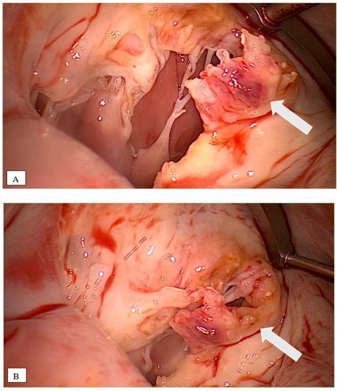
Two intraoperative clinical photographs (labeled A and B) demonstrating the gross pathology of infective endocarditis (IE) on the mitral valve. White arrows indicate large, friable, irregular vegetations attached to the valve leaflets. These vegetations exhibit a classic cauliflower-like morphology with a mottled appearance, featuring light tan tissue interspersed with dark reddish-purple areas suggestive of hemorrhage or focal necrosis. The surrounding mitral valve anatomy shows significant inflammatory disruption, including thickening of the leaflets and involvement of the subvalvular apparatus. These images illustrate the surgical presentation of destructive infective endocarditis, often associated with pathogens like Coxiella burnetii or Streptococcus gordonii, necessitating valve replacement. The clinical relevance highlights the transition from transesophageal echocardiography (TEE) findings to direct surgical visualization for assessing the extent of tissue destruction.
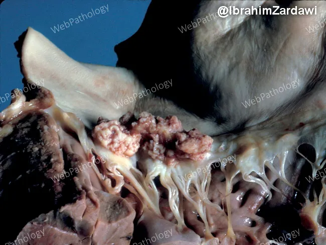
Gross pathology photograph of an explanted heart demonstrating left-sided infective endocarditis with mitral valve involvement. The image highlights a shaggy vegetative mass on the mitral valve, with thickened chordae tendineae and irregular nodularity of the cusp just to the right of the vegetation. The vegetations are friable and tan-pink, with adjacent valvular tissue showing edema and thickening. The chords appear markedly elongated and edematous, consistent with valvular destruction and tethering. The cusp shows focal thickening and rime-like nodules indicative of inflammatory deposition and possible microabscess formation. This gross morphology correlates with acute/subacute infective endocarditis, often due to bacterial pathogens such as Staphylococcus aureus or viridans streptococci, though fungal infections can also be implicated. Clinically, such findings signal bacteremia, embolic risk, and potential heart failure from valvular insufficiency. The image is valuable for education in cardiac pathology, surgical pathology, and infectious disease teaching; it demonstrates typical mitral valve vegetations, chordal involvement, and cusp irregularity useful for differential diagnosis with Libman-Sacks endocarditis, rheumatic disease, or marantic endocarditis. In practice, correlation with blood cultures, echocardiography, and systemic evaluation is essential for diagnosis and management. Documentation of vegetative morphology aids correlating radiographic and microbiological findings, informs prognosis, and guides rheology of therapeutic interventions management.
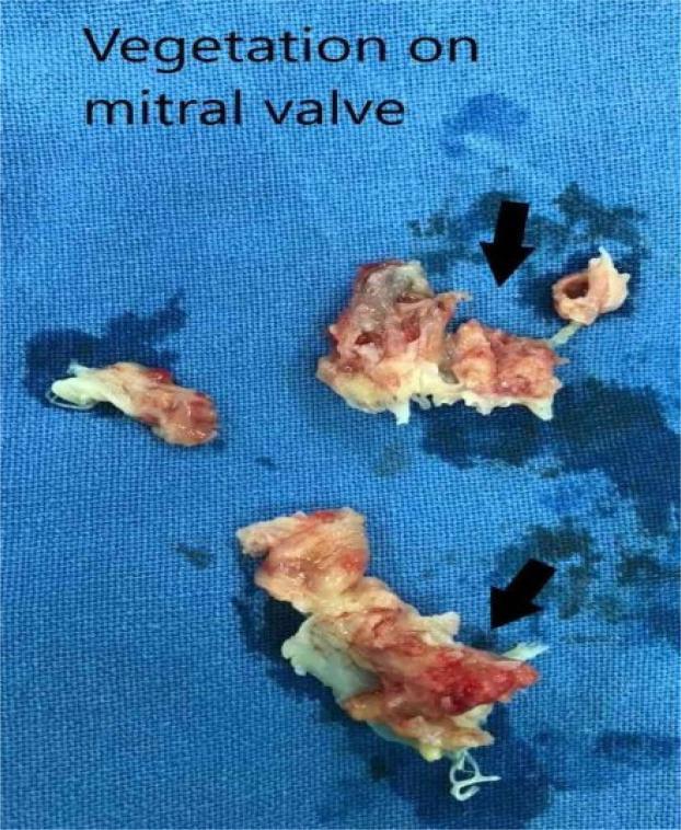
A clinical gross pathology photograph of surgically excised vegetations and destroyed mitral valve leaflets, placed on a blue surgical drape. The specimen consists of three irregular, friable tissue fragments with a variegated appearance, ranging from creamy white/yellowish tissue to deep red hemorrhagic areas. The texture is markedly rough, lobulated, and fragmented. Black arrows indicate specific areas of destroyed valvular tissue and large vegetations typical of infective endocarditis. Fine, white, filamentous chordae tendineae-like structures are visible attached to the lower tissue mass. The image demonstrates the extensive macroscopic damage and bulk of vegetation that can lead to valvular insufficiency and high embolic risk, requiring surgical intervention such as valve replacement. This visual is representative of advanced infective endocarditis affecting the mitral valve apparatus.
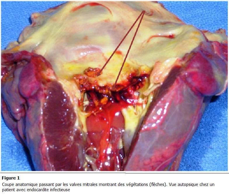
This clinical photograph displays a gross anatomical autopsy section of a human heart, focusing on the mitral valve apparatus. The image serves as a pathological specimen demonstrating infective endocarditis. Key findings include the presence of friable, irregular, brownish-red masses, identified as vegetations (indicated by arrows), attached to the mitral valve leaflets. The leaflets themselves show significant structural disruption, ulceration, and non-uniform thickening. Surrounding tissues exhibit signs of acute pathology, including hemorrhagic areas (deep red), inflammatory changes, and yellowish epicardial or valvular fat deposits. The underlying myocardium appears congested. This visual illustrates the hallmark gross features of valvular destruction and vegetation formation in bacterial endocarditis, highlighting the clinical relevance of the condition in causing valvular insufficiency and potential embolic events. The content is suitable for advanced medical education in pathology and cardiology, specifically regarding the morphological manifestations of endocardial infections.
Infective Endocarditis (IE)
Definition
Classification: Acute vs. Subacute
| Feature | Acute IE | Subacute IE |
|---|---|---|
| Organism virulence | High (e.g., S. aureus) | Low (e.g., S. viridans) |
| Valve affected | Normal or deformed | Usually deformed |
| Onset | Rapid, stormy | Insidious |
| Course | Severe, destructive | Protracted (weeks-months) |
| Treatment | Antibiotics + often surgery | Usually antibiotics alone |
| Valvular destruction | Severe | Less severe |
| Microscopy (base of vegetation) | Active inflammation | Granulation tissue (healing) |
A clear delineation between the two is not always possible; many cases fall along the spectrum.
Pathogenesis
Predisposing Cardiac Conditions
- Mitral valve prolapse - now the leading preexistent risk factor (as rheumatic disease has declined)
- Rheumatic heart disease (historically the major antecedent)
- Bicuspid aortic valve (calcified or not)
- Degenerative calcific valvular stenosis
- Prosthetic (artificial) heart valves - account for 10-20% of all IE cases
- Congenital heart defects
- Sterile platelet-fibrin deposits at pacemaker lines, indwelling vascular catheters, or endocardium damaged by flow "jets"
Host Risk Factors
- Neutropenia, immunodeficiency, malignancy
- Diabetes mellitus
- Alcohol use disorder
- Intravenous drug use (major risk - tricuspid valve involvement)
Causative Organisms
| Organism | Setting | Type of IE |
|---|---|---|
| Streptococcus viridans | Oral flora; community-acquired; damaged/deformed valves | Subacute; 50-60% of community cases |
| Staphylococcus aureus | Skin flora; healthcare settings; IV drug users; healthy or deformed valves | Acute; most common cause overall in high-income countries |
| Enterococci | Community and hospital | Variable |
| HACEK group (Haemophilus, Actinobacillus, Cardiobacterium, Eikenella, Kingella) | Oral commensals | Subacute |
| Gram-negative bacilli, Fungi | Rare; immunocompromised | Acute, severe |
| Culture-negative (~10%) | Prior antibiotics, difficult to isolate | - |
- Within 1-2 months of surgery: skin flora (S. aureus, S. epidermidis)
- More than 1 year post-surgery: streptococci and S. aureus
Portal of Entry (Bacteremia Sources)
- Dental or surgical procedures (transient bacteremia)
- Obvious infection elsewhere
- IV drug injection with contaminated needles
- Occult breaks in gut, oral cavity, or skin epithelium
- Trivial injuries
Morphology
- Fibrin
- Inflammatory cells
- Microorganisms
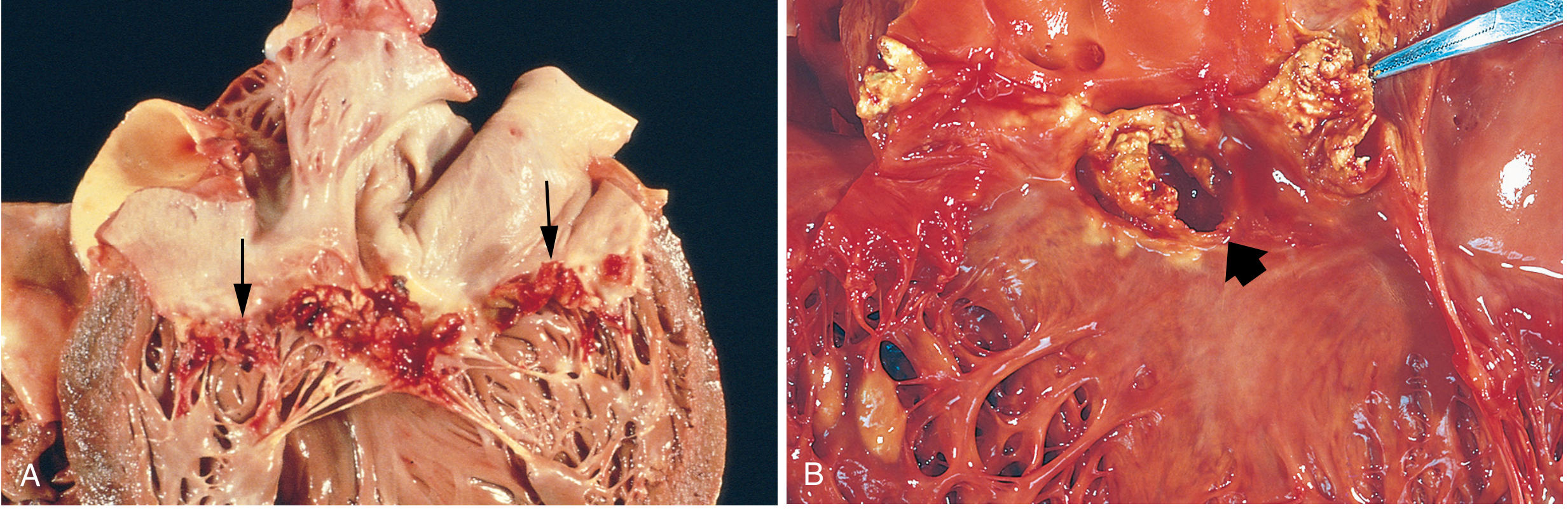
Sites of involvement
- Aortic and mitral valves - most common (left-sided)
- Tricuspid valve - frequent in IV drug users (right-sided)
Key morphological features
- Vegetations may be single or multiple
- Can involve more than one valve simultaneously
- Can erode into underlying myocardium producing an abscess cavity (ring abscess)
- Vegetations are prone to embolization due to their friable nature
- Embolic fragments containing organisms lead to:
- Septic infarcts at sites of lodgment
- Mycotic aneurysms (bacterial infection of arterial walls)
Microscopic distinction
- Subacute IE: granulation tissue at the base of vegetations (indicative of healing); with time, fibrosis, calcification, and chronic inflammatory infiltrate
- Acute IE: more active destruction, less healing response at base
Clinical Features
Systemic Features
- Fever - most consistent sign (may be absent or low-grade in subacute/elderly)
- Chills, weakness, lassitude
- Fatigue, weight loss, flu-like syndrome (subacute)
- Splenomegaly (common in subacute)
Cardiac
- Murmurs in 90% of patients with left-sided lesions (from new valvular defect or preexisting abnormality)
Peripheral Stigmata (embolic/immunologic)
| Finding | Description | Mechanism |
|---|---|---|
| Petechiae | Small skin/mucous membrane hemorrhages | Microemboli |
| Splinter (nail bed) hemorrhages | Linear hemorrhages under nails | Microemboli |
| Roth spots | Oval retinal hemorrhages with pale centers | Microemboli/immune |
| Janeway lesions | Small, erythematous/hemorrhagic, macular, non-tender lesions on palms and soles | Septic embolic events |
| Osler nodes | Small, tender subcutaneous nodules in pulp of digits; persist hours to days | Immune complex deposition |
Memory tip: Janeway = non-tender (J = Just painless); Osler = Oh-sore (tender)
Complications
- Glomerulonephritis - glomerular trapping of antigen-antibody complexes → hematuria, albuminuria, renal failure
- Septicemia / sepsis
- Arrhythmias - extension to underlying myocardium and conduction system
- Systemic embolization - stroke, splenic infarct, renal infarct, limb ischemia
- Mycotic aneurysms - weakening of arterial walls by infected emboli
- Ring abscess - erosion into myocardium at valve annulus
- Heart failure - from valvular destruction
Diagnosis: 2023 Modified Duke Criteria (Duke-ISCVID)
Pathologic Criteria (definitive)
- Microorganisms demonstrated by culture or histologic examination in a vegetation, embolic fragment, explanted prosthetic valve, or cardiac implantable electronic device (CIED)
Clinical Criteria
- Blood culture, nucleic acid-based technique, or immunofluorescence positive for a characteristic organism OR persistently positive for an unusual organism
- Echocardiographic/CT/PET imaging showing valve-related or implant-related oscillating mass, abscess, or partial separation of artificial valve
- New valvular regurgitation on imaging
- Direct inspection during heart surgery
- Predisposing heart lesion, prosthetic valve, CIED, or IV drug use
- Fever
- Vascular lesions (major arterial emboli, septic pulmonary infarcts, mycotic aneurysm, intracranial hemorrhage, conjunctival hemorrhages, Janeway lesions)
- Immunological phenomena (glomerulonephritis, Osler nodes, Roth spots, rheumatoid factor)
- Microbiologic evidence (single positive culture for unusual organism)
Prognosis
| Organism | Cure Rate |
|---|---|
| S. viridans (low virulence) | ~98% with antibiotics |
| Enterococci / S. aureus | 60-90% |
| Gram-negative bacilli / Fungi | ~50% survival |
| Prosthetic valve IE | Uniformly worse; valve re-replacement often needed |
- Left untreated, IE is generally fatal
- With appropriate long-term antibiotic therapy (6 weeks or more) and/or valve replacement, mortality is substantially reduced
- Adverse prognostic factors: septicemia, arrhythmias, systemic embolization
Comparison: Forms of Vegetative Endocarditis
| Feature | IE (Infective) | NBTE (Nonbacterial Thrombotic) | RHD (Rheumatic) | Libman-Sacks (SLE) |
|---|---|---|---|---|
| Vegetation size | Large, bulky, friable | Small (1-5 mm), bland | Small, warty | Small-medium |
| Location on valve | Cusps, can extend to chordae | Line of closure | Line of closure | Either/both sides of leaflets |
| Destructive | Yes | No | Minimal | No |
| Organisms | Yes | None (sterile) | None | None |
| Cause | Microbial infection | Hypercoagulable states (malignancy, DIC) | Strep-immune response | SLE autoimmune |
Sources: Robbins & Kumar Basic Pathology, p. 368-369; Robbins, Cotran & Kumar Pathologic Basis of Disease, pp. 528-529