
Surgical notes of tah with bso
Reading File
TAH-BSO stands for Total Abdominal Hysterectomy with Bilateral Salpingo-Oophorectomy - removal of the uterus, cervix, both fallopian tubes, and both ovaries via an abdominal approach.
Searching sources...
Finding Sources
Finding Sources
Reading File
Finding Sources
Reading File
Finding Sources
I now have comprehensive textbook content. Let me compile the complete surgical notes.
Loading Image
Here are comprehensive surgical notes for TAH-BSO (Total Abdominal Hysterectomy with Bilateral Salpingo-Oophorectomy):
Surgical Notes: TAH-BSO
Definition
Total Abdominal Hysterectomy with Bilateral Salpingo-Oophorectomy (TAH-BSO) = removal of the uterine corpus + cervix + both fallopian tubes + both ovaries via an open abdominal approach.
Indications
- Endometrial carcinoma
- Ovarian carcinoma
- Endometriosis with adnexal involvement
- Uterine fibroids (when vaginal/laparoscopic routes not suitable)
- Cervical carcinoma (as part of radical surgery)
- High-risk BRCA1/2 mutation carriers (risk-reduction surgery)
- Pelvic inflammatory disease with tubo-ovarian abscess unresponsive to treatment
Pre-operative Preparation
- Informed consent (including menopause, infertility, HRT discussion)
- Blood group & crossmatch
- Bowel preparation if colonic involvement anticipated
- DVT prophylaxis: TED stockings, LMWH
- Broad-spectrum antibiotic prophylaxis at induction
- Foley catheter insertion
- Patient positioned supine; Trendelenburg tilt may be used
Incision
Two options based on pathology, body habitus, and prior surgical history:
- Pfannenstiel (low transverse) - preferred for benign disease; cosmetically superior
- Vertical midline - preferred for malignancy, large uterus, need for greater exposure
Step-by-Step Operative Technique
Step 1 - Entry and Exploration
- Enter peritoneum; pack bowel superiorly
- Abdominal exploration to assess uterus, adnexa, and peritoneum
- Place self-retaining retractor (e.g., Balfour)
Step 2 - Uterine Elevation and Exposure
- Grasp the uterine fundus bilaterally with straight Kocher clamps or Lahey thyroid forceps
- Elevate the uterus out of the pelvis
Step 3 - Round Ligament Division
- Identify the round ligament on each side
- Clamp, cut, and ligate (suture ligation) the round ligament bilaterally
- This opens the anterior and posterior leaves of the broad ligament
Step 4 - Broad Ligament Dissection
- Incise the anterior leaf of the broad ligament toward the bladder fold
- Incise the posterior leaf of the broad ligament inferiorly
- This exposes the retroperitoneal space and allows ureteral identification
Step 5 - Bladder Dissection (Vesicouterine Pouch)
- Identify and incise the vesicouterine (uterovesical) peritoneal fold
- Dissect the bladder off the lower uterine segment and cervix using sharp dissection (Metzenbaum scissors) or electrocautery
- Retract the bladder inferiorly - this protects the bladder and ureters during subsequent steps
Step 6 - BSO: Division of Infundibulopelvic (IP) Ligament
- Critical step for BSO: Identify the ureter on the medial leaf of the broad ligament as it courses to the bladder
- Doubly clamp the infundibulopelvic (IP) ligament (contains the ovarian vessels) with curved Heaney or Zeppelin clamps
- Divide between the clamps with curved Mayo scissors
- Doubly ligate both pedicles (proximal and distal) to prevent back-bleeding
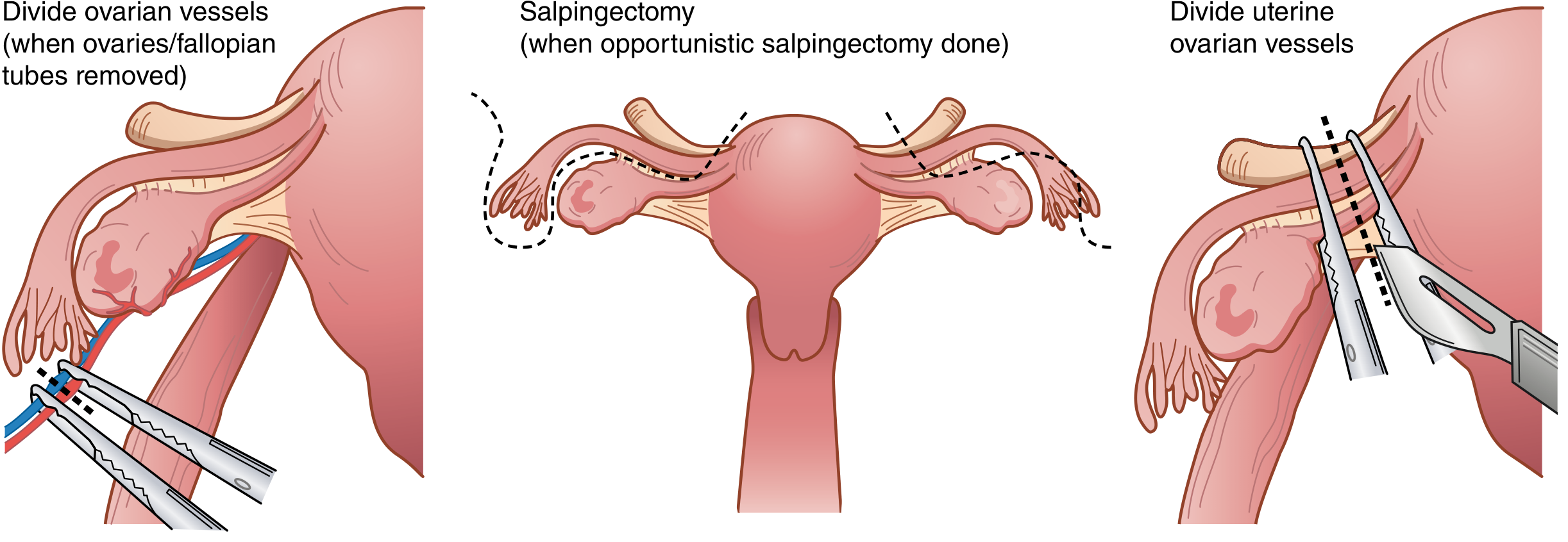
Step 7 - Skeletonization of Uterine Vessels
- Incise the remaining leaves of the posterior broad ligament inferiorly
- Skeletonize the uterine vessels as they travel along the lateral edge of the uterus
- The posterior broad ligament is cleared down to the uterosacral ligaments
Step 8 - Uterine Vessel Ligation
- Place a curved Heaney or Zeppelin clamp across the uterine vessels at the level of the internal cervical os, perpendicular to the uterus
- Cut and suture ligate the pedicle (transfixion stitch)
- Repeat on the opposite side
Step 9 - Cardinal and Uterosacral Ligament Division
- Further dissect the bladder completely below the level of the cervix
- Apply straight Heaney or Zeppelin clamps across the cardinal ligaments and uterosacral ligaments in serial steps bilaterally
- Cut and suture ligate each pedicle
- Continue until the level of the external cervical os is reached on both sides
Step 10 - Colpotomy (Vaginal Cuff Excision)
Two methods:
Method A (Clamp technique):
- Place curved clamps across the vagina medial to the prior pedicles, just below the cervix from both sides meeting in the midline
- Cut just above the clamps with curved scissors to separate the uterus and cervix from the vagina
Method B (Direct sharp incision):
- Make a direct sharp incision into the vagina just below the cervix
- Extend the incision around the cervix with curved scissors until the cervix is completely freed
- Grasp vaginal edges with Allis clamps for visualization
Step 11 - Specimen Removal
- The uterus, cervix, both fallopian tubes, and both ovaries are removed as one specimen
- Send for histopathology
Step 12 - Vaginal Cuff Closure
- Heaney transfixion stitches placed beneath the two vaginal clamps to ligate vaginal corners
- Vagina closed in the midline with:
- Interrupted figure-of-eight sutures, OR
- Running suture, OR
- Interrupted sutures
- Lateral apex sutures incorporate the ipsilateral uterosacral ligament for apical support (prevents vault prolapse)
Step 13 - Hemostasis and Closure
- Inspect all pedicles for hemostasis
- Check bladder integrity (may fill bladder with methylene blue if injury suspected)
- Irrigate the pelvis
- Visualize ureters for peristalsis
- Close the abdomen in layers (peritoneum optional, fascia with looped PDS or Vicryl, subcutaneous tissue, skin)
Specimen
| Structure | Removed |
|---|---|
| Uterine corpus | Yes |
| Cervix | Yes |
| Both fallopian tubes | Yes |
| Both ovaries | Yes |
| Parametrium | No (unlike radical/Wertheim's) |
Important Anatomical Danger Points
- Ureters - most common structure at risk; injury most likely at IP ligament ligation and uterine vessel clamp
- Bladder - during vesicouterine dissection
- Uterine vessels / iliac vessels - hemorrhage
- Rectum - during uterosacral ligament division in cases with endometriosis/adhesions
Post-operative Care
- Foley catheter for 24 hours
- Early mobilization and DVT prophylaxis
- NSAIDS + opioid analgesia as required
- Monitor urine output, hemoglobin
- HRT counselling (surgical menopause if pre-menopausal)
- Discharge typically Day 3-5
Complications
Intraoperative:
- Hemorrhage
- Ureteric injury
- Bladder injury
- Bowel injury
Early post-operative:
- Wound infection/dehiscence
- DVT/PE
- Urinary tract infection
- Ileus
Late:
- Vault prolapse
- Urinary dysfunction (urge/stress incontinence)
- Sexual dysfunction
- Premature menopause (if pre-menopausal) - risk of cardiovascular disease, osteoporosis
- Lymphoedema (if lymphadenectomy performed with malignancy cases)
Source: Sabiston Textbook of Surgery - The Biological Basis of Modern Surgical Practice, Chapter 120 (Hysterectomy section)
This is a shared conversation. Sign in to Orris to start your own chat.