
Hypopituitarism
Reading File
Now let me search the medical library for hypopituitarism content in parallel.
Finding Sources
Finding Sources
Searching PubMed
"hypopituitarism" AND "diagnosis" AND "treatment"
Good results. Now let me read the most comprehensive sources - Harrison's and Goldman-Cecil (the primary internal medicine textbooks) - in parallel.
Reading File
Reading File
Reading File
Excellent content gathered. Let me now read the Goldman-Cecil causes table and Harrison's diagnostic/treatment section to complete the picture.
Reading File
Reading File
Let me read the diagnostic testing and management sections from Harrison's.
Reading File
Reading File
Let me read the treatment table and hormone replacement details.
Reading File
Reading File
Let me get the hormone replacement table from Goldman-Cecil for completeness.
Reading File
Let me get the hormone replacement therapy table from Goldman-Cecil.
Reading File
Reading File
I now have comprehensive content from Harrison's 22E and Goldman-Cecil Medicine. Let me also fetch the hypothalamic-pituitary axis diagram to include visually.
Loading Image
I now have all the information needed to write a comprehensive answer. Let me compile everything.
Hypopituitarism
Hypopituitarism is a deficiency of one or more anterior pituitary hormones, resulting from damage to or dysfunction of the pituitary gland itself or the hypothalamus. When all pituitary hormones are deficient, it is called panhypopituitarism.
Prevalence: 290-455 cases per million; incidence ~42 per million per year.
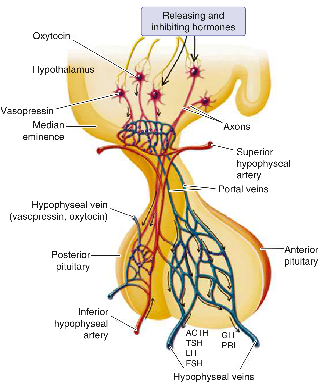
Etiology
Congenital / Developmental
| Category | Examples |
|---|---|
| Midline defects | Septo-optic dysplasia, anencephaly, cleft lip/palate, basal encephalocele |
| Genetic transcription factor mutations | HESX1, SOX2/3, LHX3/4, PROP1, PIT1, OTX2, GLI2, PAX6 (>50 genes implicated) |
| Specific gene mutations | PROP1 (GH + PRL + TSH + gonadotropin deficiency); PIT-1 (GH + PRL + TSH); TPIT (ACTH deficiency); Kallmann syndrome (GnRH defect + anosmia) |
| Pituitary aplasia/hypoplasia | Absent or rudimentary gland on MRI |
Acquired
| Category | Examples |
|---|---|
| Tumors (most common) | Pituitary macroadenoma (most common), craniopharyngioma, germinoma, meningioma, glioma, Rathke's cleft cyst, metastases (breast, lung) |
| Iatrogenic | Surgery, radiotherapy (can present years later) |
| Vascular | Sheehan syndrome (postpartum ischemic necrosis), pituitary apoplexy, subarachnoid hemorrhage, internal carotid aneurysm, sickle cell disease |
| Traumatic | TBI (increasingly recognized), surgical trauma |
| Infiltrative / Inflammatory | Lymphocytic hypophysitis (most common autoimmune - associated with pregnancy and anti-CTLA-4 therapy), sarcoidosis, histiocytosis X, granulomatous hypophysitis, IgG4-related hypophysitis |
| Infectious | TB, syphilis, meningitis, fungal infections (histoplasmosis), toxoplasmosis |
| Metabolic | Hemochromatosis (iron deposition), amyloidosis, critical illness, anorexia nervosa |
| Drug-induced | CTLA-4 inhibitors, PD-1/PD-L1 inhibitors (immune checkpoint therapy) |
| Structural | Empty sella syndrome (~10% have clinically apparent hypopituitarism) |
| Idiopathic | Some sporadic and familial cases |
Key pathologic note (Sheehan syndrome): During pregnancy, the anterior pituitary enlarges due to prolactin cell hyperplasia, but blood supply from the low-pressure hypophyseal portal system does not increase proportionately. The gland is therefore vulnerable to ischemic injury from obstetric hemorrhage. The posterior pituitary receives direct arterial supply and is much less susceptible. - Robbins & Kumar Basic Pathology
Sequence of Hormone Loss
When pituitary compression or destruction occurs progressively, trophic hormone failure follows a characteristic sequence:
GH → FSH → LH → TSH → ACTH
- GH is lost first and most commonly (present in up to 60% of hypopituitary patients)
- ACTH deficiency is a late and potentially life-threatening complication
- In children, growth failure is the presenting feature; in adults, hypogonadism is usually the earliest symptom
Clinical Manifestations
Each hormone deficiency produces distinct features:
| Deficient Hormone | Clinical Effects |
|---|---|
| GH | Children: growth retardation, short stature. Adults: abnormal body composition (↑fat, ↓muscle mass), fatigue, decreased exercise tolerance |
| LH/FSH (Gonadotropins) | Women: amenorrhea, infertility, loss of secondary sexual characteristics. Men: decreased libido, erectile dysfunction, infertility, loss of pubic/axillary hair |
| TSH | Secondary hypothyroidism: fatigue, cold intolerance, constipation, weight gain (features milder than primary hypothyroidism) |
| ACTH | Secondary adrenal insufficiency: hypocortisolism, fatigue, hypotension, hypoglycemia. Mineralocorticoid production is relatively preserved (unlike primary Addison's). Skin pallor (loss of MSH from POMC) |
| PRL | Failure of postpartum lactation |
| AVP (posterior pituitary) | Cranial diabetes insipidus: polyuria, polydipsia (indicates hypothalamic involvement) |
Hyperprolactinemia (paradoxically) can occur in hypothalamic lesions - interruption of dopaminergic inhibition disinhibits prolactin release. Diabetes insipidus in the setting of hypopituitarism almost always points to hypothalamic origin rather than isolated pituitary disease.
General features (often subtle for years, especially in men and post-reproductive women): fatigue, decreased muscle strength, increased body fat, skin pallor, and nondescript symptoms.
Diagnosis
Biochemical
Diagnosis is made by demonstrating low trophic hormone + low/inappropriately normal target organ hormone:
- Low free T4 + low or normal TSH → secondary hypothyroidism
- Low testosterone/estradiol + low or normal LH/FSH → hypogonadotropic hypogonadism
- Low morning cortisol + low/normal ACTH → secondary adrenal insufficiency
If two or more hormones are deficient, panhypopituitarism is likely, and all axes should be tested.
Provocative / Stimulation Tests (Goldman-Cecil Table 205-2)
| Hormone | Test | Normal Response |
|---|---|---|
| GH | Insulin tolerance test (ITT): Regular insulin 0.05-0.15 U/kg IV; draw GH at 0, 30, 45, 60, 90 min | GH >5 μg/L when glucose <40 mg/dL |
| GH | Arginine-GHRH test | GH >4.1 μg/L |
| GH | Glucagon test (1 mg IM) | GH >3 μg/L |
| GH | Macimorelin (oral ghrelin agonist) | Newer alternative |
| ACTH | ITT (as above, cortisol arm) | Cortisol rise >7 μg/dL or peak >20 μg/dL |
| ACTH | Cosyntropin (ACTH₂₄) 0.25 mg IM/IV | Cortisol >18 μg/dL at 30-60 min |
| ACTH | CRH test (1 μg/kg IV) | ACTH 2-4 fold rise; cortisol 20-25 μg/dL |
| TSH | Basal free T₃, free T₄, TSH | Low free T₄ + low/normal TSH |
The ITT is the gold standard for assessing both GH and ACTH reserve simultaneously but is contraindicated in active coronary artery disease and seizure disorders.
Imaging
MRI of the pituitary (with and without gadolinium contrast) is the preferred modality. Sagittal and coronal views are most useful. Normal pituitary enhances more than tumors on post-contrast T1. CT can be used when MRI is contraindicated.
Treatment
Treatment has two components: (1) replace deficient hormones and (2) address the underlying cause.
Hormone Replacement Therapy (Goldman-Cecil Table 205-3)
| Axis | Replacement | Dose |
|---|---|---|
| GH | Recombinant GH (SC) | Children: 0.25 mg/kg/day. Adults: 0.3-1.2 mg/day (titrate to IGF-1 in mid-upper normal range) |
| ACTH/Cortisol | Hydrocortisone (PO) | 10-15 mg AM + 5-10 mg PM; or prednisone 2.5 mg AM + 2.5 mg PM. Stress dosing: hydrocortisone 50-75 mg IV q8h |
| TSH/Thyroid | L-thyroxine (PO) | 0.075-0.15 mg daily (always replace cortisol FIRST to avoid precipitating adrenal crisis) |
| LH/FSH - Women | Conjugated estrogens 0.625-1.25 mg (days 1-25) + medroxyprogesterone 5-10 mg (days 15-25); or combined OCP | For fertility: exogenous FSH + LH (or hCG) |
| LH/FSH - Men | Testosterone enanthate 100-300 mg IM q1-3 weeks; or testosterone gel 5-10 g/day | For fertility: hCG ± FSH to induce spermatogenesis |
| AVP (DI) | Desmopressin (DDAVP) | 5-20 μg intranasally once or twice daily; or 0.1-0.4 mg PO q8-12h |
| Prolactin | No replacement available | - |
Critical warning: Always replace glucocorticoids before initiating thyroid hormone replacement. Starting thyroxine first in a patient with undiagnosed adrenal insufficiency can precipitate an adrenal crisis by increasing cortisol metabolism.
Underlying Cause Management
- Pituitary adenoma: Transsphenoidal surgery (also for compressive mass effect)
- Radiation: Used postoperatively for residual tumor; pituitary function must be monitored long-term
- Lymphocytic hypophysitis: Physiologic glucocorticoids; surgery only for biopsy or significant mass effect; spontaneous resolution is possible
- Perioperative: Stress-dose hydrocortisone 50-75 mg IV q8h
Prognosis
- Patients with untreated or inadequately treated hypopituitarism have significantly increased mortality, primarily from cardiovascular and cerebrovascular disease
- Previous head/neck irradiation is an additional determinant of increased cerebrovascular mortality
- With appropriate hormone replacement, most patients can maintain satisfactory clinical homeostasis
Sources:
- Harrison's Principles of Internal Medicine 22E (2025), Chapter 391 - Hypopituitarism (Melmed & Jameson)
- Goldman-Cecil Medicine, Chapter 205 - Table 205-1 (Etiology), Table 205-2 (Diagnostic Tests), Table 205-3 (Hormone Replacement)
- Robbins & Kumar Basic Pathology and Robbins, Cotran & Kumar Pathologic Basis of Disease - Sheehan syndrome and pituitary necrosis
This is a shared conversation. Sign in to Orris to start your own chat.