
Tenis corporis
Tenia corporis
tinea corporis ringworm skin lesion
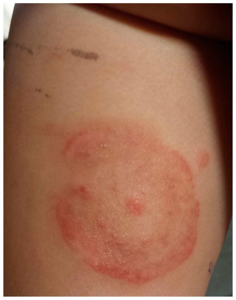
This clinical photograph displays a dermatological presentation of tinea corporis (ringworm) in a pediatric patient. The primary feature is an annular, erythematous plaque located on the trunk or proximal limb. The lesion is characterized by a well-demarcated, raised red border with evidence of mild scaling or vesiculation, indicating active peripheral growth. The center of the plaque shows partial clearing with a lighter pink hue, though it remains occupied by a few scattered inflammatory papules. Above the primary lesion, incidental linear hyperpigmented marks are visible, likely representing excoriations or minor superficial trauma. The image serves as a classic educational example of a fungal skin infection, specifically demonstrating the centrifugal spread and central clearing characteristic of Microsporum canis dermatophytosis.
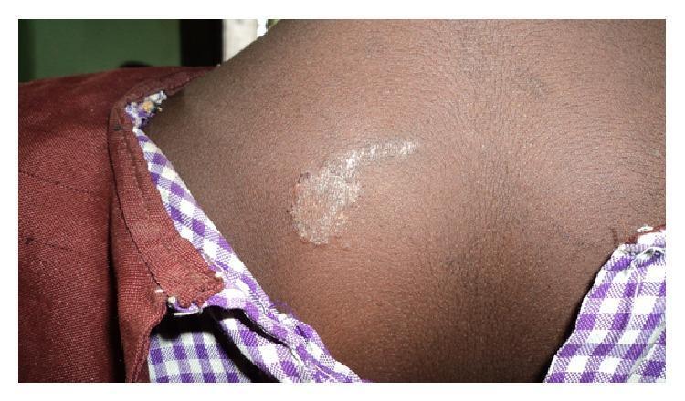
Clinical photograph of a skin lesion on the upper back of a pediatric patient with a dark skin tone, consistent with Tinea corporis (ringworm). The image demonstrates a hypopigmented, well-circumscribed patch with an irregular, slightly raised, and scaly border. Centrally, the lesion shows characteristic clearing but maintains a whitish, fine-textured scaling. A linear extension of the scaling is visible, trailing away from the primary circular focus. This presentation illustrates a common superficial fungal infection (dermatophytosis) of the glabrous skin. The anatomical location is the posterior trunk near the right scapular region. The visual features are diagnostic of a fungal etiology, highlighting the annular morphology and peripheral scale distribution typically seen in localized tinea infections.
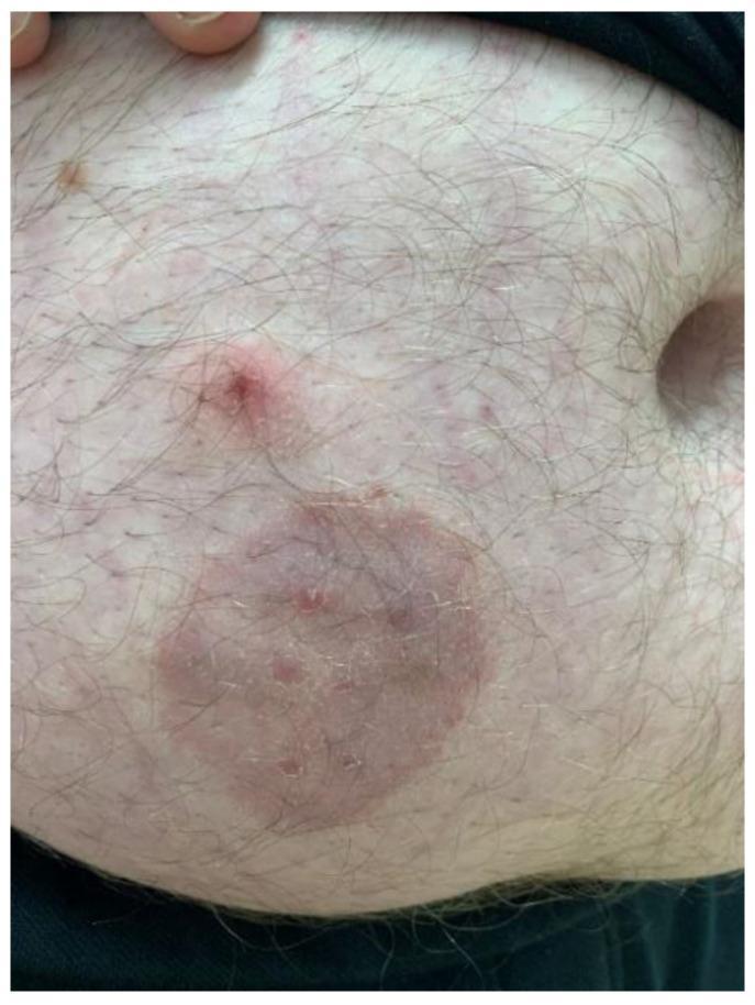
This clinical photograph displays a dermatological presentation of tinea corporis (zoonotic dermatophytosis) on the abdomen of an adult male. The primary feature is a well-circumscribed, annular, erythematous plaque approximately 4-5 cm in diameter. The lesion exhibits a classic 'ringworm' morphology with a slightly raised, scaly inflammatory border and a partially cleared central area containing subtle papular elements. Superior to the primary plaque is a secondary, smaller, raised erythematous papule with a central punctum or crust. The surrounding skin shows mild diffuse erythema and scattered smaller inflammatory macules. Hair follicles are visible throughout the affected and unaffected regions. This image serves as an educational example of fungal infection (likely Trichophyton mentagrophytes) transmitted from a pet (guinea pig) to a human host, emphasizing the visual diagnostic features of annular inflammatory skin lesions.
tinea corporis dermatophytosis treatment
Tinea Corporis
Etiology
| Genus | Species | Notes |
|---|---|---|
| Trichophyton | T. rubrum | Most common overall; may cause polycyclic/psoriasiform lesions |
| Trichophyton | T. mentagrophytes / T. interdigitale | Common; zoonotic strains from bamboo rats can cause epidemics |
| Trichophyton | T. indotineae | Emerging species, previously classified as T. mentagrophytes var. Vill |
| Trichophyton | T. tonsurans | "Tinea gladiatorum" in wrestlers |
| Microsporum | M. canis | Zoophilic; from dogs and cats, especially in children |
| Epidermophyton | E. floccosum | Less common |
- Dermatology 2-Volume Set 5e, p. 1604; Andrews' Diseases of the Skin, p. 347
Epidemiology
-
Transmitted by direct contact with infected humans or animals, via fomites, or by autoinoculation (e.g., from tinea pedis reservoirs).
-
Children are more prone to zoophilic pathogens (M. canis) from pets.
-
Occlusive clothing and humid climates worsen frequency and severity.
-
Tinea gladiatorum: seen in wrestlers; caused mainly by T. tonsurans; affects head, neck, and arms due to skin-to-skin contact and mat burns.
-
Fitzpatrick's Dermatology, p. 2974
Clinical Features
- A well-demarcated, raised, erythematous, scaly advancing border that spreads centrifugally
- Central clearing (the hallmark that gives it the name "ringworm")
- Scaling across the entire active border; the border may be vesicular
- Polycyclic plaques - multiple overlapping rings, especially in immunosuppressed patients (T. rubrum)
- Psoriasiform - mimics psoriasis, also typically T. rubrum
- Tinea incognita - atypical appearance with no raised border due to treatment with topical steroids or calcineurin inhibitors; may lack central clearing and look eczematous
- Bullous tinea - vesicles and bullae, particularly on the feet
- Majocchi granuloma - deeper follicular involvement; follicular papules and nodules in an annular arrangement; seen in women after shaving or after topical steroid use, and in immunocompromised patients; caused most often by T. rubrum
Diagnosis
- KOH preparation: Skin scrapings from the active border are examined under the microscope for branching hyphae. The most common artifact is "mosaic false hyphae" at junctions of normal keratinocytes. Cotton/synthetic fibers may also mimic hyphae.
- Fungal culture: On Sabouraud dextrose agar; growth within 1-2 weeks; identifiable to genus/species level by colony morphology.
- Skin biopsy: For chronic, refractory cases; may reveal tinea incognita.
- Andrews' Diseases of the Skin, p. 347
Differential Diagnosis
| Category | Conditions |
|---|---|
| Most likely | Erythema annulare centrifugum, nummular eczema, psoriasis, tinea versicolor, subacute cutaneous lupus erythematosus, cutaneous candidiasis |
| Consider | Contact dermatitis, atopic dermatitis, pityriasis rosea, seborrheic dermatitis |
| Rule out | Mycosis fungoides, parapsoriasis, secondary syphilis |
- Fitzpatrick's Dermatology, p. 2974
Treatment
Topical Therapy (first-line for localized disease)
| Class | Agents | Frequency | Duration |
|---|---|---|---|
| Allylamines | Terbinafine (Lamisil), Naftifine (Naftin), Butenafine (Mentax) | Once or twice daily | 1-2 weeks (terbinafine can be 1 week) |
| Imidazoles | Clotrimazole, Miconazole, Econazole, Ketoconazole, Oxiconazole, Sulconazole | Once or twice daily | 2-4 weeks |
| Other | Ciclopirox olamine, Tolnaftate | Twice daily | 2-4 weeks |
- Most topical agents are effective; choice should be guided by cost-effectiveness.
- Avoid combination products with potent corticosteroids (e.g., clotrimazole/betamethasone) - these cause widespread tinea and fungal folliculitis.
Systemic Therapy (for extensive or follicular disease)
| Drug | Notes |
|---|---|
| Terbinafine | Preferred for Trichophyton spp.; shorter courses |
| Griseofulvin | Better for Microsporum spp. |
| Itraconazole | Effective for T. rubrum, T. tonsurans, T. mentagrophytes |
| Fluconazole | Also effective |
- Andrews' Diseases of the Skin, p. 347-348; Fitzpatrick's Dermatology, p. 2974
Special Forms Summary
| Form | Key Feature |
|---|---|
| Tinea gladiatorum | Wrestlers; head, neck, arms; T. tonsurans |
| Tinea imbricata | Concentric rings; T. concentricum; geographically restricted |
| Tinea incognita | Atypical morphology from steroid/calcineurin inhibitor suppression |
| Majocchi granuloma | Deep follicular involvement; after shaving or steroid use |
| Bullous tinea | Vesicles/bullae; often on feet |
Treatment
tinea corporis treatment antifungal
Treatment of Tinea Corporis
Step 1 - Decide: Topical vs. Systemic
| Scenario | Approach |
|---|---|
| Localized plaques, glabrous skin, no follicular involvement | Topical (first-line) |
| Widespread/extensive disease | Systemic oral |
| Follicular involvement (Majocchi granuloma) | Systemic oral (mandatory) |
| Tinea incognita | Stop topical steroids + systemic oral |
| Poor response after 2 weeks of topical therapy | Switch to systemic |
| Immunocompromised patients | Systemic oral |
Topical Antifungals (Localized Disease)
| Class | Drug | Concentration | Frequency | Min Age |
|---|---|---|---|---|
| Allylamine | Terbinafine (Lamisil) | 1% cream | Once daily | ≥12 y |
| Allylamine | Naftifine (Naftin) | 2% cream | Once daily | ≥12 y |
| Benzylamine | Butenafine (Mentax) | 1% cream | Once daily | ≥12 y |
| Imidazole | Clotrimazole (Lotrimin) | 1% cream/solution | Twice daily | All ages |
| Imidazole | Miconazole (Monistat) | 2% cream | Twice daily | ≥2 y |
| Imidazole | Econazole (Spectazole) | 1% cream | Once or twice daily | All ages |
| Imidazole | Ketoconazole (Nizoral) | 2% cream | Once daily | All ages |
| Imidazole | Oxiconazole (Oxistat) | 1% cream | Once or twice daily | All ages |
| Imidazole | Luliconazole | 1% cream | Once daily | ≥2 y |
| Imidazole | Sulconazole (Exelderm) | 1% cream/solution | Once or twice daily | Adults only |
| Other | Ciclopirox (Loprox) | 0.77% cream | Twice daily | ≥10 y |
| Other | Tolnaftate | 1% cream/solution | Twice daily | All ages |
-
Allylamines and benzylamines may give a more sustained clinical cure compared to azoles.
-
Terbinafine course can be shortened to 1 week for tinea corporis.
-
Econazole and ciclopirox have additional antibacterial coverage - useful if bacterial superinfection is suspected.
-
Choice should be guided by cost-effectiveness and age appropriateness.
-
Goodman & Gilman's Pharmacological Basis of Therapeutics, p. 1506; Red Book 2021, p. 1169-1170
What NOT to Use
Combination topical antifungal + corticosteroid products (e.g., clotrimazole/betamethasone) are CONTRAINDICATED in tinea corporis.
- Widespread dissemination of tinea (tinea incognita)
- Fungal folliculitis and Majocchi granuloma
- Higher relapse rates
- Increased cost and corticosteroid side effects
Systemic (Oral) Antifungals (Widespread or Follicular Disease)
| Drug | Notes | Species Preference |
|---|---|---|
| Terbinafine | Preferred for Trichophyton spp.; fewer drug interactions; short courses possible | Trichophyton > Microsporum |
| Itraconazole | Effective; significant P450 drug interactions; avoid in CHF/ventricular dysfunction | Broad spectrum |
| Fluconazole | Effective; approved in children ≥6 months for other indications; no FDA approval specifically for tinea corporis | Broad spectrum |
| Griseofulvin | Traditional option; better for Microsporum spp.; approved by FDA for dermatophyte skin/hair/nail infection; common side effects: GI distress, headache, urticaria | Microsporum > Trichophyton |
-
Trichophyton spp. - prefer terbinafine (shorter courses)
-
Microsporum spp. - respond better to griseofulvin
-
Harrison's Principles of Internal Medicine 22e (2025), p. N/A; Andrews' Diseases of the Skin, p. 348; Goodman & Gilman's, p. 1506
Special Situations
| Situation | Treatment |
|---|---|
| Majocchi granuloma | Oral antifungal mandatory (topical insufficient); terbinafine or itraconazole |
| Tinea incognita | Discontinue topical steroids/calcineurin inhibitors + systemic antifungal |
| Tinea gladiatorum | Oral terbinafine or griseofulvin; treat contacts; decontaminate mats |
| Immunocompromised patient | Systemic therapy preferred; longer courses |
| Recurrent tinea corporis | Treat concurrent onychomycosis (tinea unguium) and tinea pedis to reduce source |
Monitoring & Follow-Up
-
Reassess at 2 weeks - if no significant improvement, reconsider diagnosis or switch to systemic therapy.
-
Topical therapy: continue for 1 week beyond clinical resolution to prevent relapse.
-
Oral agents: monitor liver function (hepatotoxicity risk for all oral antifungals).
-
Address predisposing factors: reduce moisture, avoid occlusive clothing, treat concurrent tinea at other sites.
-
Red Book 2021 (Committee on Infectious Diseases), p. 1170; Harrison's 22e, p. N/A