
Short notes on esophageal perforation
esophageal perforation Boerhaave syndrome chest CT pneumomediastinum
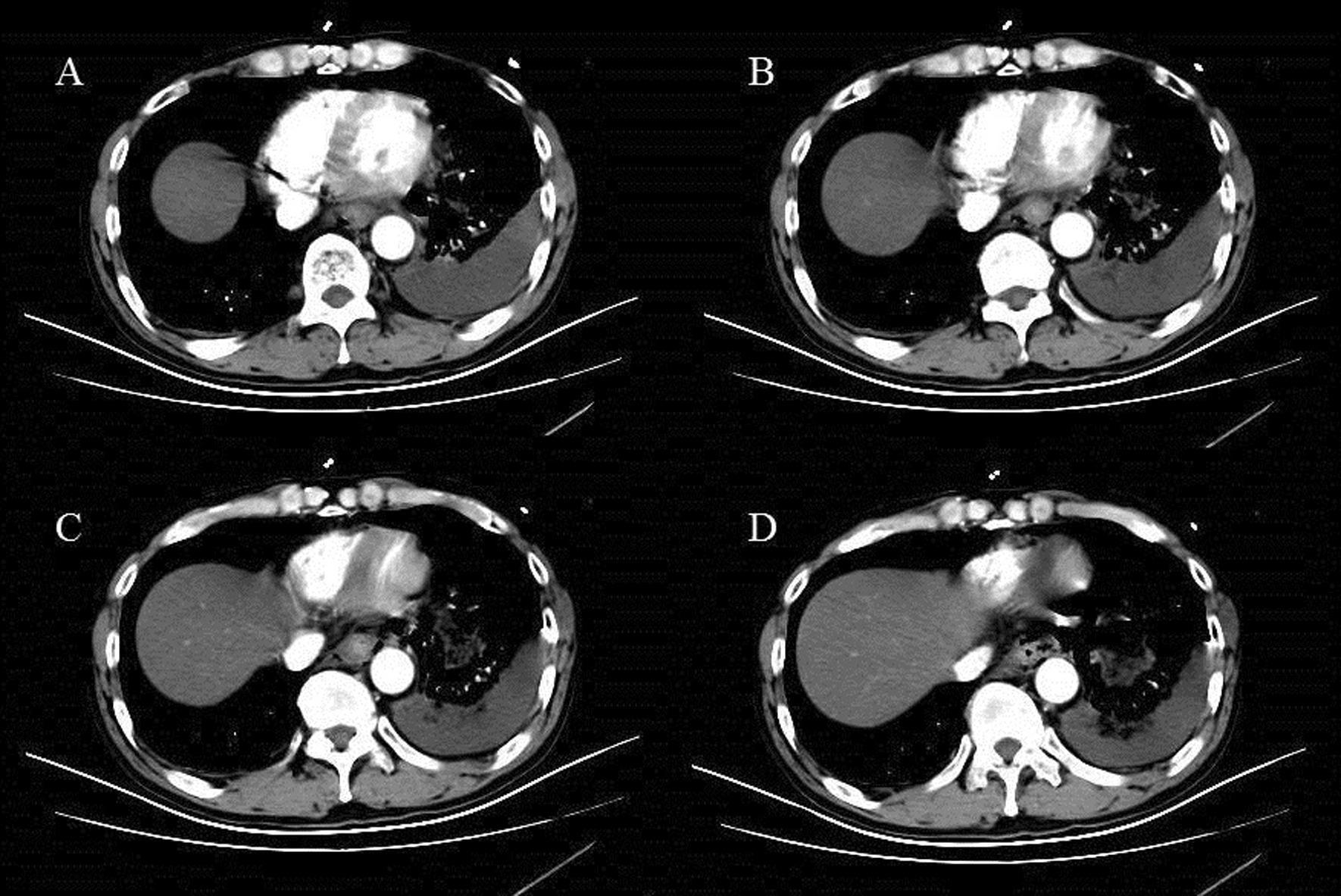
Axial contrast-enhanced CT scans of the chest (labeled A-D) demonstrating hallmark features of Boerhaave’s syndrome (spontaneous esophageal rupture). The images reveal significant pneumomediastinum, characterized by pockets of free extraluminal air tracking around the heart, distal esophagus, and descending aorta. There is clear evidence of air dissecting laterally toward the left pleural space. A large, heterogeneous left-sided pleural effusion is present, causing compressive atelectasis of the adjacent left lower lobe and a mild rightward shift of the mediastinal structures. Small amounts of subcutaneous emphysema are visible in the anterior chest wall. These findings illustrate the typical radiographic progression of esophageal perforation where air and gastrointestinal contents leak into the mediastinum and subsequently into the pleural cavity, leading to hydropneumothorax or complex effusion.
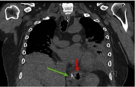
This non-contrasted coronal CT scan of the chest and upper abdomen demonstrates diagnostic features of Boerhaave's Syndrome (spontaneous esophageal perforation). A prominent red arrow points to an extraluminal pocket of gas (pneumomediastinum) located in the lower mediastinum, positioned left-lateral to the distal esophagus. Adjacent to this air collection, a green arrow identifies a nasogastric (NG) tube situated within the esophageal lumen, providing a landmark for the esophageal anatomy. The mediastinal air is seen near the diaphragmatic hiatus, a common site for pressure-induced rupture. The surrounding lung fields show partial opacification and are somewhat obscured by mediastinal structures, though no large pleural effusions are immediately apparent in this view. This imaging is critical for differentiating esophageal rupture from other causes of acute chest pain, such as aortic dissection or myocardial infarction, particularly when clinical history involves forceful vomiting or retching.
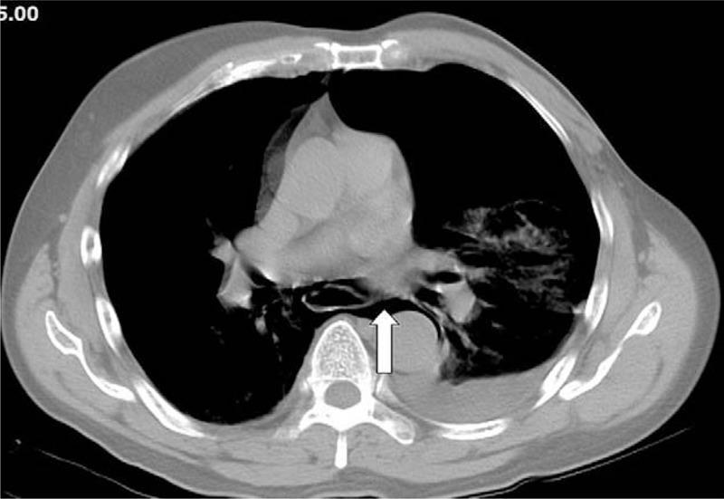
Axial contrast-enhanced thoracic CT scan at the level of the pulmonary arteries. The image demonstrates a prominent pneumomediastinum, characterized by extraluminal air (low-attenuation/black pockets) dissecting through the mediastinal planes and encompassing the peri-esophageal space (indicated by a white arrow). This finding is highly suggestive of Boerhaave syndrome (spontaneous esophageal perforation) given the clinical context of post-emetic chest pain. Additionally, there is a significant left-sided pleural effusion/hemothorax appearing as a hyperdense fluid collection in the dependent portion of the left hemithorax, with associated compressive atelectasis of the adjacent left lower lobe parenchyma. The right hemithorax appears clear. This diagnostic image is intended for medical education regarding the imaging hallmarks of esophageal rupture and secondary mediastinal air and pleural complications.
Esophageal Perforation - Short Notes
Definition
Historical Note - Boerhaave's Syndrome
Etiology / Causes
| Category | Examples |
|---|---|
| Iatrogenic (most common) | Endoscopy, esophageal dilation, variceal sclerotherapy, nasogastric tube placement, intraoperative injury |
| Spontaneous (Boerhaave's) | Forceful vomiting, seizure, straining at stool, weight lifting, childbirth, coughing |
| Trauma | Penetrating (stab/gunshot), blunt (rare) |
| Foreign body | Ingested sharp objects, button batteries, caustic ingestion |
| Tumor | Intrinsic or extrinsic cancer eroding into esophagus |
| Aortic pathology | Aortic aneurysm, aberrant right subclavian artery |
| Miscellaneous | Barrett's esophagus, Zollinger-Ellison syndrome, infection (rare) |
Key fact: Iatrogenic perforation is the most common overall cause. Boerhaave's syndrome accounts for ~15% of cases but carries the highest mortality because massive mediastinal contamination is nearly instantaneous.
Boerhaave epidemiology: >80% of cases are middle-aged men after alcohol ingestion and large meals.
Pathophysiology
- The esophagus is the only GI organ lacking a serosal layer - this allows direct contamination of adjacent spaces once the wall is breached.
- Cervical perforations enter the retropharyngeal space; fascial planes extend from the skull base to the tracheal bifurcation.
- Mid/lower esophageal perforations enter the mediastinum directly; only the thin mediastinal pleura prevents free access to the pleural cavity.
- Negative respiratory pressure promotes drainage from the GI tract into the mediastinum and pleural space, worsening contamination.
- Results in: fulminant necrotizing mediastinitis, chemical and bacterial pneumonitis, empyema, and septic shock.
- Cricopharyngeal muscle (esophageal introitus)
- Level of left main bronchus and aortic arch
- Gastroesophageal junction
Clinical Features
- Vomiting / retching
- Chest pain (acute, severe, unrelenting)
- Subcutaneous emphysema
- Severe, diffuse chest pain - radiating to back and shoulders; exacerbated by swallowing
- Dysphagia, odynophagia
- Dyspnea, hematemesis, cyanosis
- Fever, tachycardia, tachypnea - appear early
- Abdominal rigidity with hypotension
- Cervical subcutaneous emphysema - common with cervical perforations
- Hamman's crunch (mediastinal crunch) - crunching sound on auscultation synchronous with the heartbeat, caused by mediastinal air
- Pleural effusion (left-sided in Boerhaave's) - from direct contamination or sympathetic reaction
- Pneumothorax
One-third of cases are atypical - a careful history is essential; esophageal perforation should be considered in any differential that includes aortic aneurysm, PE, perforated peptic ulcer, MI, pancreatitis, or pneumonia.
Diagnosis
Chest X-Ray (Initial study)
- Pneumomediastinum (mediastinal air) ± subcutaneous emphysema
- Left-sided pleural effusion
- Pneumothorax, hydropneumothorax
- Widened mediastinum
- Lateral cervical view: air/fluid in retropharyngeal space (characteristic of cervical perforation)
CT Chest (Most sensitive)
- CT with IV contrast is the most sensitive modality for detecting mediastinal air, periesophageal fluid collections, and contrast extravasation.
- Detects pneumomediastinum, pleural effusion, and free perforation.
Esophagram (Contrast Swallow)
- Water-soluble contrast (Gastrografin) is used first - does not obscure subsequent endoscopy and avoids barium-induced mediastinal reaction.
- If Gastrografin is negative but suspicion remains high, thin barium is used next (more sensitive for small leaks).
- Confirms perforation and characterizes: location, containment, and drainage pattern.
Esophagoscopy (Flexible)
- Sensitivity 95-100%, specificity 90-100% in experienced hands.
- Allows direct visualization of mucosal integrity, perforation characterization, and identification of underlying disease.
- Use with minimal air insufflation (risk of worsening perforation or tension pneumothorax).
- Can be therapeutic (stent placement) in the same sitting.
Pittsburgh Severity Score (PSS)
| Variable | Points |
|---|---|
| Age >75 years | 1 |
| Tachycardia (>100 bpm) | 1 |
| Leukocytosis (elevated WBC) | 1 |
| Pleural effusion | 1 |
| Perforation not diagnosed within 24h | 1 |
| Malignancy | 1 |
| Fever | 2 |
| Periesophageal fluid collection | 2 |
| Free contrast extravasation | 2 |
| Respiratory compromise | 2 |
- All survivors had PSS ≤3.3; all deaths had PSS ≥6.5
- Lower scores correlate with successful nonoperative management and shorter hospital stay.
Management
Initial resuscitation (all patients)
- IV fluid resuscitation
- Broad-spectrum IV antibiotics (e.g., vancomycin 15 mg/kg + piperacillin-tazobactam 3.375 g) ± antifungal coverage
- Proton pump inhibitor (PPI)
- NPO with head of bed elevated
- Surgical ICU admission
- Early surgical consultation
1. Nonoperative (Conservative) Management
- Early diagnosis (small, contained leak)
- Perforation draining back into the esophagus
- No significant mediastinal contamination
- No sepsis or multiorgan failure
- No associated malignancy or obstruction
- NPO, IV antibiotics for 72 hours, PPI
- Repeat imaging at 72-96 hours - if no free perforation, liquid diet initiated
- Close monitoring; deterioration (fever, leukocytosis, tachypnea) mandates re-imaging and possible escalation
2. Endoscopic Stenting + VATS Drainage
- Covered self-expanding metal stent (SEMS) placed endoscopically to seal the perforation
- Followed by VATS for mediastinal and pleural debridement/drainage (within 1-2 days)
- Contrast esophagram at 48 hours post-stent to confirm sealing
- Stent removal: typically at 4-6 weeks (range 10 days to 8 weeks depending on perforation size)
- Stent migration is the most common complication (8-40% of cases)
- Endoscopic clips: for small defects (<1 cm, through-the-scope; up to ~1.3 cm, over-the-scope)
3. Open Surgical Repair
- Primary repair within 24 hours is the traditional gold standard for free perforations.
- Technique: two-layer closure with muscle flap reinforcement (intercostal, pleural, or diaphragmatic flap - "Grillo wrap" with pleura).
- After 24 hours: repair is still attempted but may be supplemented by T-tube drainage or esophageal exclusion in heavily contaminated fields.
- Esophagectomy and diversion (resection with cervical esophagostomy and gastrostomy) is reserved for the most dire cases - perforated carcinoma or severely contaminated, unrepairable defects.
Complications
- Mediastinitis (necrotizing) - most serious
- Empyema / pleural empyema
- Sepsis, septic shock, MODS
- Esophageal stricture (late)
- Tracheoesophageal fistula
- Aortoesophageal fistula (rare, catastrophic)
Prognosis / Mortality
| Factor | Effect on Mortality |
|---|---|
| Cervical perforation | Lowest mortality |
| Thoracic perforation | Highest mortality |
| Diagnosis <24 hours | Lower mortality |
| Diagnosis >24 hours | Significantly higher |
| Boerhaave's | ~35-40% overall |
| Iatrogenic | ~10-20% |
Key Differentials (Mimics of Esophageal Perforation)
- Spontaneous pneumomediastinum
- Aortic dissection / aneurysm
- Pulmonary embolism
- Perforated peptic ulcer
- Acute MI
- Pancreatitis
- Pneumonia / Pneumothorax
Dysphonia plica ventricularis
false vocal fold phonation ventricular dysphonia laryngoscopy
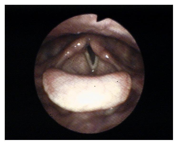
This diagnostic fiberoptic laryngoscopy image provides a superior view of the larynx during attempted phonation. The primary finding is unilateral left vocal cord paralysis. While the right vocal cord appropriately adducts toward the midline, the left vocal cord remains fixed in a more lateral, paramedian position. This asymmetry results in an incomplete glottic closure, manifesting as a visible V-shaped gap (glottal insufficiency) between the true vocal folds. Anatomical landmarks include the epiglottis in the foreground, the false vocal folds, and the arytenoid cartilages posteriorly. The left vocal fold appears relatively flaccid and lacks the tension seen on the contralateral side. This clinical finding is characteristic of recurrent laryngeal nerve neuropraxia or injury, typically presenting with symptoms of dysphonia or hoarseness due to the air leak during phonatory efforts.
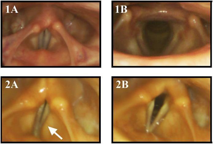
This composite of four flexible endoscopic laryngoscopy images demonstrates vocal fold motion impairment (VFMI) in Multiple System Atrophy (MSA). Figures 1A and 1B serve as healthy controls. During phonation (1A), the true vocal folds show full adduction to the midline; during the sniffing maneuver (1B), they show wide, symmetrical abduction, opening the glottis. In contrast, figures 2A and 2B depict a patient with MSA. During phonation (2A), there is insufficient adduction of the true vocal folds, resulting in a persistent glottal gap, accompanied by compensatory medial activation of the vestibular (false) folds (marked by a white arrow). During the sniffing maneuver (2B), the MSA patient demonstrates incomplete abduction (abductor restriction), where the vocal folds fail to open to the full extent seen in the control. These images illustrate the laryngeal manifestations of neurodegenerative disease, specifically the impairment of both adductor and abductor functions which can lead to dysphonia and stridor.
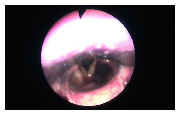
This clinical photograph shows a fiberoptic laryngoscopy view of the human larynx, specifically highlighting the glottis and surrounding supraglottic structures. The true vocal folds are clearly visible, appearing symmetrical and pale-white with normal morphology, and are positioned centrally without displacement. The ventricular folds (false vocal cords) are situated laterally to the true folds. A localized area of slight rosaceous or erythematous coloration is visible on the left ventricular fold, representing a minor residual inflammatory or vascular change. The overall anatomical placement of the larynx is midline and stable, indicating the resolution of any previous lateral pharyngeal wall bulging or endolaryngeal displacement. This image serves as a follow-up diagnostic tool to assess the laryngeal recovery and structural integrity in a patient post-treatment for cervical or parapharyngeal pathologies.
Dysphonia Plica Ventricularis (Ventricular Dysphonia)
Definition
Anatomy - The False Vocal Cords
- The ventricular folds (false cords) are paired mucosal folds situated above and lateral to the true vocal folds, separated from them by the laryngeal ventricle (sinus of Morgagni).
- They are composed of connective tissue and seromucinous glands - they are not designed for phonation.
- They contain no specialized vibratory microarchitecture (no lamina propria layers), making phonation through them inefficient and acoustically abnormal.
- Normally, the false folds assist in airway protection (coughing, Valsalva, swallowing) but do not vibrate during normal speech.
Classification (Relation to Muscular Tension Dysphonia)
| Grade | Description |
|---|---|
| Class I | Posterior glottal gap, elevated larynx, palpable neck tension; breathy or strident voice |
| Class II | Lateral-to-medial constriction; false folds adducted; greater vocal fatigue. Severe form = plica ventricularis (false folds used for phonation) |
| Class III | Anterior-to-posterior supraglottic constriction; epiglottis and arytenoids occlude ≥50% of laryngeal aditus |
| Class IV | Epiglottis and arytenoids contact each other; lateral constriction also present |
Dysphonia plica ventricularis is the most severe form of Class II MTD.
Etiology / Causes
1. Functional / Hyperfunctional (most common)
- Compensatory mechanism when the true vocal folds cannot function normally
- Overzealous or maladaptive laryngeal muscle activity
2. Common Trigger Situations
- Post-laryngitis - After acute inflammation, the patient develops a habit of false fold phonation to "protect" the swollen true folds; the habit persists after recovery
- Post-laryngeal surgery - Compensatory behavior following surgical trauma to the true vocal folds
- True vocal fold pathology: paralysis, nodules, polyps, granuloma, scarring, papillomatosis
- Psychogenic dysphonia - In some psychogenic voice disorders, the ventricular folds adduct alongside the true folds (mixed glottal-ventricular phonation); associated with high life stress, emotional upheaval, or personality factors
- Habitual misuse - Long-term vocal hyperfunction / abuse
- Neurological disorders - False fold overcompensation in vocal fold paresis or paralysis (e.g., seen in Multiple System Atrophy, RLN injury)
Voice Quality Characteristics
- Low-pitched, rough, strained voice - due to the larger, thicker mass of the ventricular folds vibrating at a lower frequency
- Hoarse and harsh quality
- Reduced loudness and pitch range
- Vocal fatigue
- The sound has been compared to the singing technique of Louis Armstrong (gravelly, rough baritone) - a recognized cultural reference in voice literature
- Pain or discomfort in the throat/neck during phonation is common
Clinical Features
- Usually adult onset; more common in females (for psychogenic forms)
- History of a preceding upper respiratory infection, laryngeal surgery, or voice trauma
- Sudden or gradual onset
- Persistent hoarseness unresponsive to conservative treatment for laryngitis
- Neck and throat discomfort or pain while speaking
- Vocal fatigue with prolonged use
- Voice breaks or inconsistency during connected speech
Diagnosis
1. Laryngoscopy / Videostroboscopy (Gold Standard)
- Flexible transnasal laryngoscopy is ideal - allows examination during connected speech and singing without tongue traction
- Findings:
- False folds medialized / adducted during phonation, partially or completely obscuring the view of the true vocal folds
- True vocal folds may appear normal (functional form) OR may show underlying pathology (organic trigger)
- Supraglottic hyperactivity with anterior-to-posterior and lateral constriction
- Videostroboscopy reveals true fold vibratory pattern is absent or replaced by the mass movement of the false folds
2. Voice Analysis (Acoustic Assessment)
- Lowered fundamental frequency
- Increased aperiodicity (jitter, shimmer)
- Reduced maximum phonation time
- Abnormal high transglottal airflow
3. Psychosocial Assessment
- In psychogenic cases: evaluation for anxiety, depression, somatization, life stressors
- Psychiatric or psychological referral if appropriate
Differential Diagnosis
| Condition | Distinguishing Feature |
|---|---|
| Spasmodic dysphonia (adductor) | Strained, strangled voice with breaks; neurological; botulinum toxin responsive |
| Vocal fold paralysis | Breathy voice; clear unilateral fold fixation on laryngoscopy |
| Reinke's edema | Diffuse polypoid degeneration of true vocal folds; smoking history |
| Puberphonia (mutational falsetto) | High-pitched voice in post-pubertal male; different mechanism (falsetto register) |
| Laryngeal malignancy | Progressive; risk factors (smoking, alcohol); irregular lesion on laryngoscopy |
| Paradoxical vocal fold motion | Episodic respiratory distress; inspiratory stridor |
Treatment
1. Voice Therapy (First-line)
- Laryngeal relaxation techniques - reduce excessive extrinsic and intrinsic laryngeal muscle tension (manual circumlaryngeal massage, yawn-sigh techniques)
- Resonance voice therapy / forward focus - redirects phonatory effort away from the larynx
- Confidential voice technique - reduces hyperfunctional drive
- Elimination of vocal misuse behaviors (excessive talking, shouting, throat clearing)
- Vocal hygiene education - hydration, avoidance of irritants, acid reflux management
- Respiratory retraining - proper breath support for phonation
- Biofeedback with videoendoscopy - for obligate false vocal fold phonation and intractable cases; real-time visualization helps patients correct aberrant laryngeal posture consciously (Cummings Otolaryngology Head and Neck Surgery)
2. Treatment of Underlying Cause
- If driven by true vocal fold pathology (e.g., nodule, polyp, paralysis): treat the primary lesion first
- Antireflux therapy (PPI) if LPR is a contributing factor
3. Psychiatric / Psychological Support
- In psychogenic cases: psychotherapy, counseling, stress management
- Especially relevant when onset correlates with emotional trauma or psychiatric comorbidity
4. Botulinum Toxin Injection (selected cases)
- Occasionally used in refractory cases to reduce false fold hypersensitivity / excessive tension
- Not a first-line treatment
Prognosis
- Functional cases respond well to voice therapy - often dramatic improvement within a few sessions once the patient learns to re-engage the true vocal folds
- Psychogenic cases may require longer therapy with psychological support
- Organic cases depend on resolution of the underlying pathology
- If untreated long-term, the compensatory false fold pattern can become deeply habitual and harder to extinguish
Key Points Summary
| Feature | Detail |
|---|---|
| Also called | Ventricular dysphonia; false cord phonation |
| Phonating structure | False (ventricular) folds instead of true vocal folds |
| Voice quality | Low-pitched, rough, hoarse, strained |
| Most common cause | Compensatory / functional after laryngitis or laryngeal surgery |
| Gold standard diagnosis | Flexible laryngoscopy / videostroboscopy |
| First-line treatment | Voice therapy with speech-language pathologist |
| Relation to MTD | Severe end of Class II Muscular Tension Dysphonia (Koufman) |