
Graves disease
Graves disease exophthalmos thyroid goiter clinical features
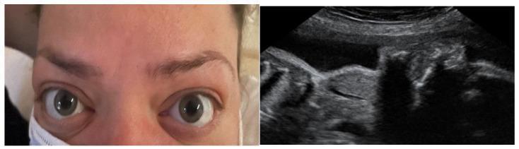
This dual-panel image demonstrates maternal and fetal manifestations of Graves' disease. The left panel is a clinical photograph of a pregnant woman's face, showing classic signs of thyroid-associated orbitopathy (TAO). Key features include bilateral exophthalmos (proptosis), lid retraction with visible superior scleral show, and periorbital edema, indicative of active Graves' ophthalmopathy. The right panel displays an obstetric ultrasound image focusing on the fetal neck. The ultrasound reveals a significant hyperechoic mass in the anterior cervical region, consistent with a fetal goiter. This finding in the context of maternal Graves' disease suggests fetal thyroid dysfunction caused by the transplacental passage of maternal thyroid-stimulating hormone receptor antibodies (TRAb). The image serves as a clinical illustration of the correlation between maternal autoimmune thyroid activity and potential neonatal hyperthyroidism or goitrous development during pregnancy.
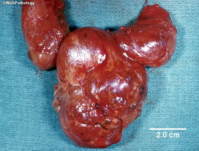
Gross pathology photograph of a total thyroidectomy specimen from a patient with multinodular goiter. The gland is markedly enlarged and lobulated, measuring approximately 12 cm by 10 cm. The external surface is irregular with multiple discrete nodules of varying size that coalesce into a bulky, reddish-brown mass. The cut surface would typically reveal variable nodule architecture with fibrous septa and colloid-filled follicles, consistent with hyperplastic nodular thyroid tissue (multinodular goiter). The overall appearance aligns with sporadic goiter most common in the United States, often euthyroid clinically, though isolated nodules may be hyperfunctioning. Graves disease features such as diffuse hyperplasia with exophthalmos are not observed. No grossly evident malignant cortex features are apparent in this image, although definitive diagnosis requires histology and clinical correlation. Differential considerations include benign nodular disease such as colloid goiter, follicular adenoma, or follicular carcinoma; clinical and radiologic correlation and fine-needle aspiration are recommended if nodules are suspicious. This specimen demonstrates the classic gross appearance of non-toxic multinodular goiter: enlarged gland with nodularity, variable texture, and preserved capsule. This image is valuable for surgical pathology education, medical student teaching, and differential diagnosis demonstrations in endocrinology and thyroid pathology; it supports correlations with thyroid function tests and imaging studies for comprehensive patient management.
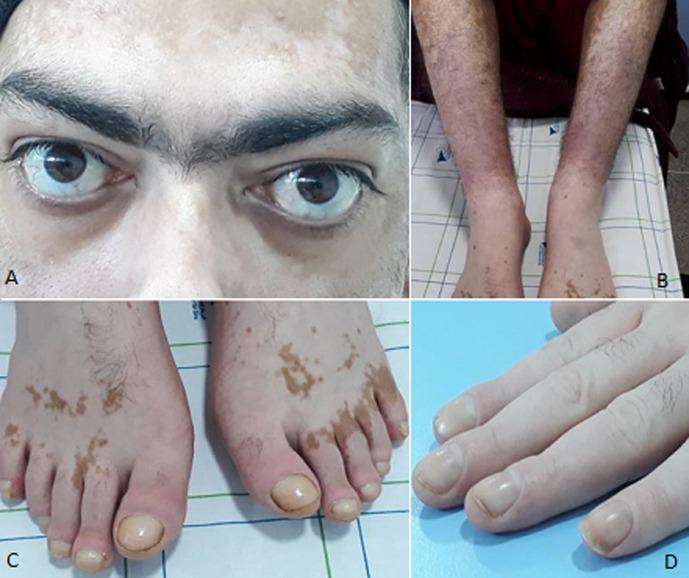
This composite clinical photograph illustrates the classic triad of extra-thyroidal manifestations associated with Graves' disease (Basedow's disease). Panel A shows bilateral exophthalmos (proptosis) and scleral show, along with periocular depigmentation consistent with vitiligo. Panel B demonstrates pretibial myxedema (thyroid dermopathy), characterized by diffuse, brawny, brownish-red induration and hyperpigmentation of the lower legs. Panels C and D display thyroid acropathy, a rare manifestation of Graves' disease. Notable features include digital clubbing (hippocratisme digital) of both fingers and toes, visible as bulbous swelling of the distal phalanges and increased convex curvature of the nail plates. Additionally, the skin on the dorsal aspect of the feet shows patchy, well-defined hyperpigmentation and hypopigmentation. This image serves as a comprehensive educational reference for the systemic dermatological and ophthalmological complications of autoimmune hyperthyroidism, highlighting the characteristic triad of exophthalmos, dermopathy, and acropathy.
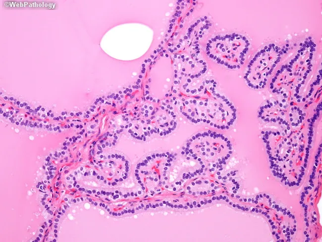
This is a brightfield, hematoxylin and eosin stained histology slide of thyroid tissue examined by light microscopy at high magnification. The specimen is thyroid gland parenchyma showing diffuse follicular hyperplasia characteristic of Graves disease. Follicles appear crowded with epithelial cells lining irregular, papillary-like folds protruding into the lumen, creating pseudo-papillary architectures that could mimic papillary thyroid carcinoma if evaluated in isolation. The nuclei within the thyrocytes are hyperplastic and dense but lack the distinctive nuclear features of papillary carcinoma, such as crowding with nuclear grooves, inclusions, Orphan Annie eye clearing, and powdery chromatin. Colloid is variably present or scant; stroma is vascular, and the overall architecture is polyfollicular with ingrowths into luminal spaces. This pattern reflects autoimmune-driven stimulation by TSH receptor autoantibodies (e.g., thyroid-stimulating immunoglobulin), resulting in diffuse goiter with hyperthyroid state. Clinically, Graves disease presents with weight loss, tachycardia, heat intolerance, and sometimes exophthalmos; histology aids diagnosis when correlated with serology and uptake studies. Key diagnostic significance: avoid mislabeling as papillary thyroid carcinoma; rely on clinical context and absence of malignant nuclear features. Potential educational uses include differential diagnosis training for thyroid pathology, immune-mediated thyroid hyperplasia vs neoplasia, and radiology-pathology correlation. This image supports teaching clinics and pathology review conferences.
"Graves disease"[MeSH Terms] AND management
Graves Disease
Overview
- Hyperthyroidism with diffuse thyroid enlargement (goiter)
- Infiltrative ophthalmopathy (exophthalmos/proptosis)
- Localized infiltrative dermopathy (pretibial myxedema) - present in a minority
Pathogenesis
- Thyroid-Stimulating Immunoglobulin (TSI) - the most common, found in ~90% of patients. This IgG antibody binds and activates the TSH receptor, mimicking TSH action - stimulating adenyl cyclase and increasing T3/T4 synthesis and release continuously.
- TSH receptor-blocking antibodies - also present in some patients; their coexistence with stimulating antibodies can produce alternating episodes of hyperthyroidism and hypothyroidism.
- Concordance in monozygotic twins: 30-40% (vs <5% in dizygotic twins)
- Associated polymorphisms: CTLA4, PTPN22, IL2RA (immune regulation genes)
- GWAS implicates variants in the TSHR gene itself
- HLA allele associations also contribute
- Stimulate fibroblast proliferation and glycosaminoglycan (hyaluronic acid, chondroitin sulfate) synthesis in the retro-orbital space
- Cause mononuclear infiltration (predominantly T cells) into orbital connective tissue
- Lead to edema and swelling of extraocular muscles
- Promote fatty infiltration (increased adipocytes)
- Net result: increased retro-orbital volume → proptosis (exophthalmos)
Morphology
Gross
- Thyroid: symmetrically enlarged, smooth, soft, intact capsule. May exceed 80 g. Cut surface: soft, beefy red-brown parenchyma.
Histology (untreated)
- Follicular epithelial cell hypertrophy and hyperplasia - tall, columnar cells lining follicles
- Pseudopapillary projections into follicle lumens (lack fibrovascular cores - important distinction from papillary carcinoma)
- Pale, scalloped colloid - actively being resorbed by hyperactive epithelium
- Lymphoid infiltrates (predominantly T cells, some B cells, plasma cells) throughout interstitium with scattered germinal centers
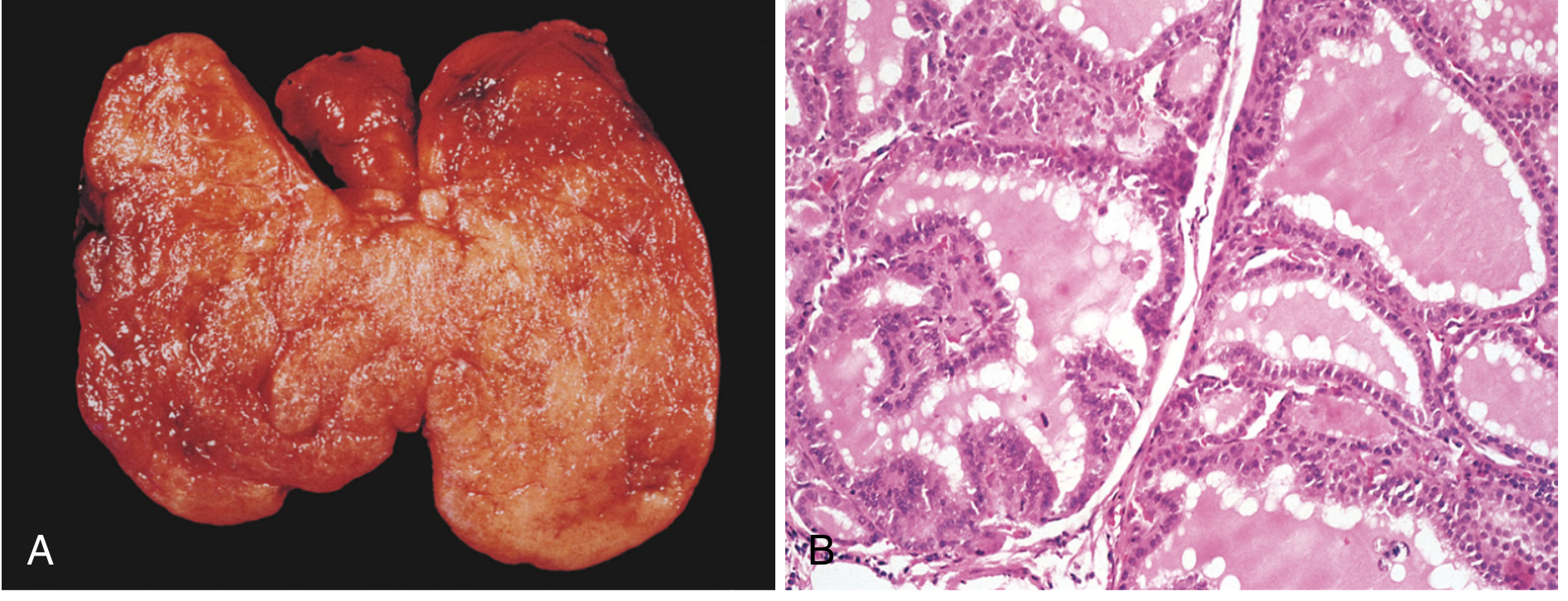
Effect of preoperative therapy on morphology:
- Iodine → epithelial involution, colloid accumulation
- PTU/thionamides → exaggerate hypertrophy/hyperplasia (by stimulating TSH secretion)
Extrathyroidal changes:
- Generalized lymphoid hyperplasia (thymic enlargement in younger patients)
- Cardiac hypertrophy; ischemic changes in pre-existing coronary artery disease
- Orbit: edema from mucopolysaccharide deposition + T cell infiltration + fibrosis
- Dermis (pretibial): glycosaminoglycan deposition + lymphocytic infiltration → skin thickening
Clinical Features
General thyrotoxicosis features:
- Weight loss despite increased appetite
- Heat intolerance, excessive sweating
- Nervousness, anxiety, tremor, hyperactivity
- Tachycardia, palpitations, atrial fibrillation
- Diarrhea, frequent bowel movements
- Oligomenorrhea in women
Graves-specific features:
| Feature | Details |
|---|---|
| Diffuse goiter | Symmetrically enlarged; audible thyroid bruit (increased blood flow) |
| Ophthalmopathy | Exophthalmos (proptosis), wide staring gaze, lid lag, lid retraction, periorbital edema, diplopia (weak extraocular muscles), corneal injury in severe cases, optic neuritis/blindness |
| Pretibial myxedema | Scaly, brawny, brownish-red induration/thickening of skin over shins |
| Thyroid acropachy | Rare - digital clubbing + soft tissue swelling of hands/feet |
- Ophthalmopathy can persist or worsen even after successful treatment of hyperthyroidism
- The extra-thyroidal manifestations can occur even in the absence of clinically evident thyroid disease
- Associated increased risk of other autoimmune diseases: SLE, pernicious anemia, type 1 diabetes, vitiligo
Diagnosis
| Investigation | Finding |
|---|---|
| TSH | Suppressed (low/undetectable) |
| Free T3, Free T4 | Elevated |
| TSI / TRAb (TSHR antibody) | Positive in ~90-95%; highly specific |
| Radioactive iodine uptake (RAIU) | Diffusely increased uptake |
| Thyroid scan | Diffuse, homogeneous uptake |
| Anti-TPO, anti-TG antibodies | Often positive but less specific |
Natural History
- Some patients have a single self-limiting episode (spontaneous remission within months to years)
- Others have lifelong thyrotoxicosis
- Many experience repeated remissions and relapses
- Well-differentiated thyroid cancer is ~2x more prevalent in Graves disease. Palpable hypofunctional nodules in Graves disease carry ~45% malignancy risk and must be evaluated aggressively.
Management
1. Antithyroid Drugs (Thionamides)
- Inhibit thyroid peroxidase-mediated oxidation, organification of iodine, and coupling of iodotyrosines
- PTU additionally inhibits peripheral T4 → T3 conversion
- Both drugs concentrate in the thyroid gland
- Both have immunosuppressive actions - reduce intrathyroidal T cells, inhibit lymphocyte function and autoantibody production → may contribute to remission
2. Radioactive Iodine (¹³¹I)
- Preferred treatment in the US for most adults; used in children when thionamides fail
- Single dose usually sufficient; reduces thyroid size to normal in most patients
- Thionamides stopped 3 days before and after therapy
- Effective in 2-4 months
- Complications:
- Transient thyrotoxicosis exacerbation (1-2 weeks post-treatment, from radiation thyroiditis)
- Rarely precipitates thyroid storm
- Hypothyroidism is near-inevitable (40-80% with higher doses within 1 year)
- Absolute contraindication: pregnancy (crosses placenta, destroys fetal thyroid)
- Caution in adolescents/children
3. Surgery (Thyroidectomy)
- Subtotal thyroidectomy is the classic procedure - narrow margin of tissue left in superolateral thyroid to preserve recurrent laryngeal nerve entry point and parathyroid blood supply
- Indications: Large goiters, failed medical therapy, contraindication to ¹³¹I, suspicious thyroid nodule, patient preference
- Effective and expeditious
- Lugol's iodine (potassium iodide solution, 5-10 drops several times daily) given preoperatively to reduce vascularity and bleeding risk
Adjuncts:
- Beta-blockers (propranolol, esmolol): control sympathetic symptoms (tachycardia, tremor, sweating, nervousness); useful pre-treatment and before surgery. Contraindicated in severe thyrotoxic cardiomyopathy with heart failure, but can benefit atrial fibrillation with heart failure
- Cholestyramine: added to thionamides + beta-blockers for faster thyroid hormone reduction
- Lithium carbonate: antithyroid effect similar to iodine; 300 mg TID-QID
- Inorganic iodine (Lugol's solution): inhibits thyroglobulin proteolysis, iodine transport, oxidation, and organification - used pre-op
Thyroid Storm (Thyrotoxic Crisis)
- Infection, surgery, trauma, labor, iodine load, ¹³¹I treatment
- Features: extreme tachycardia, hyperpyrexia, agitation, delirium, vomiting, diarrhea, cardiac failure
- Management: PTU (preferred over methimazole - also blocks T4→T3), iodine (1 hour after PTU), beta-blockers, hydrocortisone, supportive care
Comparison with Hashimoto Thyroiditis
| Feature | Graves | Hashimoto |
|---|---|---|
| Predominant antibody | TSI (stimulating TSHR-Ab) | Anti-TPO, Anti-TG |
| Thyroid function | Hyperthyroid | Hypothyroid (eventual) |
| Histology | Hyperplasia, scalloped colloid | Hurthle cell change, dense lymphocytic infiltrate, germinal centers |
| Genetic overlap | CTLA4, PTPN22, HLA | CTLA4, PTPN22, HLA |
- Robbins, Cotran & Kumar Pathologic Basis of Disease, Chapter 24
- Robbins & Kumar Basic Pathology, Chapter 18
- Cummings Otolaryngology Head and Neck Surgery, Chapter on thyroid
- Textbook of Family Medicine, 9e