
SAAG value and causes
- SAAG ≥ 1.1 g/dL = portal hypertension-related ascites (97% accuracy)
- SAAG < 1.1 g/dL = non-portal hypertension causes
SAAG serum ascites albumin gradient causes table
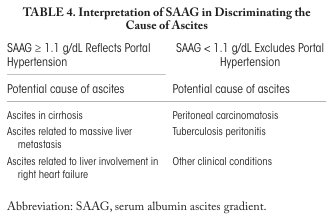
<table> <tr> <th colspan="2">TABLE 4. Interpretation of SAAG in Discriminating the Cause of Ascites</th> </tr> <tr> <th>SAAG ≥ 1.1 g/dL Reflects Portal Hypertension</th> <th>SAAG < 1.1 g/dL Excludes Portal Hypertension</th> </tr> <tr> <td> Potential cause of ascites </td> <td> Potential cause of ascites </td> </tr> <tr> <td> Ascites in cirrhosis<br> Ascites related to massive liver metastasis<br> Ascites related to liver involvement in right heart failure </td> <td> Peritoneal carcinomatosis<br> Tuberculosis peritonitis<br> Other clinical conditions </td> </tr> <tr> <td colspan="2">Abbreviation: SAAG, serum albumin ascites gradient.</td> </tr> </table>
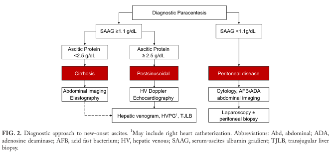
Summary : This flowchart outlines the diagnostic approach to new-onset ascites, using serum-ascites albumin gradient (SAAG) and ascitic protein levels to differentiate between cirrhosis, postsinusoidal, and peritoneal disease etiologies, and guides subsequent investigations. flowchart: # Nodes : • Diagnostic Paracentesis (rectangle) • SAAG ≥1.1 g/dL (rectangle) • Ascitic Protein <2.5 g/dL (rectangle) • Cirrhosis (red rectangle) • Abdominal imaging / Elastography (rectangle) • Ascitic Protein ≥2.5 g/dL (rectangle) • Postsinusoidal (red rectangle) • HV Doppler / Echocardiography (rectangle) • Hepatic venogram, HVPG¹, TJLB (rectangle) • SAAG <1.1 g/dL (rectangle) • Peritoneal disease (red rectangle) • Cytology, AFB/ADA, abdominal imaging (rectangle) • Laparoscopy ± peritoneal biopsy (rectangle) # Connectors : • Diagnostic Paracentesis splits into two branches: SAAG ≥1.1 g/dL and SAAG <1.1 g/dL. • SAAG ≥1.1 g/dL splits into Ascitic Protein <2.5 g/dL and Ascitic Protein ≥2.5 g/dL. • Ascitic Protein <2.5 g/dL leads to Cirrhosis, then to Abdominal imaging / Elastography. • Ascitic Protein ≥2.5 g/dL leads to Postsinusoidal, then to HV Doppler / Echocardiography. • Both Abdominal imaging / Elastography and HV Doppler / Echocardiography connect to Hepatic venogram, HVPG¹, TJLB. • SAAG <1.1 g/dL leads to Peritoneal disease, then to Cytology, AFB/ADA, abdominal imaging, and finally to Laparoscopy ± peritoneal biopsy. # Layout : • The flowchart is organized in a top-down manner, starting with Diagnostic Paracentesis at the top. • The first split is based on SAAG value (≥1.1 g/dL vs <1.1 g/dL). • The SAAG ≥1.1 g/dL branch further splits by ascitic protein level (<2.5 g/dL vs ≥2.5 g/dL). • Each branch leads to a red box indicating the main disease category, followed by recommended diagnostic tests. • The SAAG <1.1 g/dL branch leads directly to Peritoneal disease and its diagnostic steps. # Analysis : • The flowchart provides a clear, stepwise approach for evaluating new-onset ascites, using SAAG and ascitic protein to stratify patients. • Cirrhosis is suggested by SAAG ≥1.1 g/dL and low ascitic protein, while postsinusoidal causes are suggested by high ascitic protein. • Peritoneal disease is considered when SAAG is low (<1.1 g/dL). • Each pathway leads to specific diagnostic modalities, ensuring targeted evaluation based on initial laboratory findings.
<table> <tr> <th>Trial</th> </tr> <tr> <td>Effect of Albumin Administration in Hypoalbuminemic Hospitalized Patients With Community-Acquired Pneumonia (<a href="https://clinicaltrials.gov">ClinicalTrials.gov</a> Identifier: NCT04071041)</td> </tr> <tr> <td>Albumin Replacement Therapy in Septic Shock (<a href="https://clinicaltrials.gov">ClinicalTrials.gov</a> Identifier: NCT03869385)</td> </tr> <tr> <td>Albumin in Cardiac Surgery Australian (Postoperative 20% Albumin vs Standard Care and Acute Kidney Injury After High-Risk Cardiac Surgery) (Australian New Zealand Clinical Trials Registry Identifier: ACTRN1261900135516703)</td> </tr> <tr> <td>Effects of Long-Term Administration of Human Albumin in Subjects With Decompensated Cirrhosis and Ascites (<a href="https://clinicaltrials.gov">ClinicalTrials.gov</a> Identifier: NCT03451292)</td> </tr> <tr> <td>Albumin to Enhance Recovery After Acute Kidney Injury (<a href="https://clinicaltrials.gov">ClinicalTrials.gov</a> Identifier: NCT04705896)</td> </tr> </table>
SAAG - Serum-Ascites Albumin Gradient
Formula
- Both specimens must be drawn on the same day
- It is a subtraction (not a ratio)
- Accuracy for detecting portal hypertension: ~97%
Interpretation
| SAAG | Meaning |
|---|---|
| ≥ 1.1 g/dL | Portal hypertension-related ascites |
| < 1.1 g/dL | Non-portal hypertension cause |
Causes by SAAG Value
HIGH SAAG (≥ 1.1 g/dL) - Portal Hypertension-Related
| Cause | Notes |
|---|---|
| Cirrhosis | Most common overall cause of ascites |
| Alcohol-associated hepatitis | |
| Cardiac ascites | Right heart failure, constrictive pericarditis |
| Budd-Chiari syndrome | Hepatic vein thrombosis |
| Portal vein thrombosis | |
| Massive liver metastases | |
| Sinusoidal obstruction syndrome | (formerly veno-occlusive disease) |
| Fatty liver of pregnancy | |
| Acute liver failure (ALF) | |
| "Mixed" ascites | Portal HTN + another cause (e.g., infection) |
LOW SAAG (< 1.1 g/dL) - Non-Portal Hypertension
| Cause | Notes |
|---|---|
| Peritoneal carcinomatosis | Most common malignant cause |
| Tuberculous peritonitis | Lymphocyte-predominant fluid |
| Nephrotic syndrome | Low serum albumin, no portal HTN |
| Pancreatic ascites | High amylase in fluid |
| Biliary ascites | Ruptured bile duct |
| Bowel obstruction or infarction | |
| Serositis (connective tissue diseases) | SLE, RA |
| Postoperative lymphatic leak |
Diagnostic Flowchart
Key Clinical Notes
- SAAG does not diagnose the cause - it only indicates whether portal hypertension is present or absent
- Ascitic total protein adds further refinement:
- SAAG ≥ 1.1 + ascitic protein < 2.5 g/dL → Cirrhosis (sinusoidal cause)
- SAAG ≥ 1.1 + ascitic protein ≥ 2.5 g/dL → Post-sinusoidal cause (cardiac, Budd-Chiari)
- False elevations of SAAG can occur with hyperalbuminemia or chylous ascites
- SAAG is superior to the older exudate/transudate classification for ascites