
anterior knee pain evaluation cause etiology investigation management
Reading File
Anterior knee pain is one of the most common musculoskeletal complaints, especially in young active individuals. The main causes include patellofemoral pain syndrome (PFPS), patellar tendinopathy, Osgood-Schlatter disease (adolescents), prepatellar/infrapatellar bursitis, and plica syndrome. Evaluation involves a structured history, physical exam (patellar grind, Clarke's test, apprehension test), and imaging when needed. Management is largely conservative - physiotherapy, VMO strengthening, and activity modification.
Searching sources...
Finding Sources
Finding Sources
Searching PubMed
anterior knee pain patellofemoral management
Good results. Now let me read the key textbook sections in parallel.
Reading File
Reading File
Reading File
Excellent content. Now let me get more detail from the rheumatology text (on plica, maltracking) and Rockwood/Green's on evaluation, plus search for relevant images.
Reading File
Reading File
Searching Images
patellofemoral pain syndrome anatomy evaluation
Let me fetch those textbook images and also search for more specific content on evaluation/investigation.
Loading Image
Loading Image
Finding Sources
Good. Let me get the patellar tendinitis section from Tintinalli and also look for plica syndrome and bursitis content.
Reading File
Finding Sources
I now have enough material for a comprehensive answer. Let me compile this into a well-structured clinical overview.
Anterior Knee Pain: Evaluation, Causes, Investigation & Management
Anterior knee pain (AKP) is one of the most common musculoskeletal presentations in primary care and emergency medicine, affecting athletes, adolescents, and older adults alike. It is not a single diagnosis - it is a symptom complex with a structured differential.
1. Causes and Etiology
A useful framework organizes causes by anatomical structure:
Patellofemoral Causes (most common)
| Condition | Key Features |
|---|---|
| Patellofemoral Pain Syndrome (PFPS) | Most common cause of knee pain overall; gradual onset, no trauma, worse with stairs/prolonged sitting ("movie theater sign"); women > men, typically 20s-30s |
| Chondromalacia Patellae | Softening and fragmentation of patellar articular cartilage from impact loading and excessive shear stress; crepitation on exam; worse descending stairs |
| Patellar Maltracking / Subluxation | Abnormal patella tracking due to bony anatomy, soft tissue laxity, muscular imbalance, or rotational deformity; common in adolescent females |
As described in Rheumatology (Elsevier, 2022): anterior knee pain can be caused by chondromalacia patellae, patellofemoral maltracking, and medial plica syndrome, and a careful history is essential to establish the exact site and aggravating/relieving factors.
Tendon Pathology
| Condition | Key Features |
|---|---|
| Patellar Tendinopathy ("Jumper's Knee") | Runners, high jumpers, basketball/volleyball players; point tenderness at distal pole of patella or proximal patellar tendon; worse with standing, jumping, or running uphill |
| Quadriceps Tendinopathy | Less common; tenderness at superior pole of patella |
Per Tintinalli's Emergency Medicine: Patellar tendinitis (jumper's knee) presents with pain at the patellar tendon worsened by activity. Steroid injections should be avoided as they predispose to tendon rupture.
Bursitis
| Condition | Key Features |
|---|---|
| Prepatellar Bursitis | Swelling directly over the kneecap; common in carpet layers and people who kneel frequently ("housemaid's knee"); superficial to the lower half of patella |
| Infrapatellar Bursitis | Common in long-distance runners; can mimic patellar tendinitis |
Adolescent-Specific
| Condition | Key Features |
|---|---|
| Osgood-Schlatter Disease | Traction apophysitis at the patellar tendon insertion on the tibial tubercle; pain, tenderness, swelling at tibial tubercle; exacerbated by exercise; resolves with apophyseal fusion |
| Sinding-Larsen-Johansson Syndrome | Similar mechanism at inferior pole of patella |
Per Bailey and Love's Surgery (28th Ed): the extensor mechanism of the knee is a common site of pain in adolescents during growth spurts.
Other Causes
- Plica syndrome - thickened medial synovial fold causing snapping and medial anterior pain
- Fat pad impingement (Hoffa syndrome) - infrapatellar fat pad caught between femur and tibia
- Referred pain - hip pathology (e.g., slipped upper femoral epiphysis) can present as knee pain - always examine the hip
- Post-IM nailing - anterior knee pain occurs in 19-73% after tibial intramedullary nailing, likely multifactorial (fat pad scarring, infrapatellar nerve injury, nail prominence)
- Patellofemoral osteoarthritis - older patients with anterior joint line tenderness and crepitus
2. Evaluation
History
- Onset: gradual (PFPS, tendinopathy) vs. acute (trauma, dislocation)
- Location: peripatella, patellar tendon, tibial tubercle
- Aggravating factors: stairs (PFPS/chondromalacia), prolonged sitting (PFPS), jumping (tendinopathy), kneeling (bursitis)
- Age and activity level: adolescent active male (Osgood-Schlatter), young female athlete (PFPS/maltracking), middle-aged runner (tendinopathy)
- Bilateral vs. unilateral: bilateral more unusual - consider systemic causes
Physical Examination
| Test | Finding | Suggests |
|---|---|---|
| Clarke's test / Patellar grind | Pain on compressing and asking patient to contract quads | PFPS / chondromalacia |
| Patellar apprehension test | Apprehension/pain on lateral patellar displacement | Patellar subluxation/instability |
| Single leg squat | Reproduction of anterior pain | PFPS |
| Patellar glide/tilt | Reduced medial glide, increased lateral tilt | Lateral retinacular tightness |
| J-sign | Lateral deviation of patella at end of extension | Maltracking |
| Tibial tubercle tenderness | Point tenderness at tibial tubercle | Osgood-Schlatter |
| Patellar tendon palpation | Tenderness at inferior patellar pole | Patellar tendinopathy |
| Prepatellar swelling | Fluctuant swelling anterior to patella | Prepatellar bursitis |
| VMO assessment | Wasting of vastus medialis oblique | PFPS, maltracking |
| Hip examination | Abductor strength, rotation | Hip pathology referred pain; gluteal weakness as PFPS risk factor |
Rosen's Emergency Medicine notes: gait abnormalities may be present; medial and lateral patellar tenderness is prominent when due to subluxation/dislocation; single-leg squat pain is suggestive of PFPS. Risk factors include gluteal weakness, quadriceps weakness, patellar subluxation, prepatellar bursitis, arthritis, meniscal tears, and quadriceps/patellar tendinopathy.
3. Investigation
Imaging
X-rays (first-line):
- AP, lateral, and skyline (axial/sunrise) view of the patella
- Skyline view assesses patellofemoral joint space, patellar tilt, and lateral subluxation (see image below)
- Lateral view allows measurement of Insall-Salvati ratio (patellar tendon length : patellar length; >1.2 = patella alta, a risk factor for maltracking)
- Standard knee X-rays assess for osteoarthritis, loose bodies, and tibial tubercle changes
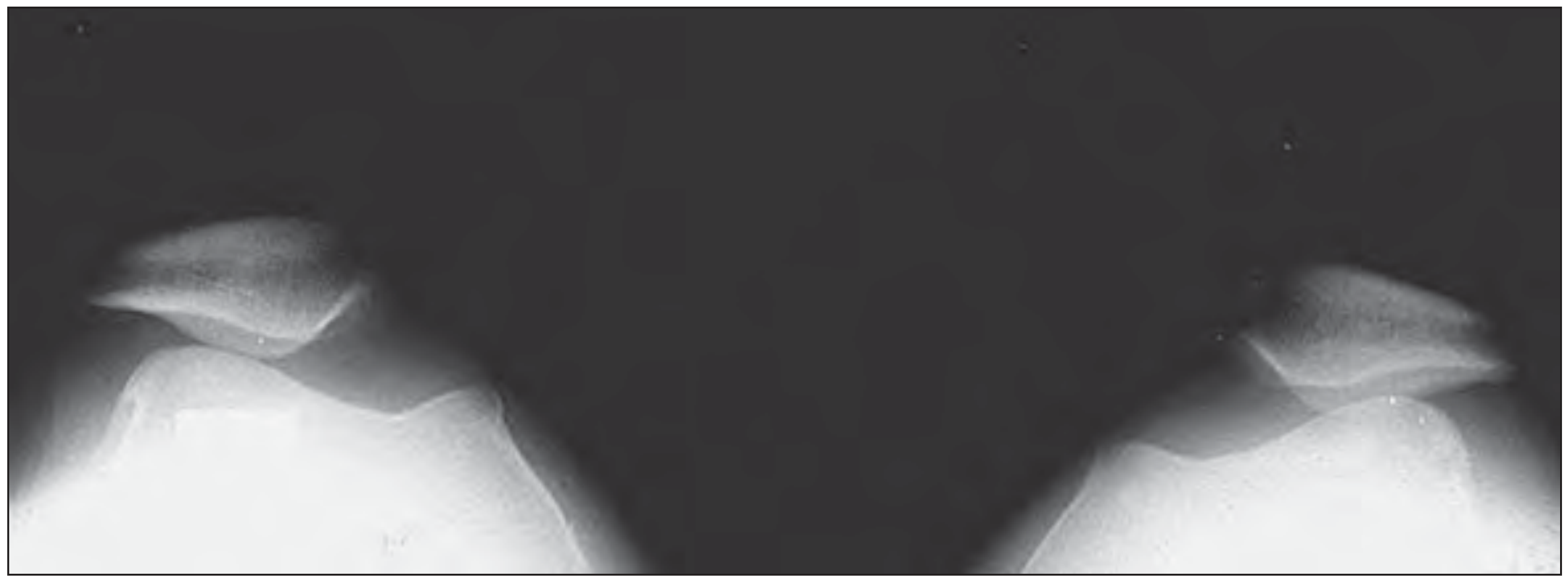
Skyline X-ray showing bilateral patellofemoral joints. Lateral subluxation of the patella is assessed on this view. - Rheumatology (Elsevier, 2022)
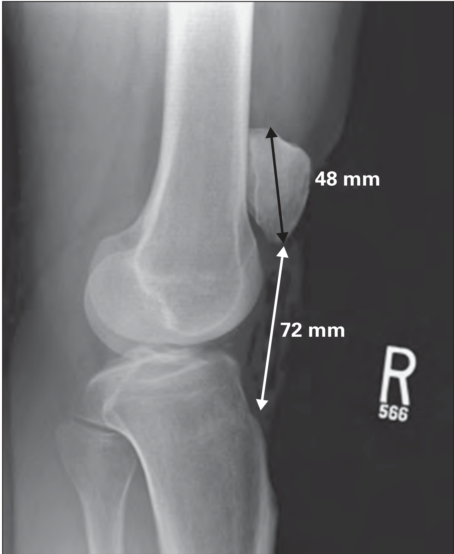
Lateral knee X-ray with Insall-Salvati ratio measurement (patellar length 48mm / patellar tendon length 72mm = 0.67 - within normal range; ratio >1.2 indicates patella alta). - Rheumatology (Elsevier, 2022)
MRI:
- Indicated when: X-rays are negative but significant symptoms persist, suspected cartilage pathology, soft tissue abnormalities (plica, fat pad), or diagnosis is unclear
- Best modality for chondromalacia grading, MPFL injury after dislocation, tendon tears
Ultrasound:
- Useful for real-time assessment of patellar/quadriceps tendons
- Can detect bursitis, guide aspiration, and assess tendon integrity
- Preferred for evaluating tendinopathy and bursitis
Note: PFPS is a clinical diagnosis. Imaging is used to exclude other causes and assess structural abnormalities, not to confirm PFPS itself.
4. Management
Conservative (First-Line for Most Causes)
Physiotherapy - cornerstone of treatment:
- VMO strengthening (vastus medialis oblique) to correct lateral tracking
- Hip abductor and external rotator strengthening - 2025 meta-analysis (PMID 39934098) confirms that combined hip + knee strengthening is superior to knee strengthening alone for PFPS
- Knee extensor training - systematic review (PMID 40861009) supports its role in PFPS
- Hamstring and quadriceps stretching
- Activity modification and load management
Analgesics:
- NSAIDs for short-term pain relief to facilitate physiotherapy initiation
- Avoid NSAIDs long-term; avoid corticosteroid injections around patellar tendon (rupture risk)
Orthotic support:
- Patellar taping (McConnell technique) to correct lateral tracking
- Foot orthotics for overpronation
- Patellar tracking braces - some benefit, though efficacy not clearly established per Rosen's
- Activity modification: avoid prolonged knee flexion, modify sitting position
Osgood-Schlatter: relative rest and analgesia; resolves with apophyseal fusion.
Bursitis: NSAIDs, activity modification, aspiration if large/septic.
Surgical (for refractory cases)
- Lateral retinacular release - for severe lateral tightness / maltracking
- Tibial tubercle osteotomy (Fulkerson procedure) - anteromedialization of tibial tubercle for chondromalacia and maltracking
- Trochleoplasty / MPFL reconstruction - for recurrent patellar dislocation
- Arthroscopic debridement - for unstable chondral fragments in chondromalacia patellae
- Plica excision - for symptomatic medial plica
Per Bailey and Love's: "Many operations improve patellar tracking and these include options for realignment of the extensor mechanism both proximally and distally."
Emerging / Adjunct Treatments
A 2025 systematic review (PMID 41009572) on PRP for patellofemoral arthritis and anterior knee pain found some evidence of benefit, though its role remains investigational and not yet standard of care.
Summary Algorithm
Anterior Knee Pain
       |
   History & Exam
       |
   Adolescent? ──Yes──> Check tibial tubercle → Osgood-Schlatter
       |                Check inferior patellar pole → Sinding-Larsen
       |
   Young adult, active?
       |
   Gradual, peripatella, worse stairs/sitting ──> PFPS (clinical dx)
       |
   Tendon point tenderness ──> Patellar tendinopathy
       |
   Anterior swelling ──> Bursitis
       |
   Trauma, dislocation ──> Maltracking/subluxation, X-ray/MRI
       |
   X-ray: AP + Lateral + Skyline
   ± MRI if needed
       |
   Management:
   Physiotherapy (VMO + hip abductors) → NSAIDs → Taping/Orthotics
   → Surgery if conservative fails (>6 months)
Sources:
- Rosen's Emergency Medicine, Concepts and Clinical Practice - PFPS: foundations, clinical features, diagnostic testing, and management
- Bailey and Love's Short Practice of Surgery, 28th Ed - anterior knee pain in adolescents, extensor mechanism
- Rheumatology, 2-Volume Set (Elsevier, 2022) - chondromalacia, patellofemoral maltracking, imaging
- Tintinalli's Emergency Medicine, Comprehensive Study - patellar tendinitis
- Goldman-Cecil Medicine - bursitis classification
- Recent PubMed: Hip + knee strengthening meta-analysis 2025 | Knee extensor training SR 2025 | PRP for PFPS SR 2025
This is a shared conversation. Sign in to Orris to start your own chat.