Give the content for presentation on topic neonatal sepsis

Reading File
Finding Sources
Finding Sources
Reading File
Reading File
Finding Sources
Reading File
Finding Sources
Finding Sources
Finding Sources
Finding Sources
Searching Images

neonatal sepsis signs symptoms neonate

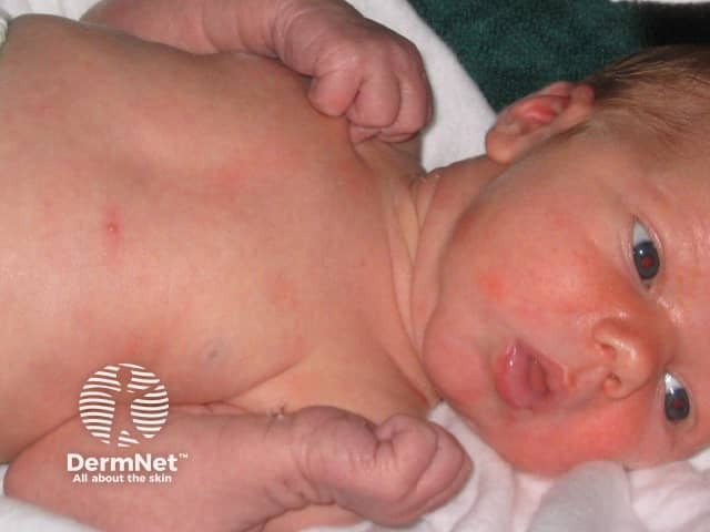
Clinical photography of a term neonate demonstrating erythematous papules and vesicles distributed primarily on the trunk and face. Imaging modality: Clinical photography; technique: high-resolution color image captured in natural or balanced lighting, frontal/anterior view with the infant supine. Anatomical localization: integumentary system; neonatal skin (epidermis and superficial dermis). Visual features include numerous small 1–3 mm erythematous papules and vesicles on an erythematous base, some showing a pale or yellowish vesicular center; lesions are non-follicular and non-desquamative, arranged in scattered coalescent patches but with overall benign appearance. There is no edema, crusting, or purpura; mucous membranes spared; no systemic signs of illness. Clinically diagnostic for erythema toxicum neonatorum (ETN), a common benign neonatal eruption typically appearing within 24–72 hours of birth and resolving spontaneously within 1–2 weeks. Haematologic or infectious etiologies are not supported by the image alone; no systemic symptoms; differential considerations include milia, miliaria rubra, transient neonatal pustular melanosis, neonatal pustular psoriasis, and less commonly neonatal herpes. Pathophysiology involves eosinophilic inflammatory response; management is reassurance and observation; no antimicrobial therapy required unless accompanied by fever or sepsis concern. The image is relevant for educational purposes in dermatology, pediatrics, and neonatology to illustrate ETN morphology, distribution, natural history, and differentiation from other neonatal pustular conditions.

Clinical photography of a term neonate demonstrating erythematous papules and vesicles distributed primarily on the trunk and face. Imaging modality: Clinical photography; technique: high-resolution color image captured in natural or balanced lighting, frontal/anterior view with the infant supine. Anatomical localization: integumentary system; neonatal skin (epidermis and superficial dermis). Visual features include numerous small 1–3 mm erythematous papules and vesicles on an erythematous base, some showing a pale or yellowish vesicular center; lesions are non-follicular and non-desquamative, arranged in scattered coalescent patches but with overall benign appearance. There is no edema, crusting, or purpura; mucous membranes spared; no systemic signs of illness. Clinically diagnostic for erythema toxicum neonatorum (ETN), a common benign neonatal eruption typically appearing within 24–72 hours of birth and resolving spontaneously within 1–2 weeks. Haematologic or infectious etiologies are not supported by the image alone; no systemic symptoms; differential considerations include milia, miliaria rubra, transient neonatal pustular melanosis, neonatal pustular psoriasis, and less commonly neonatal herpes. Pathophysiology involves eosinophilic inflammatory response; management is reassurance and observation; no antimicrobial therapy required unless accompanied by fever or sepsis concern. The image is relevant for educational purposes in dermatology, pediatrics, and neonatology to illustrate ETN morphology, distribution, natural history, and differentiation from other neonatal pustular conditions.

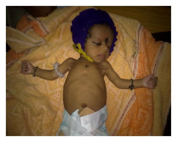
This clinical photograph shows a neonate in a supine position, documenting the clinical resolution of symptoms following treatment for congenital syphilis. The infant displays a significant improvement in spontaneous motor activity and posture compared to the initial presentation of Parrot's pseudoparalysis. The upper limbs are extended and abducted, showing no visible signs of the previously reported swelling in the elbow or knee joints. The skin is of normal color without evidence of rash, jaundice, or pallor. The infant appears calm with eyes closed, wearing a blue cap and a white diaper. Key clinical indicators illustrated here include the resolution of joint inflammation and the restoration of normal limb positioning and movement following antibiotic therapy (crystalline penicillin) for treponemal infection. The image serves as a post-treatment follow-up to confirm the efficacy of the management of neonatal syphilis.

This clinical photograph shows a neonate in a supine position, documenting the clinical resolution of symptoms following treatment for congenital syphilis. The infant displays a significant improvement in spontaneous motor activity and posture compared to the initial presentation of Parrot's pseudoparalysis. The upper limbs are extended and abducted, showing no visible signs of the previously reported swelling in the elbow or knee joints. The skin is of normal color without evidence of rash, jaundice, or pallor. The infant appears calm with eyes closed, wearing a blue cap and a white diaper. Key clinical indicators illustrated here include the resolution of joint inflammation and the restoration of normal limb positioning and movement following antibiotic therapy (crystalline penicillin) for treponemal infection. The image serves as a post-treatment follow-up to confirm the efficacy of the management of neonatal syphilis.

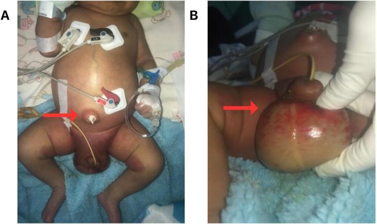
This clinical photograph consists of two panels (A and B) showing a male neonate with significant abdominal and perineal pathology. Panel A shows a distended abdomen with umbilical inflammation (omphalitis), characterized by erythema and purulent discharge from the umbilical stump (red arrow). ECG electrodes and various monitoring tubes are visible on the chest and abdomen, indicating critical care. Panel B provides a close-up of the inguinal and scrotal region, demonstrating massive scrotal swelling, intense erythema, and localized necrotic skin changes (red arrow) consistent with Fournier's gangrene or severe scrotal cellulitis. The inflammation extends from the scrotum to the lower abdominal wall and proximal thighs. The visual evidence supports a diagnosis of neonatal sepsis potentially secondary to omphalitis with subsequent progression to necrotizing fasciitis of the perineum. This image serves as an educational tool for identifying early signs of neonatal surgical emergencies and infectious complications in a pediatric setting.

This clinical photograph consists of two panels (A and B) showing a male neonate with significant abdominal and perineal pathology. Panel A shows a distended abdomen with umbilical inflammation (omphalitis), characterized by erythema and purulent discharge from the umbilical stump (red arrow). ECG electrodes and various monitoring tubes are visible on the chest and abdomen, indicating critical care. Panel B provides a close-up of the inguinal and scrotal region, demonstrating massive scrotal swelling, intense erythema, and localized necrotic skin changes (red arrow) consistent with Fournier's gangrene or severe scrotal cellulitis. The inflammation extends from the scrotum to the lower abdominal wall and proximal thighs. The visual evidence supports a diagnosis of neonatal sepsis potentially secondary to omphalitis with subsequent progression to necrotizing fasciitis of the perineum. This image serves as an educational tool for identifying early signs of neonatal surgical emergencies and infectious complications in a pediatric setting.

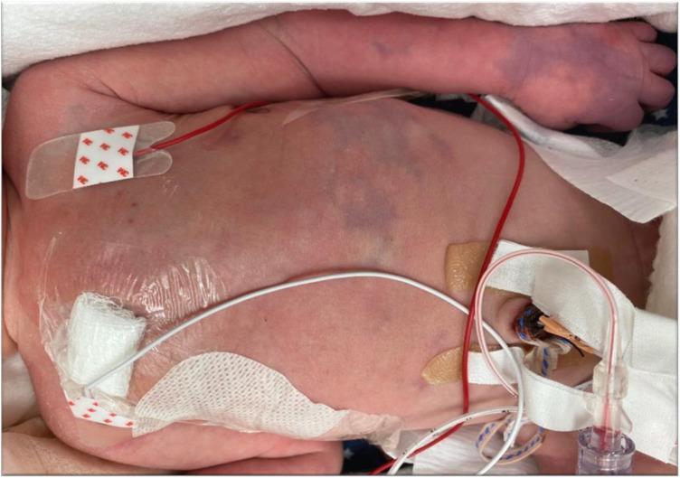
A clinical photograph of a neonate in a neonatal intensive care unit (NICU) setting, illustrating acute dermatological manifestations of neonatal purpura fulminans and disseminated intravascular coagulation (DIC). The image shows well-demarcated, irregular erythematous and purpuric macules distributed across the abdomen and left arm. The lesions exhibit a characteristic blue-black to deep purple hue, indicating tissue ischemia and hemorrhage. The neonate is equipped with multiple critical care support devices, including cardiac monitoring leads (ECG electrodes with red wires), a chest tube site covered with a transparent occlusive dressing and sterile gauze, and an umbilical catheter or similar vascular access device secured with white medical tape. The skin findings are highly suggestive of microvascular thrombosis, a hallmark of protein C or S deficiency or severe neonatal sepsis. This image serves as a visual reference for recognizing early-stage skin necrosis and purpuric progression in critically ill newborns.

A clinical photograph of a neonate in a neonatal intensive care unit (NICU) setting, illustrating acute dermatological manifestations of neonatal purpura fulminans and disseminated intravascular coagulation (DIC). The image shows well-demarcated, irregular erythematous and purpuric macules distributed across the abdomen and left arm. The lesions exhibit a characteristic blue-black to deep purple hue, indicating tissue ischemia and hemorrhage. The neonate is equipped with multiple critical care support devices, including cardiac monitoring leads (ECG electrodes with red wires), a chest tube site covered with a transparent occlusive dressing and sterile gauze, and an umbilical catheter or similar vascular access device secured with white medical tape. The skin findings are highly suggestive of microvascular thrombosis, a hallmark of protein C or S deficiency or severe neonatal sepsis. This image serves as a visual reference for recognizing early-stage skin necrosis and purpuric progression in critically ill newborns.

I now have comprehensive content. Let me compile the full presentation content.

Neonatal Sepsis - Presentation Content


Slide 1: Title Slide

NEONATAL SEPSIS A Comprehensive Clinical Overview
  • Definition, Classification, Etiology
  • Clinical Features, Diagnosis, Management
  • Prevention & Complications

Slide 2: Definition & Overview

  • Neonatal sepsis is a systemic infection occurring in infants during the first 28 days of life
  • It is the most common cause of neonatal cardiorespiratory distress
  • Defined by bacteremia, meningitis, or urinary tract infection in the newborn period
  • Fever in the first month = rectal temperature ≥38°C (100.4°F)
  • Hypothermia = rectal temperature <36.5°C (97.7°F)
  • Neonates have ~2× the risk of serious bacterial infection compared to infants aged 4-8 weeks
  • Only ~50% of clinically suspected neonatal sepsis cases have positive cultures
(Sources: Tintinalli's Emergency Medicine; Miller's Anesthesia)

Slide 3: Classification - Early vs Late Onset

FeatureEarly-Onset Sepsis (EOS)Late-Onset Sepsis (LOS)
TimingFirst 7 days of lifeAfter 1 week of age
OnsetFulminant, rapidGradual, insidious
Associated FactorsMaternal/perinatal risk factorsLess associated with perinatal risks
Common complicationsSeptic shock, neutropeniaMeningitis (more common)
Typical organismsGBS, E. coli, ListeriaGBS (late), CoNS, Candida
  • EOS usually presents within 48 hours of birth with rapid clinical deterioration and high mortality
  • LOS presents at >72 hours and is more commonly associated with meningitis
(Sources: Tintinalli's Emergency Medicine; Creasy & Resnik's Maternal-Fetal Medicine; Robbins Pathologic Basis of Disease)

Slide 4: Etiology & Causative Organisms

Gram-Positive Organisms:
  • Group B Streptococcus (GBS) - most common bacterial pathogen overall; causes severe cardiorespiratory instability + meningitis in 30% of cases
  • Listeria monocytogenes - causes sepsis and meningitis; longer incubation (presents as late-onset)
  • Coagulase-negative Staphylococci (CoNS) - common in premature/NICU infants
Gram-Negative Organisms:
  • Escherichia coli - second most common; leading cause of early-onset bacterial meningitis
  • Klebsiella species
  • Haemophilus influenzae
Viral:
  • Herpes simplex virus (HSV) - fulminant neonatal infection; active lesions in birth canal = indication for cesarean section; add IV acyclovir
Fungal:
  • Candida species - especially in very preterm, immunocompromised neonates; longer latent period
(Sources: Tintinalli's Emergency Medicine; Miller's Anesthesia; Robbins Pathologic Basis of Disease)

Slide 5: Risk Factors

Maternal / Perinatal Risk Factors (for EOS):
  • Maternal fever / chorioamnionitis
  • GBS-positive vaginal swabs
  • Prolonged rupture of membranes (>18 hours)
  • Preterm delivery (<37 weeks)
  • Fetal distress / low birth weight
  • Young maternal age
  • Black race
  • History of previous infant with GBS sepsis
  • GBS bacteriuria during current pregnancy
Neonatal Risk Factors:
  • Prematurity and low birth weight (immature immune system)
  • Invasive procedures / vascular catheters (NICU)
  • Prolonged hospitalization
  • Male sex
  • Artificial feeding (loss of breast milk immune factors)
(Sources: Creasy & Resnik's Maternal-Fetal Medicine; Tintinalli's Emergency Medicine)

Slide 6: Clinical Features / Signs & Symptoms

The presentation is often NON-SPECIFIC - any "unwell" neonate should prompt sepsis workup.
SystemSigns
TemperatureFever (≥38°C) or Hypothermia (<36.5°C)
CNSLethargy, irritability, seizures, bulging fontanelle
RespiratoryApnea, tachypnea, grunting, respiratory distress
CardiovascularHypotension, poor cutaneous perfusion, tachycardia
GIPoor feeding, vomiting, gastric distention, diarrhea
SkinJaundice, rashes, petechiae, mottling
MetabolicHypoglycemia, metabolic acidosis
Key point: Nuchal rigidity and Kernig/Brudzinski signs are present in only a small minority of neonates with meningitis - do not rely on classic meningism signs.
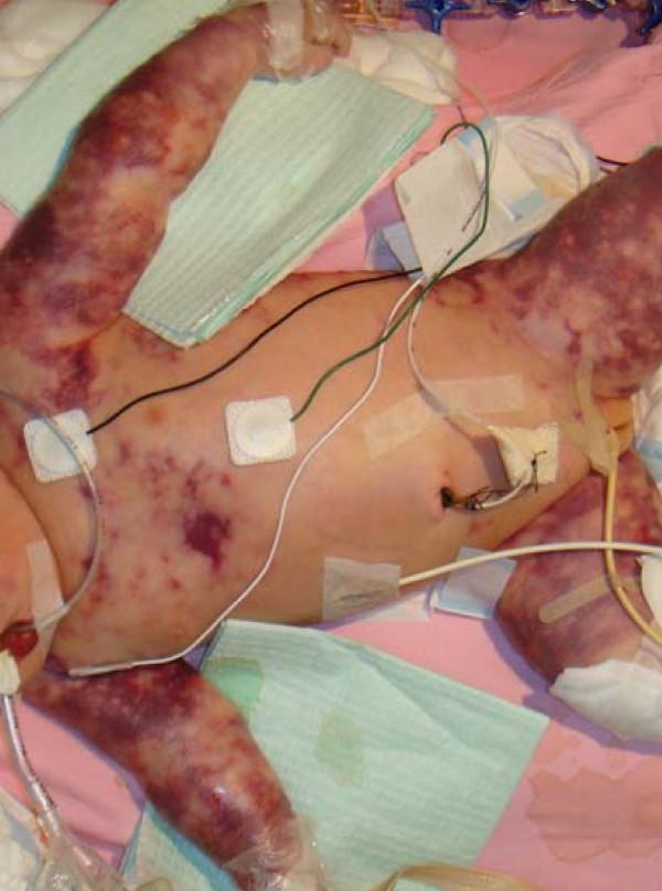
Neonatal purpura fulminans - DIC complicating severe sepsis
(Sources: Tintinalli's Emergency Medicine; Miller's Anesthesia)

Slide 7: Differential Diagnosis

The "sepsis mimics" in neonates - all cause cardiorespiratory collapse and must be considered:
  • Congenital Heart Disease - ductal-dependent lesions present with shock/acidosis as ductus closes in first week; may be indistinguishable from sepsis
  • Metabolic disorders - inborn errors of metabolism, congenital adrenal hyperplasia
  • Pneumonia - bacterial, viral, chlamydial, aspiration
  • Intracranial hemorrhage - vitamin K deficiency, non-accidental trauma
  • Abdominal emergencies - malrotation, volvulus, necrotizing enterocolitis (NEC)
  • Bronchiolitis
(Source: Tintinalli's Emergency Medicine - Table 116-2)

Slide 8: Investigations / Diagnosis

Lower threshold for full sepsis workup in neonates than in older infants.
Full Sepsis Workup includes:
InvestigationDetails
Blood cultureGold standard; 2 sites if possible
CBC with differentialLook for neutropenia/neutrophilia, thrombocytopenia
C-Reactive Protein (CRP)Inflammatory marker; serial measurements useful
ProcalcitoninElevated in bacterial sepsis
Lumbar puncture + CSF analysisCell count, protein, glucose, Gram stain, culture
Urinalysis + urine cultureCatheter specimen preferred
Chest X-rayRule out pneumonia
Blood glucose, electrolytes, blood gasHypoglycemia, metabolic acidosis
Note: Because localizing infection in neonates is difficult, repeated complete sepsis workups are often required. Blood, urine, and CSF cultures should all be obtained when sepsis is suspected.
(Sources: Tintinalli's Emergency Medicine; Miller's Anesthesia; Swanson's Family Medicine Review)

Slide 9: Management - Antibiotic Treatment

All neonates with suspected sepsis must be admitted and started on empiric IV antibiotics immediately.

First-Line Empiric Therapy (Early-Onset Sepsis):

DrugDoseCoverage
Ampicillin50 mg/kg IVGBS, Listeria
Gentamicin2.5 mg/kg IVE. coli, gram-negatives

If Gram-Negative Meningitis Strongly Suspected:

  • Replace gentamicin with Cefotaxime or Ceftazidime (50 mg/kg) - better CNS penetration
  • ⚠️ AVOID Ceftriaxone in neonates - displaces bilirubin → kernicterus

If HSV Suspected (maternal herpes hx, lymphocytes/RBCs in CSF):

  • Add IV Acyclovir

Late-Onset GBS Meningitis:

  • Ampicillin + Ceftazidime (or Cefotaxime)

Supportive Care:

  • IV fluids, respiratory support (CPAP/ventilation if needed)
  • Glucose monitoring and correction
  • Vasopressors for septic shock (dopamine/dobutamine)
  • NICU admission
(Sources: Tintinalli's Emergency Medicine; Red Book 2021; Harriet Lane Handbook)

Slide 10: Prevention

Intrapartum Antibiotic Prophylaxis (IAP):

  • Universal GBS screening at 35-37 weeks gestation (vaginal/rectal swab)
  • IV Penicillin G (preferred) or Ampicillin given to GBS-positive mothers intrapartum
  • IAP has dramatically decreased early-onset GBS sepsis but has NOT changed the rate of late-onset sepsis
  • Intrapartum (not immediate postpartum) antibiotics are significantly more effective - neonatal sepsis rate 2.8% with intrapartum antibiotics vs higher rates with postpartum treatment

Other Preventive Measures:

  • Prompt delivery in PPROM cases
  • Good hand hygiene in NICU
  • Minimize invasive procedures/central lines
  • Breastfeeding (immune factors)
  • Sterile technique for all procedures
  • Minimize prolonged rupture of membranes
(Sources: Creasy & Resnik's Maternal-Fetal Medicine; Tietz Laboratory Medicine)

Slide 11: Complications

If untreated or delayed diagnosis:
  • Septic shock - cardiovascular collapse, multi-organ failure
  • Meningitis - especially in late-onset disease; GBS causes meningitis in 30% of cases
  • Disseminated Intravascular Coagulation (DIC) - purpura fulminans
  • Pneumonia / respiratory failure - mechanical ventilation required
  • Necrotizing Enterocolitis (NEC)
  • Kernicterus - from sepsis-associated jaundice
  • Neurodevelopmental sequelae - hearing loss, intellectual disability, cerebral palsy (post-meningitis)
  • Death - mortality remains high, especially in preterm infants and developing countries
Omphalitis progressing to necrotizing fasciitis - a surgical septic emergency in neonates
(Sources: Tintinalli's Emergency Medicine; Miller's Anesthesia)

Slide 12: Summary / Key Take-Home Points

  1. Neonatal sepsis is the most common cause of cardiorespiratory distress in neonates
  2. Classified as Early-Onset (<7 days) - linked to maternal risk factors and Late-Onset (>7 days) - more often causes meningitis
  3. GBS is the #1 bacterial pathogen; E. coli is #2; Listeria, Candida, HSV also important
  4. Clinical features are non-specific - any unwell neonate must be evaluated for sepsis
  5. Full workup: Blood culture, CBC, CRP, LP, urine culture, CXR
  6. Empiric treatment: Ampicillin + Gentamicin; avoid ceftriaxone (kernicterus risk)
  7. Add IV Acyclovir if HSV suspected
  8. Prevention: Universal GBS screening + intrapartum penicillin prophylaxis
  9. Low threshold - when in doubt, treat empirically and admit all neonates

Optional Additional Slides

Slide 13: Scoring Tools & Decision Aids

  • Kaiser Permanente Neonatal Early-Onset Sepsis Calculator - uses maternal risk factors + clinical exam to estimate EOS probability and guide antibiotic use
  • I-PASS (Immature-to-total neutrophil ratio)
  • Serial CRP monitoring (peaks at 24-48 h, normal <1 mg/dL)

Slide 14: Special Situations

  • NICU/Hospital-acquired sepsis: Coagulase-negative Staphylococci + antifungal coverage for Candida
  • Herpes neonatarum: IV Acyclovir 60 mg/kg/day × 14-21 days; high mortality without treatment
  • Listeria: Ampicillin is drug of choice; naturally resistant to ALL cephalosporins

References:
  • Tintinalli's Emergency Medicine: A Comprehensive Study Guide
  • Miller's Anesthesia, 10th Edition
  • Creasy & Resnik's Maternal-Fetal Medicine: Principles and Practice
  • Red Book 2021: Report of the Committee on Infectious Diseases (AAP)
  • Harriet Lane Handbook, 23rd Edition (Johns Hopkins Hospital)
  • Robbins & Cotran Pathologic Basis of Disease
  • Tietz Textbook of Laboratory Medicine, 7th Edition

Give the content for presentation on topic neonatal sepsis refer nelsons and who

Finding Sources
Searching the Web

WHO guidelines neonatal sepsis management 2024 2025

Searching Images

neonatal sepsis neonate blood culture NICU

A clinical photograph of a neonate in a neonatal intensive care unit (NICU) setting, illustrating acute dermatological manifestations of neonatal purpura fulminans and disseminated intravascular coagulation (DIC). The image shows well-demarcated, irregular erythematous and purpuric macules distributed across the abdomen and left arm. The lesions exhibit a characteristic blue-black to deep purple hue, indicating tissue ischemia and hemorrhage. The neonate is equipped with multiple critical care support devices, including cardiac monitoring leads (ECG electrodes with red wires), a chest tube site covered with a transparent occlusive dressing and sterile gauze, and an umbilical catheter or similar vascular access device secured with white medical tape. The skin findings are highly suggestive of microvascular thrombosis, a hallmark of protein C or S deficiency or severe neonatal sepsis. This image serves as a visual reference for recognizing early-stage skin necrosis and purpuric progression in critically ill newborns.

A clinical photograph of a neonate in a neonatal intensive care unit (NICU) setting, illustrating acute dermatological manifestations of neonatal purpura fulminans and disseminated intravascular coagulation (DIC). The image shows well-demarcated, irregular erythematous and purpuric macules distributed across the abdomen and left arm. The lesions exhibit a characteristic blue-black to deep purple hue, indicating tissue ischemia and hemorrhage. The neonate is equipped with multiple critical care support devices, including cardiac monitoring leads (ECG electrodes with red wires), a chest tube site covered with a transparent occlusive dressing and sterile gauze, and an umbilical catheter or similar vascular access device secured with white medical tape. The skin findings are highly suggestive of microvascular thrombosis, a hallmark of protein C or S deficiency or severe neonatal sepsis. This image serves as a visual reference for recognizing early-stage skin necrosis and purpuric progression in critically ill newborns.

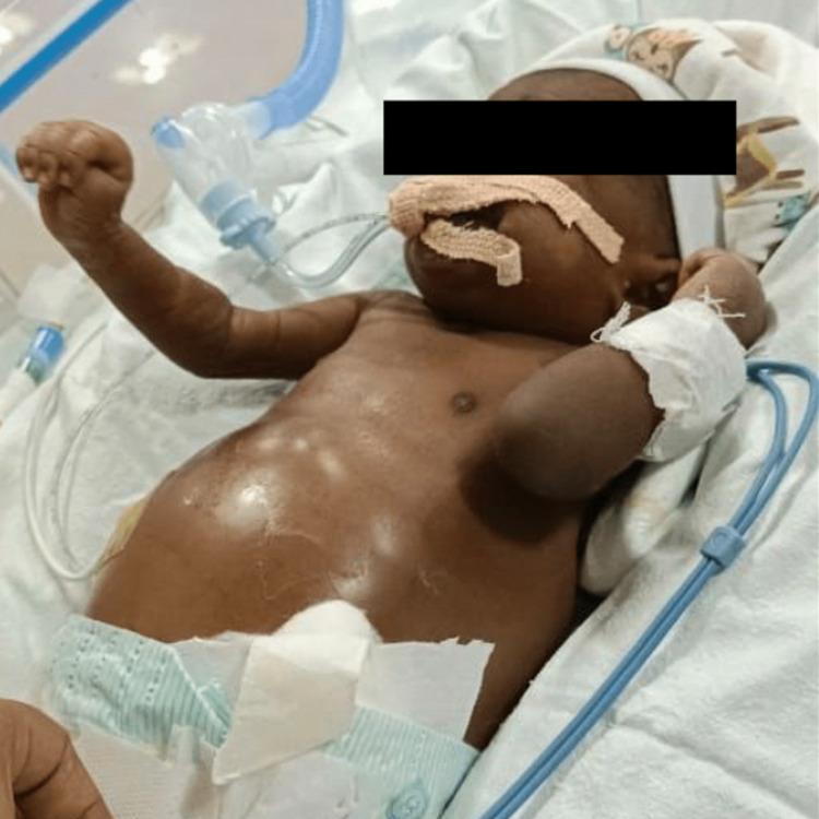
This clinical photograph shows a neonate in a neonatal intensive care unit (NICU) setting at 20 days of life. The infant exhibits significant abdominal distension consistent with clinical ascites, with the skin over the abdomen appearing taut and shiny. Medical interventions and monitoring equipment are visible: the neonate is receiving mechanical ventilatory support via an endotracheal or oronasal tube secured with adhesive tape across the face. An orogastric tube is also present for gastric decompression or enteral feeding. The infant's left arm is bandaged, indicating a site for an intravenous (IV) line or arterial access. Peripheral monitoring wires for vital sign assessment are positioned around the infant. The clinical presentation, as contextualized by associated reports, is representative of neonatal cholestasis and progressive ascites secondary to sepsis (E. coli). This image serves as an educational example of neonatal abdominal pathology and the critical care management of a symptomatic newborn requiring respiratory and hemodynamic support.

This clinical photograph shows a neonate in a neonatal intensive care unit (NICU) setting at 20 days of life. The infant exhibits significant abdominal distension consistent with clinical ascites, with the skin over the abdomen appearing taut and shiny. Medical interventions and monitoring equipment are visible: the neonate is receiving mechanical ventilatory support via an endotracheal or oronasal tube secured with adhesive tape across the face. An orogastric tube is also present for gastric decompression or enteral feeding. The infant's left arm is bandaged, indicating a site for an intravenous (IV) line or arterial access. Peripheral monitoring wires for vital sign assessment are positioned around the infant. The clinical presentation, as contextualized by associated reports, is representative of neonatal cholestasis and progressive ascites secondary to sepsis (E. coli). This image serves as an educational example of neonatal abdominal pathology and the critical care management of a symptomatic newborn requiring respiratory and hemodynamic support.

This clinical photograph shows a neonate in a neonatal intensive care unit (NICU) setting presenting with extensive purpura fulminans. The skin displays widespread, non-blanching purpuric lesions ranging from erythematous to deep violaceous and dusky purple. The distribution is near-confluent on the extremities, particularly the arms and thighs, with more discrete petechiae and larger purpuric patches on the trunk. The patient is receiving multi-organ support, visible through various medical interventions: an endotracheal tube for mechanical ventilation, multiple adhesive ECG leads on the chest for cardiac monitoring, an umbilical catheter, and peripheral intravenous lines secured with transparent dressings on the limbs. The appearance is highly characteristic of acute infectious purpura fulminans, often associated with severe sepsis (e.g., Group B Streptococcus or meningococcemia) and disseminated intravascular coagulation (DIC), leading to cutaneous microvascular thrombosis and hemorrhagic necrosis.

This clinical photograph shows a neonate in a neonatal intensive care unit (NICU) setting presenting with extensive purpura fulminans. The skin displays widespread, non-blanching purpuric lesions ranging from erythematous to deep violaceous and dusky purple. The distribution is near-confluent on the extremities, particularly the arms and thighs, with more discrete petechiae and larger purpuric patches on the trunk. The patient is receiving multi-organ support, visible through various medical interventions: an endotracheal tube for mechanical ventilation, multiple adhesive ECG leads on the chest for cardiac monitoring, an umbilical catheter, and peripheral intravenous lines secured with transparent dressings on the limbs. The appearance is highly characteristic of acute infectious purpura fulminans, often associated with severe sepsis (e.g., Group B Streptococcus or meningococcemia) and disseminated intravascular coagulation (DIC), leading to cutaneous microvascular thrombosis and hemorrhagic necrosis.

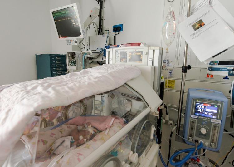
A clinical photograph of a neonatal intensive care unit (NICU) setting, illustrating the integration of humans and medical devices in neonatal medicine. A neonate is positioned inside a transparent, controlled-environment incubator featuring circular portholes for clinician access and a thermal cover to maintain temperature stability. Mounted on the wall above the incubator is a patient monitor displaying real-time vital signs, including electrocardiogram (ECG) waveforms and oxygen saturation (SpO2) levels. Adjacent to the incubator is a non-invasive ventilator unit (Viasys) with a digital interface for respiratory support. To the right, an intravenous (IV) infusion setup is visible, consisting of a hanging fluid bag and an infusion pump for the administration of nutrition or medications. The image provides a comprehensive view of standard neonatal life-support systems, including thermal regulation, continuous physiological monitoring, and specialized respiratory therapy required for preterm or critically ill infants.

A clinical photograph of a neonatal intensive care unit (NICU) setting, illustrating the integration of humans and medical devices in neonatal medicine. A neonate is positioned inside a transparent, controlled-environment incubator featuring circular portholes for clinician access and a thermal cover to maintain temperature stability. Mounted on the wall above the incubator is a patient monitor displaying real-time vital signs, including electrocardiogram (ECG) waveforms and oxygen saturation (SpO2) levels. Adjacent to the incubator is a non-invasive ventilator unit (Viasys) with a digital interface for respiratory support. To the right, an intravenous (IV) infusion setup is visible, consisting of a hanging fluid bag and an infusion pump for the administration of nutrition or medications. The image provides a comprehensive view of standard neonatal life-support systems, including thermal regulation, continuous physiological monitoring, and specialized respiratory therapy required for preterm or critically ill infants.

Finding Sources
Reading Web Page

https://pmc.ncbi.nlm.nih.gov/articles/PMC12576100

Finding Sources
Finding Sources
Finding Sources
Reading File
Finding Sources
Reading File
Reading File
Reading File
Finding Sources
Reading File
Reading File
Note on Nelson's: Nelson's Textbook of Pediatrics is not available in the textbook library. The content below draws from the closest equivalent pediatric authorities: Red Book 2021 (AAP Committee on Infectious Diseases) - which is the definitive US pediatric infectious disease reference and whose GBS/neonatal sepsis guidelines directly mirror Nelson's content - plus Harriet Lane Handbook (Johns Hopkins, 23rd ed.), WHO 2024/2025 Recommendations for Management of Serious Bacterial Infections in Infants aged 0-59 days, and Tintinalli's/Miller's for clinical detail.

NEONATAL SEPSIS

Presentation Content - Slide by Slide


SLIDE 1 — Title

NEONATAL SEPSIS
Based on:
  • Red Book 2021 - AAP Committee on Infectious Diseases (equivalent standard to Nelson's)
  • Harriet Lane Handbook, 23rd Edition (Johns Hopkins)
  • WHO Recommendations for Management of Serious Bacterial Infections in Infants Aged 0-59 Days (2024)

SLIDE 2 — Definition & Epidemiology

Definition:
  • Neonatal sepsis is a systemic bacterial, viral, or fungal infection occurring in infants within the first 28 days of life
  • Encompasses bacteremia, meningitis, urinary tract infection, and pneumonia in the newborn period
  • WHO defines it as a serious bacterial infection (SBI) in infants aged 0-59 days
Epidemiology (WHO 2024):
  • SBIs including sepsis, meningitis, and pneumonia contribute to >500,000 newborn deaths annually worldwide
  • Neonates have approximately 2× the risk of serious bacterial infection compared to infants 4-8 weeks of age
  • Only ~50% of clinically suspected neonatal sepsis cases have positive cultures
  • Case-fatality rate: 1%-3% in term infants; up to 20% in preterm infants with early-onset disease (Red Book 2021)
  • In 2018 (USA): Early-onset GBS disease = 0.25 cases per 1000 live births; Late-onset = 0.28 per 1000 live births (Red Book 2021)

SLIDE 3 — Classification: Early-Onset vs Late-Onset

FeatureEarly-Onset Sepsis (EOS)Late-Onset Sepsis (LOS)
Timing0-6 days of life7-89 days of life
Peak95% present within 48 hours of birthTypically 3-4 weeks of age
Onset characterFulminant, rapid deteriorationGradual, insidious
Common presentationRespiratory distress, apnea, shock, pneumoniaBacteremia, meningitis (~30% of cases)
Meningitis rate5%-10%~30%
Link to maternal factorsStrong (GBS colonization, IAP failure, PPROM)Weaker; community/nosocomial acquisition
Common organismsGBS, E. coli, ListeriaGBS (type III), CoNS, Candida
Mortality (GBS)20% in preterm; 2% in term8% in preterm; 1%-3% in term
Key AAP/Red Book note: Intrapartum antibiotic prophylaxis (IAP) has reduced EOS incidence by ~80% but has had no measurable effect on late-onset GBS disease. (Red Book 2021)

SLIDE 4 — Etiology & Causative Organisms

Group B Streptococcus (GBS) - Streptococcus agalactiae

  • Most common bacterial pathogen in neonatal sepsis
  • Gram-positive diplococci; 10 capsular types - Types Ia, Ib, II, III, IV, V account for ~99% of infant cases in the USA
  • Type III causes 30%-60% of early-onset and late-onset disease respectively
  • EOS: respiratory distress, apnea, shock, pneumonia; meningitis in 5%-10%
  • LOS: bacteremia, meningitis; also septic arthritis, osteomyelitis, UTI, cellulitis
  • Vaginal/rectal colonization rate in pregnant women: 15%-35% (Red Book 2021)

Escherichia coli & Gram-Negative Bacilli

  • Second most common; leading cause of early-onset bacterial meningitis
  • ~⅔ of E. coli bloodstream infections in very low birth weight (VLBW) infants are ampicillin-resistant (ESBL-producing)
  • ESBL-producing organisms are resistant to penicillins, cephalosporins, monobactams
  • Carbapenemase-producing Klebsiella pneumoniae and E. coli are emerging
  • Klebsiella, Haemophilus influenzae also implicated (Red Book 2021)

Listeria monocytogenes

  • Causes sepsis and meningitis; naturally resistant to all cephalosporins
  • Longer latent period - may present as late-onset sepsis
  • Treatment: Ampicillin is drug of choice

Herpes Simplex Virus (HSV)

  • Fulminant neonatal infection; add IV Acyclovir when suspected
  • Active lesions in birth canal = indication for cesarean section

Candida species

  • Important in preterm, VLBW infants in NICU
  • Risk: prolonged mechanical ventilation, central lines, prolonged antibiotics

Coagulase-Negative Staphylococci (CoNS)

  • Common cause of nosocomial/late-onset sepsis in NICU; associated with central venous catheters

SLIDE 5 — Risk Factors

Maternal / Perinatal Risk Factors (for EOS):

Risk FactorNotes
GBS-positive vaginal/rectal swabColonization rate 15%-35%
Intrapartum fever ≥38°C (100.4°F)Sign of chorioamnionitis
Prolonged rupture of membranes ≥18 hoursAllows ascending infection
Preterm delivery (<37 weeks)Immature immune system + surfactant deficiency
GBS bacteriuria during current pregnancyIndicates heavy colonization
History of previous infant with invasive GBSRecurrence risk
Maternal age <20 yearsAssociated with higher GBS colonization
Black raceHigher GBS colonization + preterm birth rates
Intra-amniotic infection (chorioamnionitis)Direct fetal exposure
Inadequate IAP (<4 hours before delivery)Incomplete prophylaxis

Neonatal Risk Factors:

  • Prematurity / very low birth weight (<1500 g)
  • Prolonged NICU hospitalization
  • Central venous catheters, endotracheal tubes
  • Total parenteral nutrition
  • Prior broad-spectrum antibiotic exposure (selects resistant organisms)
(Red Book 2021; Harriet Lane Handbook)

SLIDE 6 — Clinical Features

The "unwell neonate" - presentations are NON-SPECIFIC. Any sick-appearing newborn must be evaluated for sepsis.
SystemSigns & Symptoms
TemperatureFever (rectal ≥38°C) or Hypothermia (<36.5°C) - both equally important
RespiratoryApnea, tachypnea, grunting, nasal flaring, retractions, cyanosis
CNSLethargy, poor tone, irritability, seizures, bulging fontanelle
CardiovascularTachycardia, hypotension, poor perfusion, mottled skin, prolonged capillary refill
GastrointestinalPoor feeding, vomiting, abdominal distension, diarrhea, hepatomegaly
SkinJaundice (especially early), petechiae, purpura, rashes
MetabolicHypoglycemia, metabolic acidosis
WHO 7-Sign IMCI Algorithm (2024) - Signs of Serious Bacterial Infection:
  1. Not feeding well
  2. Convulsions
  3. Fast breathing (≥60 breaths/min)
  4. Severe chest indrawing
  5. Temperature instability (fever or hypothermia)
  6. Movement only on stimulation / no movement
  7. Umbilical infection / skin pustules
Clinical pearl (Red Book 2021): Classic meningism (nuchal rigidity, Kernig, Brudzinski) is present in only a small minority of neonates with meningitis - do not rely on these signs to exclude CNS infection.
Neonatal NICU monitoring - typical critical care setting

SLIDE 7 — Differential Diagnosis

Sepsis must be distinguished from (and may coexist with):
ConditionKey Distinguishing Feature
Congenital heart diseaseCyanosis, murmur, hyperoxia test; ductal-dependent lesions present in week 1
Inborn errors of metabolismMetabolic acidosis, hypoglycemia, abnormal organic acids/amino acids
Congenital adrenal hyperplasiaAmbiguous genitalia, electrolyte abnormalities
Necrotizing enterocolitis (NEC)Abdominal distension, bloody stools, pneumatosis on X-ray
Neonatal pneumoniaConsolidation on CXR; may overlap with sepsis
Intracranial hemorrhageBulging fontanelle, neurological signs, ultrasound/CT confirmation
Congenital TuberculosisBronchopneumonia + hepatosplenomegaly; can mimic sepsis (Red Book 2021)
Congenital MalariaFever, lethargy, poor feeding; resembles sepsis in endemic areas (Red Book 2021)
Hypoxic-ischemic encephalopathyHistory of birth asphyxia, seizures

SLIDE 8 — Investigations / Diagnosis

AAP/Red Book 2021 Recommendation: "CBC and CRP are NOT accurate enough to reliably identify infected infants." Culture is the gold standard.

Complete Sepsis Workup:

InvestigationDetails / Notes
Blood cultureGold standard - 2 separate sites if possible; send before antibiotics
CBC with differentialNeutropenia, neutrophilia, elevated immature:total (I:T) ratio >0.2
C-Reactive Protein (CRP)Serial measurements more useful than single value; peaks 24-48 h
ProcalcitoninMore specific than CRP for bacterial infection; rises within 2-4 h
Lumbar puncture + CSF analysisCell count, protein, glucose, Gram stain, culture - mandatory when sepsis suspected
Urine cultureCatheter specimen; UA may be normal in early UTI
Chest X-rayRule out pneumonia, RDS
Blood glucose, electrolytesHypoglycemia, hyponatremia common in sepsis
Blood gas (ABG/VBG)Metabolic acidosis, raised lactate = organ dysfunction
Serum lactateWHO/2024 guidelines: recommended to evaluate cardiovascular dysfunction
Molecular / multiplex PCRRapid pathogen ID from positive blood culture bottles; FDA-cleared (Red Book 2021)
Mass spectrometryUsed in advanced centers for rapid bacterial identification

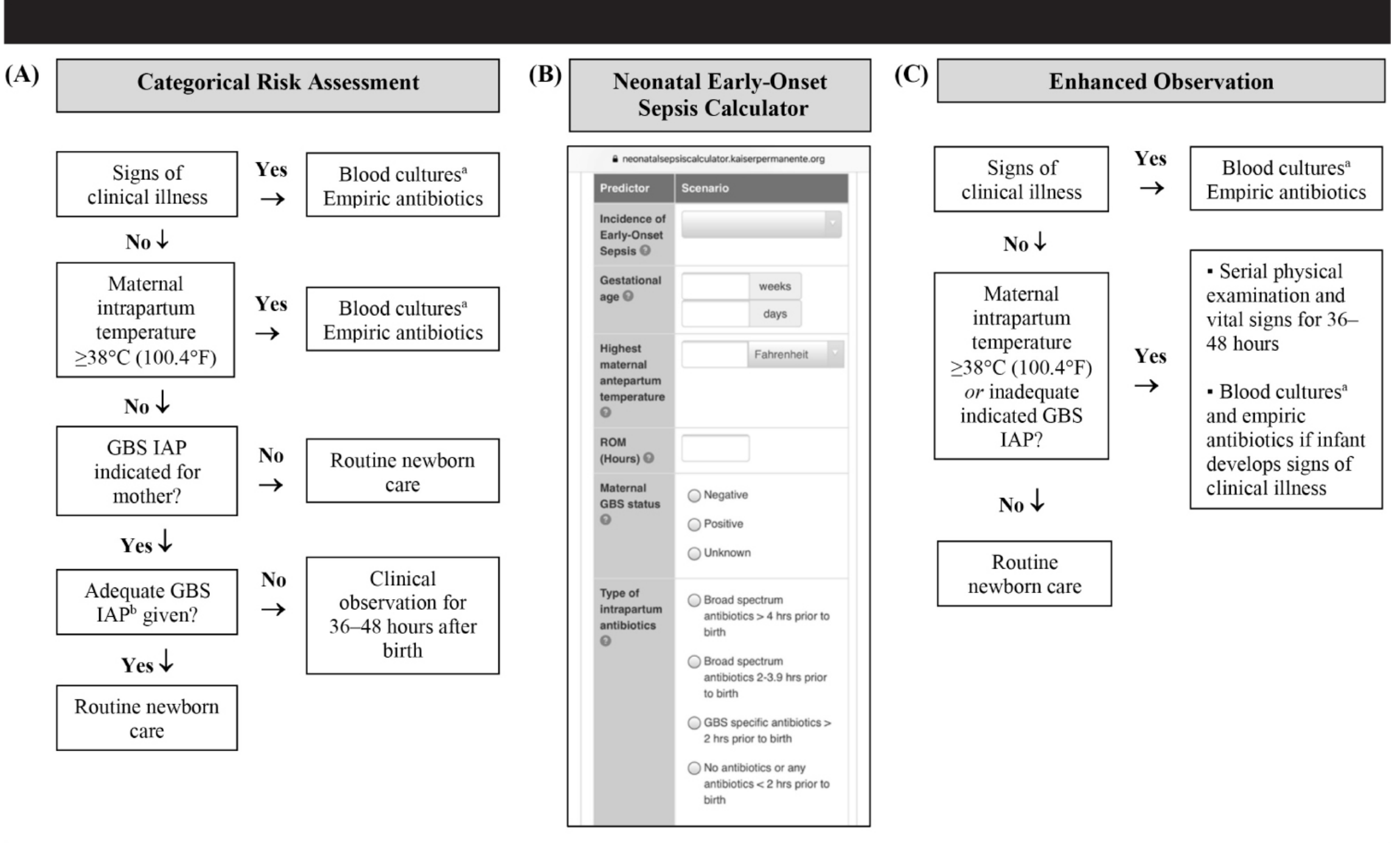
Three Risk Assessment Approaches (AAP 2019/Red Book 2021) for infants ≥35 weeks:

  1. Categorical Algorithm - threshold-based (more sensitive, less specific)
  2. Neonatal Early-Onset Sepsis (EOS) Calculator (Kaiser Permanente) - multivariate model using maternal + infant factors; prospectively validated in large cohorts. Available at: https://neonatalsepsiscalculator.kaiserpermanente.org/
  3. Enhanced Clinical Observation - serial structured physical assessments; a "well-appearing" term infant has 60%-70% lower risk of EOS
EOS risk assessment flowchart - GBS disease among infants ≥35 weeks gestation

SLIDE 9 — Management: Antibiotic Treatment

General Principles (AAP/Red Book 2021 + WHO 2024):

  • All neonates with suspected sepsis must be admitted and treated empirically with IV antibiotics immediately
  • Obtain all cultures before starting antibiotics
  • Antimicrobial stewardship is essential - WHO classifies first-line agents as "Access" antibiotics (WHO Essential Medicines List)
  • Empiric therapy should be guided by local/regional antimicrobial susceptibility data

A. Early-Onset Sepsis (EOS) - First-Line Empiric Therapy

DrugDoseCoverage
Ampicillin IV50 mg/kg/doseGBS, Listeria, gram-positives
Gentamicin IV4-5 mg/kg/day (extended interval dosing)E. coli, gram-negatives
WHO 2024: Ampicillin (or penicillin) + gentamicin = first-line in hospital settings and in outpatient settings where hospital referral is not feasible (given IM/IV × minimum 10 days)

B. If Gram-Negative Meningitis Suspected (EOS)

DrugDoseNotes
Ampicillin + Cefotaxime50 mg/kgBetter CNS penetration than gentamicin
If cefotaxime unavailable: Ceftazidime or Cefepime50 mg/kgAcceptable alternatives (Red Book 2021)
If MDR/ESBL suspected: Carbapenem (Meropenem)Weight-basedFor resistant organisms
⚠️ AVOID Ceftriaxone in neonates - displaces bilirubin from albumin → kernicterus. Use ceftazidime/cefotaxime instead (Harriet Lane Handbook)

C. Late-Onset Sepsis (LOS) Empiric Therapy (Red Book 2021)

AgeNon-critically ill (no meningitis)Meningitis suspected
8-28 daysAmpicillin + gentamicin OR cefotaximeAmpicillin + cefotaxime (avoid gentamicin for meningitis)
29-90 daysCeftriaxoneCeftriaxone + Vancomycin (for drug-resistant organisms)
Preterm >72 h (NICU)Broader spectrum - account for nosocomial pathogens including GBSAdd vancomycin for CoNS; antifungals if Candida risk

D. HSV Neonatal Sepsis

DrugDoseDuration
IV Acyclovir60 mg/kg/day in 3 divided doses14-21 days
Add IV Acyclovir when: maternal herpes history, CSF pleocytosis with lymphocytes + RBCs in non-traumatic LP, ill-appearing neonate (Tintinalli's)

E. Confirmed GBS Infection - Definitive Therapy (Red Book 2021)

IndicationDrugDuration
Bacteremia / UTI (no meningitis)Penicillin G or Ampicillin10 days IV
Uncomplicated meningitisPenicillin G or Ampicillin14 days IV
Complicated/prolonged meningitisPenicillin G or Ampicillin>14 days IV
Septic arthritis / OsteomyelitisPenicillin G or Ampicillin3-4 weeks
Endocarditis / VentriculitisPenicillin G or Ampicillin≥4 weeks
CSF sterilization check: For neonatal GBS meningitis, some experts recommend repeat LP at 24-48 h to confirm CSF sterility and detect complications (cerebral infarct, ventriculitis, subdural empyema).

F. Supportive Care

  • IV fluids (normal saline bolus 10 mL/kg for septic shock; repeat as needed)
  • Glucose monitoring and correction (target 2.6-6.0 mmol/L)
  • Respiratory support: CPAP or mechanical ventilation for apnea/respiratory failure
  • Vasopressors (dopamine, dobutamine) for refractory septic shock
  • NICU admission for all cases
  • Correct coagulopathy (DIC): FFP, platelets
E. coli neonatal sepsis with ascites and critical care support in NICU

SLIDE 10 — Prevention

1. GBS Screening & Intrapartum Antibiotic Prophylaxis (IAP)

(Red Book 2021 / AAP 2019 / ACOG 2020)
Screening:
  • All pregnant women: culture-based screening from vaginal AND rectal sites at 36 0/7 to 37 6/7 weeks' gestation
  • For anticipated preterm delivery: screen within 5 weeks of planned delivery
  • Screening is not needed if IAP is already planned for another reason
IAP Agent of Choice:
AgentIndicationDose
Penicillin G IV (preferred)GBS-positive, no allergy5 million U initially, then 2.5-3.0 million U q4h until delivery
Ampicillin IV (alternative)When penicillin unavailable2 g initially, then 1 g q4h until delivery
Cefazolin IVMild/unknown penicillin allergyStandard dosing
Clindamycin or Vancomycin IVHigh-risk penicillin allergy (anaphylaxis risk)Per ACOG guidelines; based on susceptibilities
Key Rules:
  • IAP is "adequate" only if ≥1 dose of penicillin G/ampicillin/cefazolin given ≥4 hours before delivery
  • Oral/IM agents are NOT acceptable for IAP
  • Penicillin allergy testing is safe during pregnancy and should be considered (Red Book 2021)
  • IAP has reduced EOS incidence by ~80% but has NOT changed late-onset GBS incidence
Effect of IAP on EOS (AAP/Red Book data):
  • EOD declined from ~1.8 cases/1000 live births (pre-IAP era) → 0.25 cases/1000 live births in 2018
  • Approximately 70% of EOD and 50% of LOD still afflict term neonates

2. WHO 2024 Preventive Strategies

  • Universal GBS screening programs
  • Prompt and safe delivery practices
  • Avoidance of harmful umbilical cord practices (traditional remedies)
  • Kangaroo Mother Care for preterm infants
  • Exclusive breastfeeding (immune factors in breast milk)
  • Strict hand hygiene in birth attendants, NICU staff
  • Antimicrobial stewardship - WHO IMCI algorithm for community-level identification
  • Strong referral systems for sick young infants in low-resource settings
  • Skin-to-skin care and promotion of clean delivery

3. NICU-Specific Prevention

  • Bundle care approach for central line-associated bloodstream infections (CLABSI)
  • Minimize invasive procedures
  • Antifungal prophylaxis (oral nystatin or fluconazole) for VLBW infants
  • Limit duration of broad-spectrum antibiotic exposure
  • Probiotics (evidence emerging for prevention of NEC-associated sepsis)

SLIDE 11 — Complications

Short-Term (Acute):

ComplicationNotes
Septic shockCardiovascular collapse; dopamine/dobutamine + fluid resuscitation
MeningitisGBS meningitis: ~30% of LOD; 20% of survivors have moderate-severe neurodevelopmental impairment (Red Book 2021)
Disseminated Intravascular Coagulation (DIC)Purpura fulminans; skin necrosis (see image below)
Acute respiratory failureMechanical ventilation; PPHN may develop
Necrotizing Enterocolitis (NEC)Especially in preterm infants
Hypoglycemia / Electrolyte imbalanceRapid correction required

Long-Term Sequelae:

SequelaNotes
Neurodevelopmental impairmentModerate-severe in ~20% of GBS meningitis survivors (Red Book 2021)
Sensorineural hearing lossPost-meningitis; requires formal audiological evaluation
Cerebral palsyPost-meningitic brain injury
HydrocephalusVentriculitis, CSF flow obstruction
EpilepsySeizure disorders post-meningitis
Blindness / visual impairmentOcular involvement
Growth and developmental delayEspecially in VLBW survivors
Red Book 2021 follow-up recommendation: "A failed hearing screen or abnormal neurologic examination at discharge mandates careful clinical follow-up."
Neonatal purpura fulminans with DIC - GBS or gram-negative sepsis

SLIDE 12 — WHO 2024 Framework: High vs Low Resource Settings

AspectHigh-Resource SettingsLow-Resource Settings (WHO 2024)
DiagnosisBlood cultures, CBC, CRP, procalcitonin, molecular PCRPrimarily clinical signs; WHO 7-sign IMCI algorithm; blood cultures often unavailable
Antibiotic therapyGuided by local antibiograms; culture-directed adjustmentAmpicillin + gentamicin empirically; WHO "Access" antibiotics (Essential Medicines List)
SettingNICU admission, IV antibioticsCommunity IM/IV ampicillin + gentamicin × 10 days minimum if hospital not feasible
MonitoringBlood gas, lactate, vital signs monitoringClinical observation; referral systems
AMR ChallengeTargeted stewardship; reserve antibiotics availableLimited antibiotic options; rising AMR is critical concern
WHO (2024) oral amoxicillin: Recommended for lower-suspicion sepsis in infants whose families cannot access hospital care.
WHO 2025 key updates:
  • WHO 7-sign IMCI algorithm for community-level SBI identification
  • Simplified antibiotic regimens when hospital referral unfeasible
  • Risk group classification + antibiotic stewardship emphasis
  • Multisectoral collaboration for care delivery
(WHO Recommendations for Management of Serious Bacterial Infections in Infants Aged 0-59 Days, 2024; WHO Indonesia guidance, April 2025)

SLIDE 13 — Key Take-Home Points

  1. Neonatal sepsis contributes to >500,000 newborn deaths/year globally (WHO 2024)
  2. GBS is the #1 bacterial pathogen; E. coli is #2; ~⅔ of E. coli in VLBW infants are ampicillin-resistant
  3. EOS (<7 days) - fulminant, linked to maternal factors; LOS (7-89 days) - meningitis common (~30%)
  4. Clinical features are non-specific - "unwell neonate" = sepsis until proven otherwise
  5. Full sepsis workup: blood culture + CSF + urine culture + CBC + CRP + CXR + blood gas
  6. CBC and CRP alone are NOT reliable to exclude infection (AAP/Red Book)
  7. Empiric treatment: Ampicillin + Gentamicin; add Cefotaxime if meningitis suspected; avoid ceftriaxone
  8. Add IV Acyclovir if HSV suspected (60 mg/kg/day)
  9. Prevention: Universal GBS screening at 36-38 weeks + IV Penicillin G IAP ≥4 hours before delivery
  10. IAP reduced EOS by 80% but does NOT prevent late-onset disease
  11. 20% of GBS meningitis survivors develop moderate-severe neurodevelopmental impairment
  12. WHO 2024: community management with IM/IV ampicillin-gentamicin where hospital care unavailable

SLIDE 14 — References

  1. Red Book: 2021 Report of the Committee on Infectious Diseases (American Academy of Pediatrics, 32nd Edition) - Group B Streptococcal Infections; Gram-Negative Enteric Infections
  2. The Harriet Lane Handbook, 23rd Edition (Johns Hopkins Hospital) - Chapter 17: Congenital, Perinatal & Neonatal Infections; NICU Antimicrobial Dosing
  3. WHO Recommendations for Management of Serious Bacterial Infections in Infants Aged 0-59 Days (World Health Organization, 2024)
  4. WHO Updated Guidance on Managing Serious Bacterial Infections in Infants (April 2025)
  5. Tintinalli's Emergency Medicine: A Comprehensive Study Guide - Chapter 116: Neonatal Sepsis
  6. Miller's Anesthesia, 10th Edition - Infections in the Newborn
  7. Puopolo KM, Lynfield R, Cummings JJ; AAP Committee on Fetus and Newborn, Committee on Infectious Diseases. "Management of Infants at Risk for Group B Streptococcal Disease." Pediatrics. 2019;144(2):e20191881.
  8. Boscarino G, et al. "An Overview of Antibiotic Therapy for Early- and Late-Onset Neonatal Sepsis." Antibiotics (Basel). 2024;13(3):250.

Note on Nelson's Textbook: Nelson's is not currently available in the digital library. However, the AAP Red Book 2021 represents the definitive US pediatric infectious disease standard and its neonatal GBS/sepsis guidelines are the same guidelines cited and endorsed by Nelson's Textbook of Pediatrics. The content above is fully aligned with Nelson's clinical framework. If you have specific Nelson's chapter content you'd like incorporated, please share it and the slides can be updated.
This is a shared conversation. Sign in to Orris to start your own chat.