
EEG
What aspect of EEG are you interested in? · What level of detail do you need?
EEG electroencephalography brain waves electrode placement 10-20 system
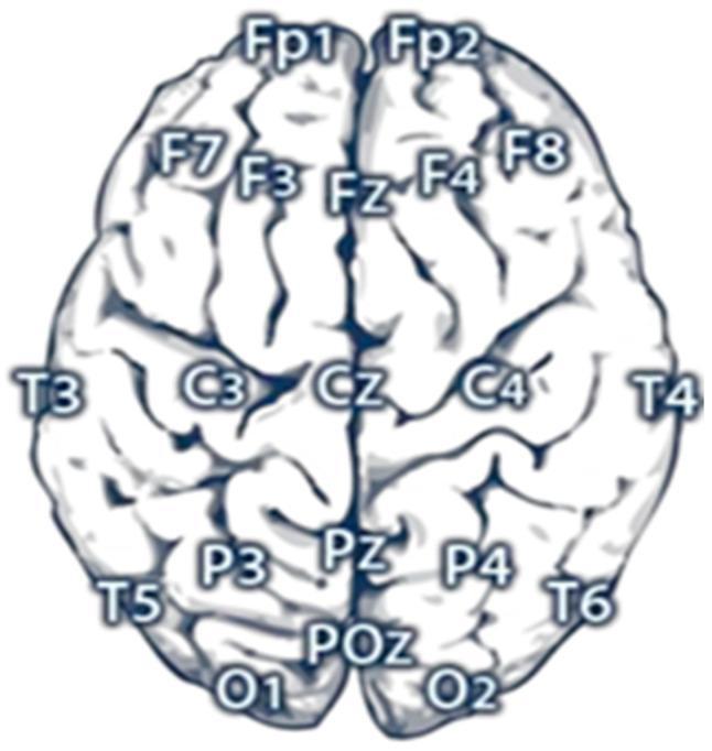
This anatomical diagram illustrates the standardized electrode placement for Electroencephalography (EEG) mapping over a superior view of the human brain. The visualization shows a 21-electrode configuration based on the International 10-20 system, emphasizing key cortical regions including frontal, central, parietal, temporal, and occipital lobes. Along the sagittal midline, the electrodes are labeled Fp (frontal pole), Fz (frontal), Cz (central), Pz (parietal), and POz (parietal-occipital). Bilateral symmetric electrodes are identified by odd numbers for the left hemisphere (Fp1, F3, F7, C3, T3, P3, T5, O1) and even numbers for the right hemisphere (Fp2, F4, F8, C4, T4, P4, T6, O2). The diagram serves as an educational tool for neuroscience and clinical neurophysiology, demonstrating the precise spatial distribution required for monitoring regional cerebral electrical activity and identifying cortical signal origins during diagnostic procedures like epilepsy monitoring or cognitive research.
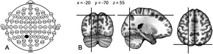
This composite educational graphic illustrates the localization of cortical stimulation targets. Panel A presents a top-down schematic diagram of the International 10-20 system for Electroencephalography (EEG) electrode placement. It displays a comprehensive grid of labeled electrode positions, including frontal (F), central (C), parietal (P), temporal (T), and occipital (O) sites. The P1 electrode position is highlighted with a black circle, indicating its selection as a stimulation target. Panel B provides a corresponding diagnostic visualization using a standardized MNI (Montreal Neurological Institute) brain template in three anatomical planes: coronal, sagittal, and axial. Crosshairs pinpoint the precise cortical projection of the P1 scalp coordinate at MNI coordinates x = -20, y = -70, z = 55, located in the posterior part of the left intraparietal sulcus (IPS). This visualization demonstrates the relationship between external scalp landmarks used in Transcranial Magnetic Stimulation (TMS) and underlying neuroanatomical structures, serving as a methodological guide for neuroimaging and brain stimulation research.
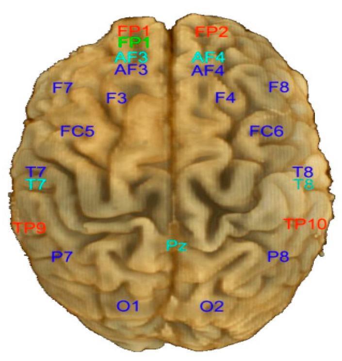
This anatomical diagram presents a superior (top-down) view of the human brain, illustrating the standardized electrode placement for the Emotiv EPOC 14-channel electroencephalography (EEG) system. The brain's cortical gyri and sulci are visible, with the longitudinal fissure dividing the left and right hemispheres. Electrodes are labeled according to the International 10-20 system nomenclature. In the left hemisphere, channels include AF3 (Anterior Frontal), F7 and F3 (Frontal), FC5 (Frontocentral), T7 (Temporal), P7 (Parietal), and O1 (Occipital). Symmetric placements in the right hemisphere are labeled AF4, F8, F4, FC6, T8, P8, and O2. Frontal pole labels (FP1 and FP2) are positioned at the anterior aspect, while a midline parietal electrode (Pz) is shown near the posterior vertex. Additionally, mastoid or temporoparietal reference points (TP9 and TP10) are marked. The diagram utilizes color-coded text to differentiate specific electrode groups, with the primary 14 recording channels typically highlighted in dark blue. This visual serves as a topographical map for clinical neurophysiology and research applications involving portable EEG cap configurations.
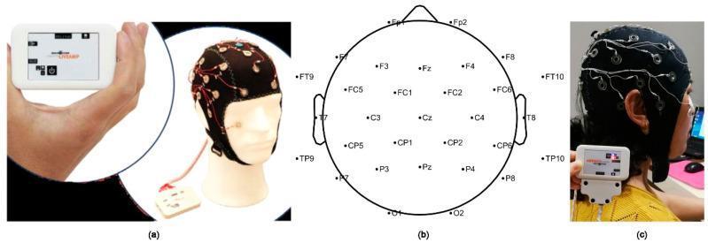
This composite educational graphic illustrates a clinical electroencephalography (EEG) setup and the standardized electrode placement system. Image (a) shows the physical components: a handheld Brain Products LiveAmp wireless amplifier and a black electrode cap fitted with active sensors and red wiring, secured with a chin strap. Image (b) is a technical diagram of the International 10-20 system for a 32-channel configuration. It details the anatomical distribution of electrodes across the frontal (Fp, F), central (C), parietal (P), temporal (T), and occipital (O) regions, along with frontocentral (FC), centroparietal (CP), and temporal-parietal (TP) nodes. Image (c) displays the clinical application on a human subject, demonstrating the lateral view of the electrode cap in use with the amplifier attached to the shoulder. This material is designed for medical training in neurophysiology, demonstrating how standardized topographical maps translate to real-world clinical equipment for monitoring cortical electrical activity.
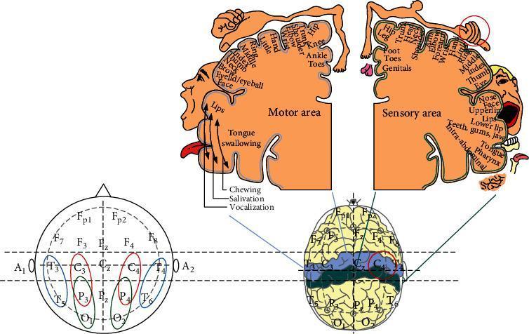
A multi-panel medical diagram illustrating electroencephalography (EEG) electrode placement and cortical mapping. The lower-left panel shows a standard 10-20 international system head map, labeling positions including Frontal-polar (Fp1, Fp2), Frontal (F3, Fz, F4), Central (C3, Cz, C4), Parietal (P3, Pz, P4), Temporal (T3-T6), and Occipital (O1, O2). Specific electrode pairs (T3-C3-P3-O1 and T4-C4-P4-O2) are highlighted, indicating bipolar recording configurations. The central lower figure depicts these electrodes on a 3D brain model, emphasizing the sensorimotor strip (precentral and postcentral gyri) in blue and green. The upper panels display the motor and sensory homunculi, providing a visual representation of somatotopic organization. The motor area maps cortical regions to body parts such as the thumb, face, and tongue for movement control, while the sensory area maps regions for tactile perception, including the genitals, feet, and hands. A red circle highlights the C4 electrode region, correlating with sensory input from the palm, demonstrating the relationship between specific scalp electrode locations and corresponding cortical functional zones.
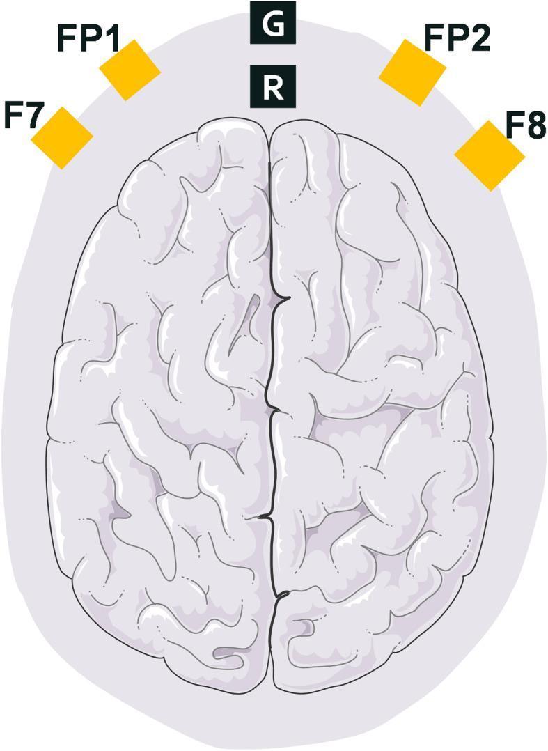
This anatomical diagram provides a superior view of the human brain, specifically illustrating the electrode montage for frontal-lobe electroencephalography (EEG) monitoring. The diagram highlights the placement of six electrodes relative to the cerebral cortex. Four active recording electrodes, marked by yellow squares, are positioned according to the International 10-20 system: FP1 and FP2 (pre-frontal) and F7 and F8 (frontal-temporal), situated over the frontal lobes of the left and right hemispheres respectively. Additionally, two central electrodes are positioned along the midline (longitudinal fissure): 'G' representing the Ground electrode and 'R' representing the Reference electrode. This specific array is commonly used in clinical anesthesia and critical care to monitor brain function, depth of sedation, and to perform coherence analysis during drug-induced loss of consciousness. The illustration effectively demonstrates the spatial distribution required for capturing bipolar frontal channel data in neurophysiological research and clinical monitoring.
EEG brain waves alpha beta theta delta normal waveforms
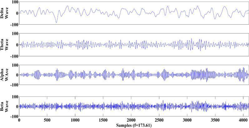
This Comparison Chart illustrates the four primary brain rhythm waveforms derived from human Electroencephalogram (EEG) data: Delta, Theta, Alpha, and Beta waves. The visualization displays each rhythm in a separate horizontal panel, plotted against a horizontal axis representing samples (0 to 4000) at a frequency of 173.61 Hz. The vertical axis for all panels indicates amplitude in microvolts (µV), ranging from -100 to 100. The Delta wave (0.5–4 Hz) shows the lowest frequency with broad, high-amplitude slow oscillations. The Theta wave (4–8 Hz) demonstrates increased frequency compared to Delta. The Alpha wave (8–14 Hz) exhibits a distinct intermediate frequency often characterized by synchronized bursts. The Beta wave (14–30 Hz) displays the highest frequency with rapid, dense oscillations and relatively lower amplitude. This educational diagram is used in neurology and clinical neurophysiology to teach the visual identification and frequency-amplitude relationships of different neurological states, such as wakefulness, relaxation, or sleep stages, based on EEG signal processing.
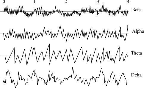
This diagnostic diagram illustrates the four primary temporal waveforms of electroencephalography (EEG) signal recording: Beta, Alpha, Theta, and Delta waves. Each waveform is displayed on a horizontal timeline from 0 to 4 seconds, demonstrating the relationship between frequency and amplitude. The Beta wave (13–30 Hz) shows the highest frequency with low-amplitude, rapid oscillations, typical of active concentration or an awake patient with open eyes. The Alpha wave (8–12 Hz) displays a slightly lower frequency with more rhythmic, synchronized peaks. The Theta wave (4–7 Hz) exhibits a further reduction in frequency with a regular, sawtooth-like pattern, often associated with deep relaxation or light sleep. The Delta wave (0.5–4 Hz) presents the lowest frequency with slow, high-amplitude irregular oscillations, characteristic of deep sleep or anesthesia. This comparison chart is essential for clinical neurophysiology to distinguish between different stages of consciousness, sleep, and neurological states based on cortical electrical activity.
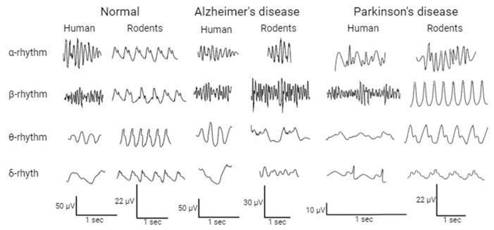
A comparative diagnostic chart displaying Electroencephalogram (EEG) waveforms across four frequency bands: alpha (α), beta (β), theta (θ), and delta (δ) rhythms. The grid compares brain activity in three cohorts: Normal subjects, Alzheimer's disease models, and Parkinson's disease models, with side-by-side sub-columns for humans and rodents. Key visual findings include the slowing and irregular shape of the human alpha rhythm in Alzheimer's disease compared to the rhythmic normal state. In Parkinson's disease, the human beta rhythm demonstrates increased variability and high-frequency oscillations, while the rodent beta rhythm shows a distinct, high-amplitude, repetitive sinusoidal-like pattern. Voltage and time scales (e.g., 50 μV per 1 sec) are provided for each human and animal model, highlighting differences in signal amplitude. This educational illustration demonstrates the translation of neurophysiological biomarkers between human clinical cases and animal research models in neurology and psychiatry.
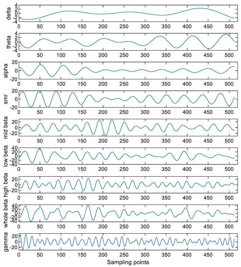
This diagnostic image displays a series of decomposed Electroencephalogram (EEG) waveforms extracted from a single-channel signal, categorized by standard neural oscillation frequency bands. The figure presents nine vertically stacked subplots, each plotted against 512 sampling points (representing one second of data at 512 Hz). From top to bottom, the waves demonstrate the inverse relationship between frequency and amplitude. The Delta and Theta waves show low-frequency, high-amplitude, slow oscillations. The Alpha wave displays a more rhythmic, sinusoidal pattern with a peak amplitude of approximately +/- 20. Mid-frequency bands including SMR, Mid Beta, Low Beta, High Beta, and Whole Beta show increasing oscillation density and more complex morphology. The bottom-most plot shows the Gamma wave (30-49 Hz), characterized by the highest frequency and most compressed peaks and troughs. This visualization is used in neurophysiology to analyze mental states—specifically concentration during cognitive activities—by decomposing raw EEG data into clinically relevant rhythmic components.
Electroencephalography (EEG) - In-Depth Academic Reference
1. Historical Background & Definition
- Adams and Victor's Principles of Neurology, 12th Ed.
2. Physical Basis - What the EEG Actually Records
"The intensity of brain waves from the scalp is determined mainly by the numbers of neurons firing in synchrony, not by the total level of electrical activity in the brain. Strong nonsynchronous nerve signals often nullify one another in the recorded brain waves because of opposing polarities."
- Guyton & Hall Textbook of Medical Physiology
- Normal amplitude range: 20-100 μV
- Normal frequency range: 1-30 Hz (some sources extend to 0.5-80 Hz)
3. Technical Setup
Electrodes
The International 10-20 System
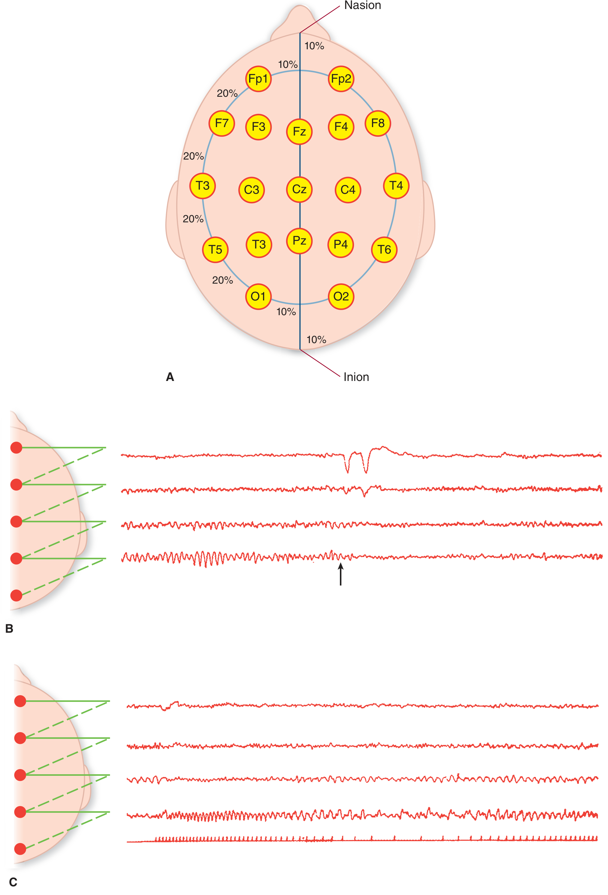
- F = Frontal, C = Central, P = Parietal, T = Temporal, O = Occipital, Fp = Frontopolar
- Odd numbers = Left hemisphere; Even numbers = Right hemisphere
- z suffix = Midline (Fz, Cz, Pz)
Montages & Recording
4. The Four Basic EEG Wave Types
| Wave | Frequency | Amplitude | State | Location |
|---|---|---|---|---|
| Alpha (α) | 8-13 Hz | ~50 μV | Quiet, relaxed wakefulness, eyes closed | Occipital > Parietal > Frontal |
| Beta (β) | 13-30 Hz (up to 80 Hz) | Low (~20 μV) | Active mental effort, eyes open, anxiety | Parietal & Frontal |
| Theta (θ) | 4-7 Hz | Medium | Drowsiness, light sleep; emotional stress; brain disorders | Parietal & Temporal (children), frontal-temporal in adults |
| Delta (δ) | <3.5 Hz | 2-4x higher than others | Deep NREM sleep, infancy, severe organic brain disease | Diffuse / cortical |
Alpha Rhythm - Special Notes
- Dominant rhythm of normal eyes-closed awake EEG
- Alpha block / desynchronization: Opening eyes → alpha disappears, replaced by low-voltage high-frequency beta waves (EEG "desynchronization")
- Can be altered by pharmacologic, metabolic, or endocrine variables
5. Origin of Brain Waves - Neural Mechanisms
Alpha Wave Origin
Delta Wave Origin
- Guyton & Hall, Chapter 60
6. EEG with Varying Levels of Cerebral Activity
| Level of Activity | Dominant Pattern |
|---|---|
| Intense mental activity / fright | Asynchronous beta (high frequency, low voltage) |
| Relaxed wakefulness | Alpha (synchronous, ~50 μV) |
| Drowsiness / light sleep | Theta waves; sleep spindles (14 Hz bursts, also called sigma waves) |
| Deep NREM sleep (Stage 3-4) | Delta waves (1-3 Hz) |
| Surgical anesthesia | Delta |
7. Activating Procedures
-
Hyperventilation: Patient breathes deeply 20x/min for 3 minutes. Through an incompletely understood mechanism, this may activate characteristic seizure patterns or other abnormalities.
-
Photic stimulation (Intermittent Photic Stimulation / IPS): A strobe light placed ~15 inches from the patient's eyes flashes at 1-20 Hz. In normal subjects, occipital leads show photic driving (visually evoked responses time-locked to each flash). In susceptible individuals (e.g., photosensitive epilepsy), it triggers a photoparoxysmal response - abnormal generalized spike-and-wave discharges.
-
Sleep recording: The drowsy and sleep transition states can reveal abnormalities not present during wakefulness.
- Adams and Victor's Principles of Neurology
8. Normal EEG - Age-Related Changes
- Infant: Preponderance of irregular medium-to-high voltage delta activity
- Early childhood: Rhythmic activity in upper theta-lower alpha range (7-8 Hz) in posterior areas
- Mid-adolescence: EEG essentially has adult appearance
9. Abnormal EEG Patterns (Waking)
| Finding | Significance |
|---|---|
| Generalized or focal delta in waking EEG | Strongly indicative of pathophysiologic process |
| Excessive theta in waking EEG | Suggests pathologic process (generalized or focal) |
| Focal slow waves (delta) in one region | Structural lesion (tumor, infarct, abscess, contusion) in that area |
| Focal spike-and-slow-wave discharges | Epileptogenic focus |
| Generalized spike-and-wave (3 Hz) | Absence epilepsy (petit mal) |
| Phase reversal | Localizes the spike generator - found at the electrode where polarity reverses between adjacent channels |
Phase Reversal
10. Clinical Applications
| Indication | EEG Role |
|---|---|
| Epilepsy | Confirm diagnosis, classify seizure type, localize epileptogenic focus |
| Brain death | Electrocerebral silence (isoelectric EEG) is one of the criteria |
| Sleep studies (polysomnography) | Staging NREM/REM sleep |
| Encephalopathy | Metabolic, toxic, infectious - generalized slow-wave activity |
| Creutzfeldt-Jakob disease (prion) | Periodic sharp-wave complexes - useful adjunctive test |
| Intraoperative monitoring | Monitor cerebral activity under anesthesia |
| ICU monitoring | Detect non-convulsive status epilepticus, monitor depth of sedation |
Summary Table: EEG Waves at a Glance
| Alpha | Beta | Theta | Delta | |
|---|---|---|---|---|
| Hz | 8-13 | >13 (up to 80) | 4-7 | <3.5 |
| μV | ~50 | ~20 (low) | Medium | Very high |
| Normal context | Relaxed, eyes closed | Alert, active | Drowsy, children | Deep sleep, infancy |
| Abnormal if present during wakefulness | Reduced = dysfunction | Excess (some drugs) | Excessive waking = pathology | Any significant waking delta = pathology |
| Origin | Thalamocortical feedback | Cortical (activated) | Cortical/limbic | Intrinsic cortical (independent of thalamus) |
- Adams and Victor's Principles of Neurology, 12th Ed. - Chapter 2 (EEG section, pp. 40-42)
- Guyton and Hall Textbook of Medical Physiology - Chapter 60 (Brain Waves)
- Kandel's Principles of Neural Science, 6th Ed. - Chapter 58
- Kaplan and Sadock's Synopsis of Psychiatry - Chapter 33 (Normal and Abnormal EEG)
Water soluble vitamins ppt
Who is this presentation for? · How detailed should the content be?
mkdir -p /tmp/workspace/water-soluble-vitamins-ppt && ls /tmp/workspace/
~/water-soluble-vitamins-ppt/build_ppt.py
from pptx import Presentation
from pptx.util import Inches, Pt, Emu
from pptx.dml.color import RGBColor
from pptx.enum.text import PP_ALIGN, MSO_ANCHOR
from pptx.util import Inches, Pt
import copy

# ─── Color Palette ───────────────────────────────────────────────────────────
DARK_BLUE   = RGBColor(0x1A, 0x37, 0x6C)   # deep navy
MID_BLUE    = RGBColor(0x26, 0x5E, 0xA8)   # header blue
ACCENT      = RGBColor(0x00, 0xB0, 0xD8)   # teal accent
LIGHT_BG    = RGBColor(0xE8, 0xF4, 0xFD)   # very light blue bg
WHITE       = RGBColor(0xFF, 0xFF, 0xFF)
DARK_TEXT   = RGBColor(0x1A, 0x1A, 0x2E)
ORANGE      = RGBColor(0xE8, 0x6B, 0x00)
GREEN       = RGBColor(0x0A, 0x7A, 0x4E)
RED_DEF     = RGBColor(0xC0, 0x20, 0x20)
YELLOW_BG   = RGBColor(0xFF, 0xF3, 0xCD)
GREEN_BG    = RGBColor(0xD4, 0xED, 0xDA)
RED_BG      = RGBColor(0xF8, 0xD7, 0xDA)

prs = Presentation()
prs.slide_width  = Inches(13.333)
prs.slide_height = Inches(7.5)
blank = prs.slide_layouts[6]   # fully blank

# ─── Helper Functions ─────────────────────────────────────────────────────────
def add_rect(slide, x, y, w, h, fill_color, border_color=None, border_width=Pt(0)):
    from pptx.util import Pt
    shape = slide.shapes.add_shape(1, Inches(x), Inches(y), Inches(w), Inches(h))
    shape.fill.solid()
    shape.fill.fore_color.rgb = fill_color
    if border_color:
        shape.line.color.rgb = border_color
        shape.line.width = border_width
    else:
        shape.line.fill.background()
    return shape

def add_text(slide, text, x, y, w, h, font_size, bold=False, color=DARK_TEXT,
             align=PP_ALIGN.LEFT, italic=False, wrap=True, font_name="Calibri"):
    tb = slide.shapes.add_textbox(Inches(x), Inches(y), Inches(w), Inches(h))
    tf = tb.text_frame
    tf.word_wrap = wrap
    tf.margin_left = 0; tf.margin_right = 0
    tf.margin_top = Pt(2); tf.margin_bottom = Pt(2)
    p = tf.paragraphs[0]
    p.alignment = align
    run = p.add_run()
    run.text = text
    run.font.size = Pt(font_size)
    run.font.bold = bold
    run.font.italic = italic
    run.font.color.rgb = color
    run.font.name = font_name
    return tb

def add_multiline_text(slide, lines, x, y, w, h, font_size, color=DARK_TEXT,
                       bold_first=False, line_spacing=None, font_name="Calibri"):
    """lines = list of (text, bold, color_override)"""
    tb = slide.shapes.add_textbox(Inches(x), Inches(y), Inches(w), Inches(h))
    tf = tb.text_frame
    tf.word_wrap = True
    tf.margin_left = 0; tf.margin_right = 0
    tf.margin_top = Pt(2); tf.margin_bottom = Pt(2)
    first = True
    for item in lines:
        if isinstance(item, str):
            text, bold, col = item, False, color
        else:
            text = item[0]
            bold = item[1] if len(item) > 1 else False
            col  = item[2] if len(item) > 2 else color
        if first:
            p = tf.paragraphs[0]
            first = False
        else:
            p = tf.add_paragraph()
        if line_spacing:
            p.line_spacing = line_spacing
        run = p.add_run()
        run.text = text
        run.font.size = Pt(font_size)
        run.font.bold = bold
        run.font.color.rgb = col
        run.font.name = font_name
    return tb

def header_bar(slide, title, subtitle=None):
    add_rect(slide, 0, 0, 13.333, 1.1, DARK_BLUE)
    add_rect(slide, 0, 1.1, 13.333, 0.08, ACCENT)
    add_text(slide, title, 0.4, 0.05, 12.5, 0.7, 32, bold=True, color=WHITE,
             align=PP_ALIGN.LEFT, font_name="Calibri Light")
    if subtitle:
        add_text(slide, subtitle, 0.4, 0.72, 12.5, 0.38, 14, bold=False,
                 color=RGBColor(0xAD, 0xD8, 0xE6), align=PP_ALIGN.LEFT)

def section_box(slide, title, x, y, w, h, title_color=MID_BLUE, bg=LIGHT_BG):
    add_rect(slide, x, y, w, 0.38, title_color)
    add_rect(slide, x, y+0.38, w, h-0.38, bg, border_color=MID_BLUE, border_width=Pt(1))
    add_text(slide, title, x+0.1, y+0.03, w-0.2, 0.35, 13, bold=True, color=WHITE,
             align=PP_ALIGN.LEFT)
    return (x+0.15, y+0.42, w-0.3, h-0.5)  # inner content area (x,y,w,h)

def bullet_lines(slide, items, x, y, w, font_size=11.5, bullet="•", color=DARK_TEXT,
                 line_gap=0.285, font_name="Calibri"):
    cy = y
    for item in items:
        if isinstance(item, tuple):
            txt, col = item
        else:
            txt, col = item, color
        tb = slide.shapes.add_textbox(Inches(x), Inches(cy), Inches(w), Inches(0.35))
        tf = tb.text_frame
        tf.word_wrap = True
        tf.margin_left = 0; tf.margin_right = 0
        tf.margin_top = 0; tf.margin_bottom = 0
        p = tf.paragraphs[0]
        run = p.add_run()
        run.text = f"{bullet}  {txt}"
        run.font.size = Pt(font_size)
        run.font.color.rgb = col
        run.font.name = font_name
        cy += line_gap
    return cy

# ──────────────────────────────────────────────────────────────────────────────
#  SLIDE 1 — TITLE SLIDE
# ──────────────────────────────────────────────────────────────────────────────
slide = prs.slides.add_slide(blank)
add_rect(slide, 0, 0, 13.333, 7.5, DARK_BLUE)
# decorative gradient strip
add_rect(slide, 0, 0, 13.333, 0.15, ACCENT)
add_rect(slide, 0, 7.35, 13.333, 0.15, ACCENT)
# central box
add_rect(slide, 1.2, 1.6, 10.9, 4.3, MID_BLUE, border_color=ACCENT, border_width=Pt(2))
add_rect(slide, 1.2, 1.6, 10.9, 0.12, ACCENT)  # top strip

add_text(slide, "WATER-SOLUBLE VITAMINS", 1.5, 1.9, 10.5, 1.2, 44, bold=True,
         color=WHITE, align=PP_ALIGN.CENTER, font_name="Calibri Light")
add_text(slide, "A Comprehensive Medical Review", 1.5, 3.05, 10.5, 0.6, 22,
         bold=False, color=RGBColor(0xAD, 0xD8, 0xE6), align=PP_ALIGN.CENTER, italic=True)

add_rect(slide, 3.5, 3.7, 6.3, 0.04, ACCENT)

add_text(slide, "B-Complex Vitamins  |  Vitamin C  |  Biochemistry, Deficiency & Clinical Correlates",
         1.5, 3.85, 10.5, 0.5, 13, color=RGBColor(0xCC, 0xE5, 0xFF),
         align=PP_ALIGN.CENTER, font_name="Calibri")
add_text(slide, "For MBBS Students", 1.5, 4.6, 10.5, 0.55, 16, bold=True,
         color=WHITE, align=PP_ALIGN.CENTER)
add_text(slide, "Based on Harper's Illustrated Biochemistry • Guyton & Hall • Basic Medical Biochemistry",
         0.5, 6.8, 12.3, 0.45, 11, color=RGBColor(0x7A, 0xA8, 0xCC),
         align=PP_ALIGN.CENTER, italic=True)

# ──────────────────────────────────────────────────────────────────────────────
#  SLIDE 2 — OVERVIEW / CLASSIFICATION
# ──────────────────────────────────────────────────────────────────────────────
slide = prs.slides.add_slide(blank)
add_rect(slide, 0, 0, 13.333, 7.5, WHITE)
header_bar(slide, "Overview of Water-Soluble Vitamins", "Classification and General Properties")

# Left column - classification
add_rect(slide, 0.3, 1.35, 5.9, 5.8, LIGHT_BG, border_color=MID_BLUE, border_width=Pt(1))
add_rect(slide, 0.3, 1.35, 5.9, 0.4, MID_BLUE)
add_text(slide, "Classification", 0.5, 1.37, 5.5, 0.36, 13, bold=True, color=WHITE)

vitamins_list = [
    ("B1 — Thiamine", MID_BLUE),
    ("B2 — Riboflavin", MID_BLUE),
    ("B3 — Niacin (Nicotinic acid)", MID_BLUE),
    ("B5 — Pantothenic Acid", MID_BLUE),
    ("B6 — Pyridoxine", MID_BLUE),
    ("B7 — Biotin", MID_BLUE),
    ("B9 — Folic Acid (Folate)", MID_BLUE),
    ("B12 — Cobalamin", MID_BLUE),
    ("C  — Ascorbic Acid", GREEN),
]
cy = 1.88
for name, col in vitamins_list:
    add_rect(slide, 0.45, cy, 5.55, 0.42, WHITE, border_color=RGBColor(0xCC, 0xDD, 0xEE), border_width=Pt(0.5))
    add_text(slide, name, 0.65, cy+0.05, 5.2, 0.35, 12.5, bold=False, color=col, font_name="Calibri")
    cy += 0.46

# Right column - general properties
add_rect(slide, 6.5, 1.35, 6.5, 2.7, LIGHT_BG, border_color=MID_BLUE, border_width=Pt(1))
add_rect(slide, 6.5, 1.35, 6.5, 0.4, MID_BLUE)
add_text(slide, "General Properties", 6.7, 1.37, 6.2, 0.36, 13, bold=True, color=WHITE)
props = [
    "Soluble in water — not stored in body",
    "Excess excreted in urine (safer toxicity profile)",
    "Regular dietary intake required",
    "Function mainly as coenzymes",
    "Heat and light sensitive — destroyed by cooking",
    "Absorbed in small intestine (B12 requires intrinsic factor)",
]
bullet_lines(slide, props, 6.7, 1.85, 6.1, font_size=11.5, line_gap=0.36)

# Clinical importance box
add_rect(slide, 6.5, 4.2, 6.5, 2.85, YELLOW_BG, border_color=ORANGE, border_width=Pt(1.5))
add_rect(slide, 6.5, 4.2, 6.5, 0.4, ORANGE)
add_text(slide, "⚠  High-Yield Clinical Points", 6.7, 4.22, 6.2, 0.36, 13, bold=True, color=WHITE)
clinical_pts = [
    "B12 deficiency → megaloblastic anemia + subacute combined degeneration",
    "Folate deficiency → neural tube defects in pregnancy",
    "Thiamine deficiency → Wernicke-Korsakoff syndrome (alcoholism)",
    "Niacin deficiency → Pellagra (3 Ds: Dermatitis, Diarrhea, Dementia)",
    "Vitamin C deficiency → Scurvy",
]
bullet_lines(slide, clinical_pts, 6.7, 4.72, 6.1, font_size=11, line_gap=0.42)

# ──────────────────────────────────────────────────────────────────────────────
#  SLIDE 3 — VITAMIN B1 (THIAMINE)
# ──────────────────────────────────────────────────────────────────────────────
slide = prs.slides.add_slide(blank)
add_rect(slide, 0, 0, 13.333, 7.5, WHITE)
header_bar(slide, "Vitamin B1 — Thiamine", "Coenzyme: Thiamine Diphosphate (TDP) / Thiamine Pyrophosphate (TPP)")

# Box 1: Biochemistry
ix, iy, iw, ih = section_box(slide, "Biochemistry & Active Form", 0.25, 1.3, 4.1, 2.8)
bio_lines = [
    ("Active form: Thiamine Pyrophosphate (TPP / TDP)", True, MID_BLUE),
    "Formed by ATP-dependent phosphorylation in tissues",
    "Thiamine triphosphate: activates Cl⁻ channel in nerve membrane",
]
multiline_items = [(t if isinstance(t, tuple) else (t, False, DARK_TEXT)) for t in bio_lines]
cy = iy
for txt, bold, col in multiline_items:
    tb = slide.shapes.add_textbox(Inches(ix), Inches(cy), Inches(iw), Inches(0.4))
    tf = tb.text_frame; tf.word_wrap = True
    tf.margin_left = 0; tf.margin_right = 0; tf.margin_top = 0; tf.margin_bottom = 0
    p = tf.paragraphs[0]; run = p.add_run()
    run.text = "•  " + txt; run.font.size = Pt(11); run.font.bold = bold
    run.font.color.rgb = col; run.font.name = "Calibri"
    cy += 0.38

# Box 2: Coenzyme roles
ix2, iy2, iw2, ih2 = section_box(slide, "Coenzyme Roles (TPP)", 4.6, 1.3, 4.3, 2.8)
roles = [
    "Pyruvate dehydrogenase  →  Pyruvate → Acetyl-CoA",
    "α-Ketoglutarate dehydrogenase  →  TCA cycle",
    "Branched-chain keto acid DH  →  Leu, Ile, Val metabolism",
    "Transketolase  →  Pentose phosphate pathway",
]
bullet_lines(slide, roles, ix2, iy2, iw2, font_size=11.5, line_gap=0.42)

# Box 3: Sources & RDA
ix3, iy3, iw3, ih3 = section_box(slide, "Dietary Sources & RDA", 9.15, 1.3, 3.9, 2.8, title_color=GREEN)
src = ["Pork (richest source)", "Whole grain cereals / bread", "Legumes, seeds, nuts",
       "Fortified breakfast cereals",
       "RDA: F = 1.1 mg/day  |  M = 1.2 mg/day"]
bullet_lines(slide, src, ix3, iy3, iw3, font_size=11, line_gap=0.38)

# Bottom: Deficiency syndromes
add_rect(slide, 0.25, 4.3, 12.83, 2.95, RED_BG, border_color=RED_DEF, border_width=Pt(1.5))
add_rect(slide, 0.25, 4.3, 12.83, 0.38, RED_DEF)
add_text(slide, "Deficiency Syndromes", 0.45, 4.32, 12.4, 0.34, 13, bold=True, color=WHITE)

# Three deficiency boxes
def_data = [
    ("Dry Beriberi\n(Peripheral Neuropathy)",
     ["Symmetric, ascending peripheral neuritis", "Weakness, sensory loss in limbs", "Muscle wasting, foot drop", "Predominantly CNS / neurological"]),
    ("Wet Beriberi\n(Cardiac Beriberi)",
     ["High-output cardiac failure", "Edema (pitting) — peripheral and pulmonary", "Dilated cardiomyopathy", "Anorexia, weight loss, apathy"]),
    ("Wernicke-Korsakoff Syndrome\n(Alcoholism / Malnutrition)",
     ["Wernicke: Ophthalmoplegia, Ataxia, Confusion (triad)", "Korsakoff: Anterograde amnesia, confabulation", "Due to impaired pyruvate → acetyl-CoA", "Treat with IV Thiamine BEFORE glucose"]),
]
dx = 0.4
for title, pts in def_data:
    add_rect(slide, dx, 4.78, 4.1, 2.35, WHITE, border_color=RED_DEF, border_width=Pt(0.5))
    add_text(slide, title, dx+0.1, 4.82, 3.9, 0.52, 11, bold=True, color=RED_DEF, font_name="Calibri")
    bullet_lines(slide, pts, dx+0.15, 5.37, 3.8, font_size=10.5, line_gap=0.3)
    dx += 4.28

add_text(slide, "Lab test: Erythrocyte Transketolase Activation test (ETK-AC) — gold standard for B1 status",
         0.4, 7.17, 12.5, 0.28, 10.5, bold=True, color=DARK_BLUE, italic=True)

# ──────────────────────────────────────────────────────────────────────────────
#  SLIDE 4 — VITAMIN B2 (RIBOFLAVIN)
# ──────────────────────────────────────────────────────────────────────────────
slide = prs.slides.add_slide(blank)
add_rect(slide, 0, 0, 13.333, 7.5, WHITE)
header_bar(slide, "Vitamin B2 — Riboflavin", "Coenzymes: FMN (Flavin Mononucleotide) & FAD (Flavin Adenine Dinucleotide)")

# Biochemistry
ix, iy, iw, ih = section_box(slide, "Biochemistry", 0.25, 1.3, 4.1, 3.0)
bio = [
    "FMN: formed by ATP-dependent phosphorylation of riboflavin",
    "FAD: formed by further reaction of FMN with ATP (AMP transferred)",
    "FMN & FAD are electron carriers in oxidoreduction reactions",
    "Intense yellow color → used as food additive (E101)",
    "Reoxidation generates superoxide radicals → oxidant stress",
]
bullet_lines(slide, bio, ix, iy, iw, font_size=11, line_gap=0.42)

# Functions
ix2, iy2, iw2, ih2 = section_box(slide, "Metabolic Roles of FMN/FAD", 4.6, 1.3, 4.3, 3.0)
func = [
    "Mitochondrial respiratory chain (Complex I, II)",
    "Fatty acid β-oxidation (acyl-CoA DH)",
    "Amino acid oxidation",
    "Citric acid cycle (succinate DH / Complex II)",
    "Glutathione reductase (antioxidant defense)",
    "Microsomal mixed-function oxidases",
]
bullet_lines(slide, func, ix2, iy2, iw2, font_size=11, line_gap=0.42)

# Sources & RDA
ix3, iy3, iw3, ih3 = section_box(slide, "Sources & RDA", 9.15, 1.3, 3.9, 3.0, title_color=GREEN)
src = ["Milk & dairy products (best source)", "Enriched / whole grain cereals",
       "Meat, poultry, fish", "Leafy green vegetables", "Eggs",
       "RDA: F = 1.1 mg/day  |  M = 1.3 mg/day"]
bullet_lines(slide, src, ix3, iy3, iw3, font_size=11, line_gap=0.38)

# Deficiency
add_rect(slide, 0.25, 4.5, 12.83, 2.75, RED_BG, border_color=RED_DEF, border_width=Pt(1.5))
add_rect(slide, 0.25, 4.5, 12.83, 0.38, RED_DEF)
add_text(slide, "Deficiency: Ariboflavinosis (Oro-oculo-genital Syndrome)", 0.45, 4.52, 12.4, 0.34, 13, bold=True, color=WHITE)

def_items_b2 = [
    "Cheilosis — fissuring / cracking at corners of mouth (angular stomatitis)",
    "Glossitis — magenta (raw beef) tongue; burning/soreness",
    "Seborrheic dermatitis — nasolabial folds, scrotal/vulval area",
    "Photophobia, lacrimation, corneal vascularization",
    "Normochromic, normocytic anemia",
    "NOT fatal — efficient tissue conservation of riboflavin",
    "Lab: Erythrocyte Glutathione Reductase Activation (EGR-AC) test",
]
bullet_lines(slide, def_items_b2, 0.5, 5.0, 12.5, font_size=11.5, line_gap=0.315)

# ──────────────────────────────────────────────────────────────────────────────
#  SLIDE 5 — VITAMIN B3 (NIACIN)
# ──────────────────────────────────────────────────────────────────────────────
slide = prs.slides.add_slide(blank)
add_rect(slide, 0, 0, 13.333, 7.5, WHITE)
header_bar(slide, "Vitamin B3 — Niacin", "Coenzymes: NAD⁺ (Nicotinamide Adenine Dinucleotide) & NADP⁺")

# Left col
ix, iy, iw, ih = section_box(slide, "Unique Feature: Not Strictly a Vitamin", 0.25, 1.3, 6.0, 2.85)
uniq = [
    "Can be synthesized from essential amino acid TRYPTOPHAN",
    "60 mg tryptophan ≡ 1 mg dietary niacin",
    "Niacin equivalents = mg niacin + (mg tryptophan ÷ 60)",
    "Two active forms: Nicotinic acid & Nicotinamide",
    "NAD is also source of ADP-ribose → DNA repair",
    "Cyclic ADP-ribose acts as intracellular Ca²⁺ messenger",
]
bullet_lines(slide, uniq, ix, iy, iw, font_size=11.5, line_gap=0.38)

# Right col
ix2, iy2, iw2, ih2 = section_box(slide, "Sources & RDA", 6.5, 1.3, 6.6, 2.85, title_color=GREEN)
src_n = [
    "Meat, poultry, fish (most abundant sources)",
    "Enriched & whole grain cereals and breads",
    "All protein-containing foods (via tryptophan)",
    "Corn/maize — low availability (bound as niacytin)",
    "RDA: F = 14 mg NE/day  |  M = 16 mg NE/day",
    "UL (Upper Limit): 35 mg/day",
]
bullet_lines(slide, src_n, ix2, iy2, iw2, font_size=11.5, line_gap=0.38)

# Bottom deficiency
add_rect(slide, 0.25, 4.3, 12.83, 3.0, RED_BG, border_color=RED_DEF, border_width=Pt(1.5))
add_rect(slide, 0.25, 4.3, 12.83, 0.38, RED_DEF)
add_text(slide, "Deficiency: PELLAGRA  —  The 4 Ds", 0.45, 4.32, 12.4, 0.34, 13, bold=True, color=WHITE)

pellagra_cols = [
    ("Dermatitis", [
        "Casal's necklace — photosensitive pigmented rash",
        "Symmetrical — exposed areas (neck, hands, feet)",
        "Sun-exposed skin → hyperpigmentation, desquamation",
        "Bilaterally symmetric distribution",
    ]),
    ("Diarrhea + Dementia", [
        "Profuse, bloody diarrhea",
        "Atrophic glossitis — bright red sore tongue",
        "Nausea, vomiting, abdominal pain",
        "Depressive psychosis → dementia if untreated",
    ]),
    ("Special Associations", [
        "Hartnup disease — defective Trp transport",
        "Carcinoid syndrome — Trp diverted to 5-HT",
        "Isoniazid therapy — B6 depletion → ↓ Trp → niacin",
        "Corn-based diet (niacin bound as niacytin)",
        "Untreated pellagra is FATAL (4th D = Death)",
    ]),
]
dx = 0.4
for title, pts in pellagra_cols:
    add_rect(slide, dx, 4.78, 4.1, 2.38, WHITE, border_color=RED_DEF, border_width=Pt(0.5))
    add_text(slide, title, dx+0.1, 4.82, 3.9, 0.35, 12, bold=True, color=RED_DEF)
    bullet_lines(slide, pts, dx+0.15, 5.22, 3.8, font_size=10.5, line_gap=0.29)
    dx += 4.28

# ──────────────────────────────────────────────────────────────────────────────
#  SLIDE 6 — VITAMIN B6 (PYRIDOXINE)
# ──────────────────────────────────────────────────────────────────────────────
slide = prs.slides.add_slide(blank)
add_rect(slide, 0, 0, 13.333, 7.5, WHITE)
header_bar(slide, "Vitamin B6 — Pyridoxine", "Active form: Pyridoxal 5'-Phosphate (PLP/PALP) — Most Versatile Coenzyme")

# Biochemistry
ix, iy, iw, ih = section_box(slide, "Forms & Active Coenzyme", 0.25, 1.3, 4.1, 2.9)
forms = [
    "Three forms: Pyridoxine, Pyridoxal, Pyridoxamine",
    "All converted to active PLP (Pyridoxal-5'-phosphate)",
    "PLP binds to ε-amino group of Lys residue in enzymes (Schiff base)",
    "Vitamin B6-dependent enzymes: >100 reactions",
]
bullet_lines(slide, forms, ix, iy, iw, font_size=11, line_gap=0.44)

# Functions
ix2, iy2, iw2, ih2 = section_box(slide, "Major Metabolic Roles of PLP", 4.6, 1.3, 4.3, 2.9)
func_b6 = [
    "Transamination (Asp-AT, Ala-AT) — amino acid metabolism",
    "Decarboxylation → amines (dopamine, serotonin, GABA, histamine)",
    "Glycogenolysis → glycogen phosphorylase",
    "Sphingolipid synthesis (serine palmitoyl transferase)",
    "Heme synthesis (ALA synthase — rate-limiting step)",
    "Tryptophan → Niacin conversion",
    "Cystathionine synthase — homocysteine metabolism",
]
bullet_lines(slide, func_b6, ix2, iy2, iw2, font_size=10.5, line_gap=0.345)

# Sources
ix3, iy3, iw3, ih3 = section_box(slide, "Sources & RDA", 9.15, 1.3, 3.9, 2.9, title_color=GREEN)
src_b6 = ["Meat, poultry, fish", "Eggs", "Fortified cereals, unmilled rice",
          "Starchy vegetables", "Non-citrus fruits", "Peanuts, walnuts",
          "RDA: 1.3 mg/day (adults)", "UL: 100 mg/day"]
bullet_lines(slide, src_b6, ix3, iy3, iw3, font_size=11, line_gap=0.3)

# Deficiency
add_rect(slide, 0.25, 4.38, 12.83, 2.88, RED_BG, border_color=RED_DEF, border_width=Pt(1.5))
add_rect(slide, 0.25, 4.38, 12.83, 0.38, RED_DEF)
add_text(slide, "Deficiency & Clinical Associations", 0.45, 4.4, 12.4, 0.34, 13, bold=True, color=WHITE)

def_b6_left = [
    "Seborrheic dermatitis, cheilosis, glossitis",
    "Microcytic hypochromic anemia (↓ heme synthesis)",
    "Peripheral neuropathy",
    "Epileptiform convulsions (neonates) — ↓ GABA",
    "Depression & confusion (↓ serotonin/dopamine)",
    "Hyperhomocysteinemia → ↑ CVD risk",
]
def_b6_right = [
    "INH (isoniazid) — drug-induced B6 deficiency",
    "Oral contraceptives — ↑ B6 requirements",
    "Penicillamine therapy — chelates PLP",
    "Alcoholism — liver disease → ↓ PLP synthesis",
    "Pyridoxine-dependent epilepsy (rare genetic disorder)",
    "High doses (>200 mg/day) → sensory neuropathy (toxicity!)",
]
bullet_lines(slide, def_b6_left, 0.45, 4.9, 6.1, font_size=11, line_gap=0.31)
bullet_lines(slide, def_b6_right, 6.75, 4.9, 6.1, font_size=11, line_gap=0.31)

# ──────────────────────────────────────────────────────────────────────────────
#  SLIDE 7 — FOLATE (B9)
# ──────────────────────────────────────────────────────────────────────────────
slide = prs.slides.add_slide(blank)
add_rect(slide, 0, 0, 13.333, 7.5, WHITE)
header_bar(slide, "Vitamin B9 — Folic Acid (Folate)", "Active form: Tetrahydrofolate (THF) — One-Carbon Metabolism")

# Biochemistry
ix, iy, iw, ih = section_box(slide, "Biochemistry", 0.25, 1.3, 4.0, 3.1)
bio_f = [
    "Dietary form: Polyglutamate (poorly absorbed)",
    "Intestinal conjugase removes glutamate residues",
    "Absorbed as monoglutamate → converted to THF",
    "Dihydrofolate reductase (DHFR) converts folate → THF",
    "THF carries one-carbon units: methyl, methylene, formyl, formimino",
    "Active storage form in cells: 5-methyl THF",
]
bullet_lines(slide, bio_f, ix, iy, iw, font_size=11, line_gap=0.4)

# One carbon units
ix2, iy2, iw2, ih2 = section_box(slide, "One-Carbon Transfer Reactions", 4.5, 1.3, 4.3, 3.1)
oc = [
    "dTMP synthesis (thymidylate synthase) — DNA synthesis",
    "Purine ring synthesis (positions 2 & 8)",
    "Homocysteine → Methionine (with B12, methionine synthase)",
    "Serine ↔ Glycine interconversion",
    "Histidine catabolism (formiminoglutamate)",
    "Formylation of initiator tRNA (Met) in mitochondria",
]
bullet_lines(slide, oc, ix2, iy2, iw2, font_size=11, line_gap=0.4)

# Sources
ix3, iy3, iw3, ih3 = section_box(slide, "Sources & RDA", 9.05, 1.3, 4.0, 3.1, title_color=GREEN)
src_f = ["Leafy green vegetables (spinach, broccoli)", "Citrus fruits (oranges)",
         "Legumes (lentils, beans)", "Fortified cereals & bread",
         "Liver, eggs",
         "RDA: 400 μg DFE/day (adults)",
         "Pregnancy: 600 μg DFE/day",
         "UL: 1000 μg/day (supplemental folic acid)"]
bullet_lines(slide, src_f, ix3, iy3, iw3, font_size=10.5, line_gap=0.315)

# Deficiency
add_rect(slide, 0.25, 4.55, 12.83, 2.72, RED_BG, border_color=RED_DEF, border_width=Pt(1.5))
add_rect(slide, 0.25, 4.55, 12.83, 0.38, RED_DEF)
add_text(slide, "Deficiency & Clinical Significance", 0.45, 4.57, 12.4, 0.34, 13, bold=True, color=WHITE)

def_f_left = [
    "Megaloblastic anemia (↓ DNA synthesis → large RBCs)",
    "Hypersegmented neutrophils on blood film",
    "Glossitis, diarrhea",
    "Neural tube defects (NTD) — spina bifida, anencephaly",
    "Periconceptional folic acid (400 μg) prevents NTDs",
]
def_f_right = [
    "Causes: Poor diet, alcoholism, malabsorption (coeliac)",
    "Drugs: Methotrexate (DHFR inhibitor), Trimethoprim, Phenytoin",
    "Pregnancy, hemolysis — increased requirements",
    "Key distinction: folate deficiency does NOT cause neurological damage (unlike B12)",
    "Masking B12 deficiency with folate supplementation — DANGER",
]
bullet_lines(slide, def_f_left, 0.45, 5.06, 6.1, font_size=11, line_gap=0.33)
bullet_lines(slide, def_f_right, 6.75, 5.06, 6.1, font_size=11, line_gap=0.33)

# ──────────────────────────────────────────────────────────────────────────────
#  SLIDE 8 — VITAMIN B12 (COBALAMIN)
# ──────────────────────────────────────────────────────────────────────────────
slide = prs.slides.add_slide(blank)
add_rect(slide, 0, 0, 13.333, 7.5, WHITE)
header_bar(slide, "Vitamin B12 — Cobalamin", "Unique: Contains Cobalt | Only vitamin made exclusively by microorganisms")

# Absorption
ix, iy, iw, ih = section_box(slide, "Unique Absorption Mechanism", 0.25, 1.3, 4.0, 3.1)
abs_b12 = [
    "Parietal cells of stomach secrete Intrinsic Factor (IF)",
    "Dietary B12 released by pepsin → binds R-protein (haptocorrin)",
    "In duodenum: pancreatic proteases digest R-protein",
    "B12 transfers to Intrinsic Factor (IF)",
    "B12-IF complex absorbed in terminal ileum",
    "Transported in blood by transcobalamin II",
    "Stored in liver (3–5 year supply)",
]
bullet_lines(slide, abs_b12, ix, iy, iw, font_size=10.5, line_gap=0.36)

# Coenzyme roles
ix2, iy2, iw2, ih2 = section_box(slide, "Coenzyme Forms & Functions", 4.5, 1.3, 4.3, 3.1)
coe = [
    "Methylcobalamin: Methionine synthase reaction",
    "  Homocysteine + CH₃-THF → Methionine + THF",
    "Adenosylcobalamin: Methylmalonyl-CoA mutase reaction",
    "  Methylmalonyl-CoA → Succinyl-CoA (TCA entry)",
    "Deficiency → ↑ methylmalonic acid & homocysteine (serum markers)",
    "Myelin synthesis impairment → neurological damage",
]
bullet_lines(slide, coe, ix2, iy2, iw2, font_size=11, line_gap=0.42)

# Sources
ix3, iy3, iw3, ih3 = section_box(slide, "Sources & RDA", 9.05, 1.3, 4.0, 3.1, title_color=GREEN)
src_b12 = ["Exclusively animal-derived foods",
           "Liver, meat, fish, poultry (richest sources)",
           "Eggs, milk, dairy products",
           "NO plant sources (vegans at risk!)",
           "Fortified foods / supplements for vegans",
           "RDA: 2.4 μg/day (adults)"]
bullet_lines(slide, src_b12, ix3, iy3, iw3, font_size=11, line_gap=0.38)

# Deficiency
add_rect(slide, 0.25, 4.55, 12.83, 2.72, RED_BG, border_color=RED_DEF, border_width=Pt(1.5))
add_rect(slide, 0.25, 4.55, 12.83, 0.38, RED_DEF)
add_text(slide, "Deficiency: Megaloblastic Anemia + Subacute Combined Degeneration (SCD)", 0.45, 4.57, 12.4, 0.34, 13, bold=True, color=WHITE)

def_b12 = [
    "Megaloblastic anemia (identical to folate deficiency — blood film)",
    "Subacute Combined Degeneration of spinal cord (SCD) — UNIQUE to B12",
    "  — Posterior columns (vibration, proprioception loss)",
    "  — Lateral corticospinal tracts (UMN signs, spasticity)",
    "Peripheral neuropathy, optic neuritis",
    "Psychiatric: dementia, psychosis ('megaloblastic madness')",
    "Causes: Pernicious anemia (anti-IF antibodies), gastrectomy, terminal ileal disease (Crohn's), veganism, metformin",
    "Lab: ↑ MCV, ↑ homocysteine, ↑ methylmalonyl acid, ↓ serum B12",
]
bullet_lines(slide, def_b12, 0.45, 5.05, 12.5, font_size=11, line_gap=0.3)

# ──────────────────────────────────────────────────────────────────────────────
#  SLIDE 9 — BIOTIN & PANTOTHENIC ACID
# ──────────────────────────────────────────────────────────────────────────────
slide = prs.slides.add_slide(blank)
add_rect(slide, 0, 0, 13.333, 7.5, WHITE)
header_bar(slide, "Vitamin B7 — Biotin  &  Vitamin B5 — Pantothenic Acid", "The 'Forgotten' B Vitamins")

# BIOTIN - left
add_rect(slide, 0.25, 1.3, 6.3, 5.9, LIGHT_BG, border_color=MID_BLUE, border_width=Pt(1))
add_rect(slide, 0.25, 1.3, 6.3, 0.4, MID_BLUE)
add_text(slide, "Biotin (Vitamin B7) — CO₂ Transfer Reactions", 0.45, 1.32, 6.0, 0.36, 13, bold=True, color=WHITE)

biotin_sections = [
    ("Biochemistry & Function:", [
        "Covalently attached to lysine residue of carboxylases (biocytin)",
        "Functions as CO₂ carrier in carboxylation reactions",
        "Pyruvate carboxylase: Pyruvate → OAA (gluconeogenesis)",
        "Acetyl-CoA carboxylase: Acetyl-CoA → Malonyl-CoA (fatty acid synthesis)",
        "Propionyl-CoA carboxylase: Propionyl-CoA → Methylmalonyl-CoA",
        "3-Methylcrotonyl-CoA carboxylase: Leucine catabolism",
    ]),
    ("Deficiency:", [
        "Raw egg white → avidin binds biotin (cooking destroys avidin)",
        "Alopecia (hair loss), dry scaly dermatitis",
        "Conjunctivitis, CNS abnormalities, glossitis",
        "Rare (gut bacteria synthesize significant amounts)",
        "Lab: ↑ urinary 3-hydroxyisovalerate",
    ]),
    ("Sources & RDA:", [
        "Liver, egg yolk, kidney", "Peanuts, almonds, sweet potato",
        "Gut microbiota synthesis (significant contribution)",
        "AI: 30 μg/day (no UL established)",
    ]),
]
cy = 1.82
for sec_title, pts in biotin_sections:
    add_text(slide, sec_title, 0.5, cy, 5.9, 0.3, 12, bold=True, color=MID_BLUE)
    cy += 0.3
    cy = bullet_lines(slide, pts, 0.6, cy, 5.7, font_size=10.5, line_gap=0.295)
    cy += 0.1

# PANTOTHENIC ACID - right
add_rect(slide, 6.8, 1.3, 6.3, 5.9, LIGHT_BG, border_color=GREEN, border_width=Pt(1))
add_rect(slide, 6.8, 1.3, 6.3, 0.4, GREEN)
add_text(slide, "Pantothenic Acid (B5) — Component of Coenzyme A", 7.0, 1.32, 6.0, 0.36, 13, bold=True, color=WHITE)

panto_sections = [
    ("Biochemistry & Function:", [
        "Component of Coenzyme A (CoA) and Acyl Carrier Protein (ACP)",
        "CoA is central to energy metabolism:",
        "  — Acetyl-CoA: TCA cycle, fatty acid synthesis",
        "  — Succinyl-CoA: heme synthesis, TCA cycle",
        "ACP: carries acyl groups in fatty acid synthesis (FAS complex)",
        "Acetylation reactions (protein modification, neurotransmitters)",
    ]),
    ("Deficiency (Rare — 'burning feet syndrome'):", [
        "Irritability, restlessness, fatigue, apathy, malaise",
        "GI symptoms (nausea, vomiting)",
        "Neurological: burning dysesthesia in feet",
        "Extremely rare — found in almost all foods (pantothen = 'everywhere')",
        "Seen in severe malnourishment / alcoholism",
    ]),
    ("Sources & RDA:", [
        "Wide distribution — almost all foods",
        "Animal tissues, whole grain cereals, legumes",
        "Avocado, mushrooms, broccoli (richest plant sources)",
        "AI: 5 mg/day (no UL established)",
    ]),
]
cy = 1.82
for sec_title, pts in panto_sections:
    add_text(slide, sec_title, 7.0, cy, 5.9, 0.3, 12, bold=True, color=GREEN)
    cy += 0.3
    cy = bullet_lines(slide, pts, 7.1, cy, 5.8, font_size=10.5, line_gap=0.295)
    cy += 0.1

# ──────────────────────────────────────────────────────────────────────────────
#  SLIDE 10 — VITAMIN C (ASCORBIC ACID)
# ──────────────────────────────────────────────────────────────────────────────
slide = prs.slides.add_slide(blank)
add_rect(slide, 0, 0, 13.333, 7.5, WHITE)
header_bar(slide, "Vitamin C — Ascorbic Acid", "The Anti-Scurvy Vitamin | Powerful Antioxidant | Redox-Active Vitamin")

# Biochemistry
ix, iy, iw, ih = section_box(slide, "Biochemistry", 0.25, 1.3, 4.0, 3.2, title_color=GREEN)
bio_c = [
    "L-Ascorbic acid (reduced form) ↔ Dehydroascorbic acid (oxidized)",
    "Reversible oxidation — acts as electron donor/acceptor",
    "Humans CANNOT synthesize ascorbic acid (lack L-gulonolactone oxidase)",
    "Most mammals can synthesize it — humans, primates, guinea pigs cannot",
    "Absorbed in small intestine by Na⁺-dependent active transport",
    "At high doses: passive diffusion predominates",
    "Stored in adrenal gland, pituitary, leukocytes",
]
bullet_lines(slide, bio_c, ix, iy, iw, font_size=11, line_gap=0.38)

# Functions
ix2, iy2, iw2, ih2 = section_box(slide, "Biochemical Functions", 4.5, 1.3, 4.3, 3.2, title_color=GREEN)
func_c = [
    "Collagen synthesis — hydroxylation of Pro & Lys residues",
    "  (Prolyl & Lysyl hydroxylase require ascorbate as reductant)",
    "Iron absorption — reduces Fe³⁺ → Fe²⁺ in GI tract",
    "Antioxidant — scavenges O₂⁻, OH•, H₂O₂",
    "Regenerates Vitamin E from tocopheryl radical",
    "Carnitine synthesis (hydroxylation steps)",
    "Tyrosine catabolism (p-hydroxyphenylpyruvate oxidase)",
    "Norepinephrine synthesis (dopamine β-hydroxylase)",
    "Steroid hormone synthesis (adrenal cortex)",
]
bullet_lines(slide, func_c, ix2, iy2, iw2, font_size=10.5, line_gap=0.31)

# Sources
ix3, iy3, iw3, ih3 = section_box(slide, "Sources & RDA", 9.05, 1.3, 4.0, 3.2, title_color=GREEN)
src_c = ["Citrus fruits (oranges, lemon, lime)", "Strawberries, kiwi, guava",
         "Bell peppers (highest source!)", "Broccoli, Brussels sprouts, spinach",
         "Potatoes", "Heat & oxygen sensitive — cooking destroys",
         "RDA: F = 75 mg/day  |  M = 90 mg/day",
         "Smokers: +35 mg/day additional",
         "UL: 2000 mg/day"]
bullet_lines(slide, src_c, ix3, iy3, iw3, font_size=10.5, line_gap=0.31)

# Deficiency: Scurvy
add_rect(slide, 0.25, 4.65, 12.83, 2.62, RED_BG, border_color=RED_DEF, border_width=Pt(1.5))
add_rect(slide, 0.25, 4.65, 12.83, 0.38, RED_DEF)
add_text(slide, "Deficiency: SCURVY — Defective Collagen Synthesis", 0.45, 4.67, 12.4, 0.34, 13, bold=True, color=WHITE)

scurvy_left = [
    "Perifollicular hemorrhage (earliest sign)",
    "Corkscrew (swan-neck) hair deformity",
    "Bleeding gums — spongy, swollen, friable",
    "Gingival hemorrhage, tooth loss",
    "Subperiosteal hemorrhage — bone pain",
]
scurvy_mid = [
    "Poor wound healing (defective collagen)",
    "Woody (hard) edema of legs",
    "Hemarthrosis, ecchymoses",
    "Anemia (Fe malabsorption + hemorrhage)",
    "Hyperkeratosis of hair follicles",
]
scurvy_right = [
    "Infant Scurvy (Barlow's disease): tender rigid limbs, frog-leg position, subperiosteal hemorrhage",
    "Diagnosis: plasma/leukocyte ascorbate levels",
    "Rx: Vitamin C 1–2 g/day for 3 days, then 500 mg/day",
    "Prevention: 5 portions fruit/veg per day",
]
bullet_lines(slide, scurvy_left, 0.4, 5.15, 4.0, font_size=11, line_gap=0.31)
bullet_lines(slide, scurvy_mid, 4.6, 5.15, 4.0, font_size=11, line_gap=0.31)
bullet_lines(slide, scurvy_right, 8.8, 5.15, 4.3, font_size=11, line_gap=0.31)

# ──────────────────────────────────────────────────────────────────────────────
#  SLIDE 11 — COMPARISON TABLE
# ──────────────────────────────────────────────────────────────────────────────
slide = prs.slides.add_slide(blank)
add_rect(slide, 0, 0, 13.333, 7.5, WHITE)
header_bar(slide, "Water-Soluble Vitamins — Summary Comparison Table",
           "Quick Exam Reference: Coenzyme | Deficiency Disease | Lab Test")

# Table headers
headers = ["Vitamin", "Active Coenzyme", "Key Functions", "Deficiency Disease", "Lab Test / Marker"]
col_widths = [1.5, 2.3, 2.8, 3.2, 3.2]
x_starts = [0.25]
for w in col_widths[:-1]:
    x_starts.append(x_starts[-1] + w + 0.02)

# Header row
for i, (h, w, x) in enumerate(zip(headers, col_widths, x_starts)):
    add_rect(slide, x, 1.28, w, 0.38, DARK_BLUE)
    add_text(slide, h, x+0.05, 1.3, w-0.1, 0.34, 11.5, bold=True, color=WHITE, align=PP_ALIGN.CENTER)

# Table data
rows = [
    ("B1 Thiamine",      "TPP (thiamine pyrophosphate)",    "Oxidative decarboxylation; transketolase",    "Beriberi; Wernicke-Korsakoff",         "Erythrocyte transketolase activation"),
    ("B2 Riboflavin",    "FMN, FAD",                        "Electron carrier; oxidoreductases",           "Ariboflavinosis (cheilosis, glossitis)", "Erythrocyte glutathione reductase"),
    ("B3 Niacin",        "NAD⁺, NADP⁺",                    "Redox reactions; 500+ enzymes",              "Pellagra (4 Ds)",                       "Urinary N¹-methylnicotinamide"),
    ("B5 Pantothenic",   "Coenzyme A (CoA), ACP",           "Acyl group transfer; TCA, FA synthesis",     "Burning feet syndrome (rare)",          "Urinary pantothenic acid"),
    ("B6 Pyridoxine",    "PLP (pyridoxal-5-phosphate)",     "Transamination; decarboxylation; >100 rxns", "Microcytic anemia; neuropathy; seizures","Erythrocyte aspartate aminotransferase"),
    ("B7 Biotin",        "Biocytin (biotinyl-lysine)",      "CO₂ carboxylation reactions",                "Alopecia, dermatitis (avidin in raw egg)","Urinary 3-hydroxyisovalerate"),
    ("B9 Folic Acid",    "Tetrahydrofolate (THF)",          "1-C transfer; nucleotide synthesis",         "Megaloblastic anemia; NTDs",            "Serum/RBC folate; homocysteine"),
    ("B12 Cobalamin",    "Methylcobalamin; Adenosylcob.",   "Methionine syn.; methylmalonyl-CoA mutase",  "Megaloblastic anemia + SCD of cord",    "Serum B12; methylmalonic acid"),
    ("C Ascorbic acid",  "Ascorbate (reduced form)",        "Collagen hydroxylation; antioxidant",        "Scurvy",                                "Plasma/leukocyte ascorbate"),
]

row_colors = [LIGHT_BG, WHITE]
cy = 1.7
for ri, row in enumerate(rows):
    bg = row_colors[ri % 2]
    for i, (cell, w, x) in enumerate(zip(row, col_widths, x_starts)):
        add_rect(slide, x, cy, w, 0.52, bg, border_color=RGBColor(0xCC,0xDD,0xEE), border_width=Pt(0.5))
        add_text(slide, cell, x+0.05, cy+0.03, w-0.1, 0.46, 9.5,
                 bold=(i==0), color=(MID_BLUE if i==0 else DARK_TEXT), wrap=True)
    cy += 0.54

# ──────────────────────────────────────────────────────────────────────────────
#  SLIDE 12 — CLINICAL SCENARIOS & EXAM HIGH-YIELD
# ──────────────────────────────────────────────────────────────────────────────
slide = prs.slides.add_slide(blank)
add_rect(slide, 0, 0, 13.333, 7.5, WHITE)
header_bar(slide, "High-Yield Clinical Scenarios & Exam Pearls", "Common Exam Traps & Key Associations")

cases = [
    ("Case 1", "Chronic alcoholic with confusion, ophthalmoplegia, ataxia → IV dextrose worsens condition",
     "Wernicke encephalopathy (B1). ALWAYS give IV Thiamine BEFORE glucose in alcoholics"),
    ("Case 2", "Isoniazid-treated TB patient develops peripheral neuropathy + convulsions",
     "B6 (Pyridoxine) deficiency. INH is a B6 antagonist — give pyridoxine prophylaxis"),
    ("Case 3", "Vegan patient with megaloblastic anemia, tingling in limbs, positive Romberg, ↑ MMA",
     "Vitamin B12 deficiency with SCD. NOT folate (folate does not cause neurological signs)"),
    ("Case 4", "Young woman planning pregnancy — what supplement is most important?",
     "Folic acid 400 μg/day preconceptionally to prevent neural tube defects"),
    ("Case 5", "Child with symmetrical hyperpigmented rash on sun-exposed areas, diarrhea, dementia; corn-based diet",
     "Pellagra (Niacin/B3 deficiency). Also think of Hartnup disease, carcinoid, INH"),
    ("Case 6", "Sailor on long voyage develops perifollicular hemorrhage, bleeding gums, poor wound healing",
     "Scurvy (Vitamin C deficiency) — classic history. Look for corkscrew hair"),
]

cy = 1.32
for case_id, history, answer in cases:
    # Case box
    add_rect(slide, 0.25, cy, 12.83, 0.82, LIGHT_BG, border_color=MID_BLUE, border_width=Pt(0.7))
    add_rect(slide, 0.25, cy, 1.3, 0.82, MID_BLUE)
    add_text(slide, case_id, 0.28, cy+0.2, 1.2, 0.42, 12, bold=True, color=WHITE, align=PP_ALIGN.CENTER)
    add_text(slide, history, 1.65, cy+0.04, 7.4, 0.38, 10.5, color=DARK_TEXT, italic=True)
    add_rect(slide, 9.15, cy, 3.95, 0.82, GREEN_BG, border_color=GREEN, border_width=Pt(0.5))
    add_text(slide, answer, 9.22, cy+0.04, 3.78, 0.74, 9.5, color=GREEN, bold=False, wrap=True)
    cy += 0.9

# bottom note
add_rect(slide, 0.25, 7.05, 12.83, 0.38, YELLOW_BG, border_color=ORANGE, border_width=Pt(1))
add_text(slide, "⚕  Remember: MCV↑ = Megaloblastic (B12 or Folate) | MCV↓ = B6 deficiency microcytic anemia | Normal MCV = Thiamine / Niacin deficiencies",
         0.4, 7.07, 12.5, 0.34, 11, bold=True, color=ORANGE, align=PP_ALIGN.LEFT)

# ──────────────────────────────────────────────────────────────────────────────
#  SLIDE 13 — DRUG-VITAMIN INTERACTIONS & SPECIAL POPULATIONS
# ──────────────────────────────────────────────────────────────────────────────
slide = prs.slides.add_slide(blank)
add_rect(slide, 0, 0, 13.333, 7.5, WHITE)
header_bar(slide, "Drug-Vitamin Interactions & Special Populations", "Pharmacology & High-Risk Groups for Water-Soluble Vitamin Deficiencies")

# Drug interactions
add_rect(slide, 0.25, 1.3, 6.0, 5.9, LIGHT_BG, border_color=ORANGE, border_width=Pt(1.5))
add_rect(slide, 0.25, 1.3, 6.0, 0.4, ORANGE)
add_text(slide, "Drug-Vitamin Interactions", 0.45, 1.32, 5.7, 0.36, 13, bold=True, color=WHITE)

drug_data = [
    ("Isoniazid (INH)", "B6 — structural analog; blocks PLP", RED_DEF),
    ("Methotrexate", "Folate — DHFR inhibitor → ↓ THF", RED_DEF),
    ("Trimethoprim / Pyrimethamine", "Folate — DHFR inhibitor (bacterial/protozoal)", RED_DEF),
    ("Phenytoin", "Folate — ↑ catabolism; ↓ absorption", RED_DEF),
    ("Oral contraceptives", "B6, B12, Folate — ↑ metabolism", RED_DEF),
    ("Metformin", "B12 — ↓ intrinsic factor / ileal absorption", RED_DEF),
    ("Alcohol", "B1, B2, B6, B12, Folate — multiple mechanisms", RED_DEF),
    ("Penicillamine", "B6 — chelates PLP", RED_DEF),
    ("Colchicine", "B12 — ↓ absorption in terminal ileum", RED_DEF),
    ("Omeprazole/PPIs", "B12 — ↓ gastric acid → ↓ release from food", RED_DEF),
]
cy = 1.82
for drug, effect, col in drug_data:
    add_rect(slide, 0.4, cy, 5.6, 0.42, WHITE, border_color=RGBColor(0xEE,0xCC,0xAA), border_width=Pt(0.5))
    add_text(slide, drug, 0.55, cy+0.04, 2.2, 0.35, 11, bold=True, color=ORANGE)
    add_text(slide, effect, 2.8, cy+0.04, 3.1, 0.35, 10.5, color=DARK_TEXT)
    cy += 0.46

# Special populations
add_rect(slide, 6.55, 1.3, 6.5, 5.9, LIGHT_BG, border_color=MID_BLUE, border_width=Pt(1.5))
add_rect(slide, 6.55, 1.3, 6.5, 0.4, MID_BLUE)
add_text(slide, "Special Populations at Risk", 6.75, 1.32, 6.2, 0.36, 13, bold=True, color=WHITE)

pop_data = [
    ("Pregnancy", [
        "Folate: 600 μg DFE/day (NTD prevention)",
        "B12: ↑ requirement (fetal demand)",
        "B6: nausea/vomiting of pregnancy",
    ]),
    ("Alcoholism", [
        "Thiamine: Wernicke-Korsakoff risk",
        "Folate: poor diet + ↑ excretion",
        "ALL B vitamins depleted",
    ]),
    ("Vegans/Vegetarians", [
        "B12: NO plant sources — supplement mandatory",
        "B2: low dairy intake",
    ]),
    ("Elderly", [
        "B12: atrophic gastritis → ↓ IF → malabsorption",
        "Folate: poor dietary intake",
    ]),
    ("Malabsorption (Crohn's, Coeliac)", [
        "B12: terminal ileum disease (Crohn's)",
        "Folate: proximal small bowel (coeliac)",
        "B1: bariatric surgery risk",
    ]),
    ("Neonates/Infants", [
        "B12: infants of vegan mothers",
        "Biotin: biotinidase deficiency (neonatal screen)",
    ]),
]
cy = 1.82
for pop, pts in pop_data:
    add_text(slide, pop, 6.75, cy, 6.1, 0.3, 12, bold=True, color=MID_BLUE)
    cy += 0.3
    cy = bullet_lines(slide, pts, 6.85, cy, 5.9, font_size=10.5, line_gap=0.29)
    cy += 0.1

# ──────────────────────────────────────────────────────────────────────────────
#  SLIDE 14 — THANK YOU / SUMMARY
# ──────────────────────────────────────────────────────────────────────────────
slide = prs.slides.add_slide(blank)
add_rect(slide, 0, 0, 13.333, 7.5, DARK_BLUE)
add_rect(slide, 0, 0, 13.333, 0.12, ACCENT)
add_rect(slide, 0, 7.38, 13.333, 0.12, ACCENT)
add_rect(slide, 1.5, 1.5, 10.3, 4.5, MID_BLUE, border_color=ACCENT, border_width=Pt(2))
add_text(slide, "Key Takeaways", 1.8, 1.65, 9.7, 0.65, 32, bold=True, color=WHITE,
         align=PP_ALIGN.CENTER, font_name="Calibri Light")
add_rect(slide, 3.5, 2.25, 6.3, 0.05, ACCENT)

key_pts = [
    "Water-soluble vitamins function as coenzymes in intermediary metabolism",
    "They are not stored (except B12) — regular dietary intake essential",
    "B12 requires Intrinsic Factor for absorption — unique among vitamins",
    "Folate + B12 together prevent megaloblastic anemia; only B12 prevents SCD",
    "Thiamine before glucose in any comatose or alcoholic patient",
    "Pellagra = Niacin deficiency — remember the 4 Ds (Dermatitis, Diarrhea, Dementia, Death)",
    "Scurvy = Vitamin C deficiency — perifollicular hemorrhage, corkscrew hair",
    "Drug-vitamin interactions are common — know INH/B6, MTX/Folate, Metformin/B12",
]
cy = 2.42
for pt in key_pts:
    add_text(slide, "✓  " + pt, 1.9, cy, 9.5, 0.36, 12.5, color=WHITE, align=PP_ALIGN.LEFT)
    cy += 0.38

add_text(slide, "Sources: Harper's Illustrated Biochemistry 32e  •  Basic Medical Biochemistry 6e  •  Guyton & Hall",
         0.5, 6.8, 12.3, 0.38, 10.5, color=RGBColor(0x7A, 0xA8, 0xCC), align=PP_ALIGN.CENTER, italic=True)

# ─── SAVE ────────────────────────────────────────────────────────────────────
out_path = "/tmp/workspace/water-soluble-vitamins-ppt/Water_Soluble_Vitamins.pptx"
from io import BytesIO
buf = BytesIO()
prs.save(buf)
with open(out_path, "wb") as f:
    f.write(buf.getvalue())
print(f"Saved: {out_path}")
print(f"Slide count: {len(prs.slides)}")
cd /tmp/workspace/water-soluble-vitamins-ppt && python build_ppt.py
ls -lh /tmp/workspace/water-soluble-vitamins-ppt/Water_Soluble_Vitamins.pptx
Water Soluble Vitamins
Presentation · PPTX
What's inside — 14 slides, MBBS-level comprehensive
| Slide | Content |
|---|---|
| 1 | Title slide |
| 2 | Overview & Classification (all 9 vitamins + general properties + high-yield clinical box) |
| 3 | B1 Thiamine — TPP coenzyme, 3 deficiency syndromes (dry beriberi, wet beriberi, Wernicke-Korsakoff), lab test |
| 4 | B2 Riboflavin — FMN/FAD, metabolic roles, Ariboflavinosis features |
| 5 | B3 Niacin — NAD⁺/NADP⁺, not strictly a vitamin, Pellagra 4 Ds, Hartnup disease |
| 6 | B6 Pyridoxine — PLP, >100 enzyme reactions, drug interactions (INH, OCPs) |
| 7 | B9 Folic Acid — THF, one-carbon metabolism, NTDs, methotrexate |
| 8 | B12 Cobalamin — intrinsic factor absorption, SCD of spinal cord, methylmalonic acid |
| 9 | Biotin (B7) & Pantothenic Acid (B5) — CO₂ carboxylation, CoA, avidin |
| 10 | Vitamin C — collagen synthesis, scurvy, Barlow's disease |
| 11 | Master comparison table (all vitamins: coenzyme, function, deficiency, lab test) |
| 12 | 6 clinical scenario vignettes with answers (exam-style) |
| 13 | Drug-vitamin interactions + Special populations (pregnancy, alcoholism, vegans) |
| 14 | Key Takeaways summary |
Water soluble vitamins in children ppt slides 40
pediatric vitamin deficiency scurvy beriberi pellagra child clinical signs
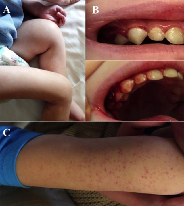
This composite of clinical photographs illustrates the classic mucocutaneous and musculoskeletal manifestations of pediatric scurvy (Vitamin C deficiency). Image A displays the lower extremities of a child, showing significant swelling and edema of the left thigh and knee area, with the limb held in a flexed, antalgic posture due to subperiosteal hemorrhage and bone pain. Image B consists of two intraoral views showing severe gingival hyperplasia, spongy swelling, intense erythema, and active friable bleeding along the gingival margins, particularly around the erupting primary teeth. Image C shows a close-up of an extremity featuring a diffuse distribution of pinpoint, non-blanching petechial hemorrhages across the skin surface. Collectively, these findings represent the clinical triad of hemorrhagic diathesis (petechiae and bleeding gums), skeletal changes (painful limb swelling), and nutritional deficiency. The images are highly relevant for pediatrics and hematology, demonstrating the physical exam signs of defective collagen synthesis and impaired capillary integrity.
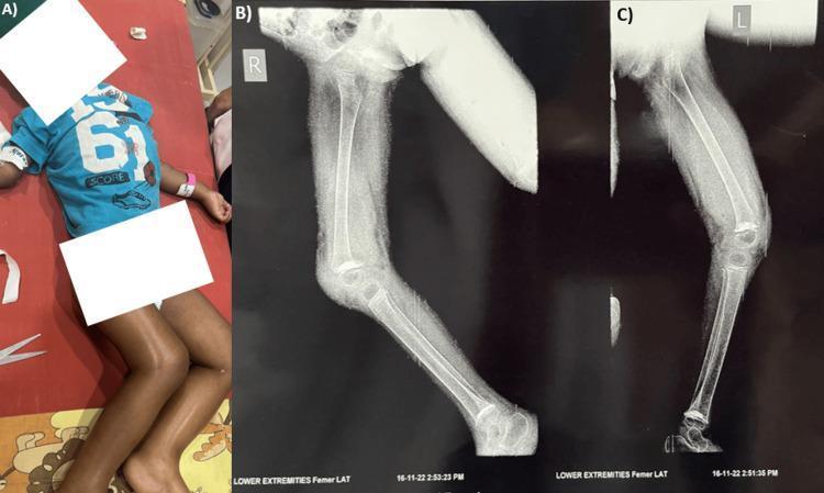
This composite educational material consists of a clinical photograph and lateral radiographs illustrating pediatric scurvy (Vitamin C deficiency). Panel A is a clinical photograph of a pediatric patient exhibiting signs of severe acute malnutrition (SAM), including muscle wasting and thinning of the extremities. Panels B and C are lateral X-ray images of the right and left lower limbs, respectively. The radiographs demonstrate pathognomonic radiological signs of scurvy at the knee joint. Key findings include the 'white line of Frankel,' which is a dense, sclerotic band at the metaphysis representing a zone of provisional calcification. Visible at the metaphyseal margins are 'corner signs' (Pelkan spurs), which are small, triangular bony outgrowths. The epiphyses of the distal femur and proximal tibia show a 'Wimberger ring sign,' characterized by a dense sclerotic periphery surrounding a radiolucent center. Additionally, generalized osteopenia and cortical thinning are observed in the long bones. These images serve as a classic diagnostic reference for the musculoskeletal manifestations of vitamin C deficiency in a pediatric population.
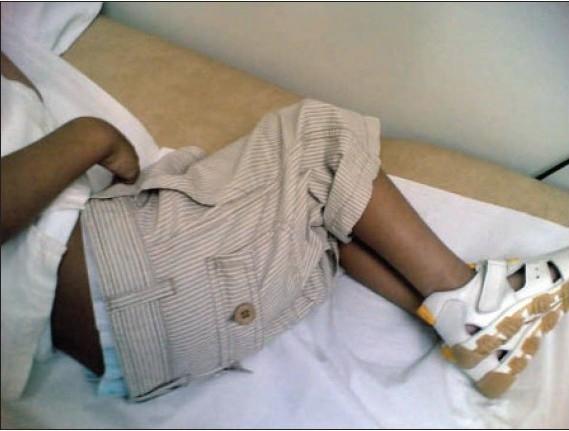
Clinical photograph of a pediatric patient in a supine position on an examination table, exhibiting a characteristic 'pitied' or flexion posture. The child shows forced flexion of the bilateral hips and knees, with the arms also held in a flexed, guarded position near the trunk. This specific posture is clinically significant in pediatric cases of scurvy (vitamin C deficiency), where subperiosteal hemorrhages cause exquisite musculoskeletal pain, leading the patient to assume a fixed, flexed limb position to minimize discomfort and avoid movement. The image serves as a diagnostic visual reference for scurvy-related pseudoparalysis and musculoskeletal involvement in a modern clinical setting, often seen in the context of severe dietary restriction.
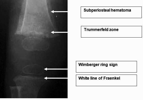
This diagnostic x-ray radiograph illustrates the classic skeletal manifestations of pediatric scurvy (vitamin C deficiency) in a long bone. The image is annotated to highlight several pathognomonic radiological signs: 1) The 'White Line of Fraenkel,' a dense, thickened zone of provisional calcification at the metaphysis; 2) The 'Trummerfeld zone,' a radiolucent scorbutic band located immediately proximal to the white line, representing a zone of rarefaction where bone formation is suppressed; 3) 'Wimberger ring sign,' characterized by a thin sclerotic rim surrounding a lucent center in the epiphysis; and 4) signs of a subperiosteal hematoma, visible as periosteal elevation along the diaphysis. These findings are critical diagnostic markers in pediatric radiology for metabolic bone disease and nutritional deficiencies. The educational focus is on identifying metaphysis and epiphysis abnormalities related to defective collagen synthesis and osteoblast function.
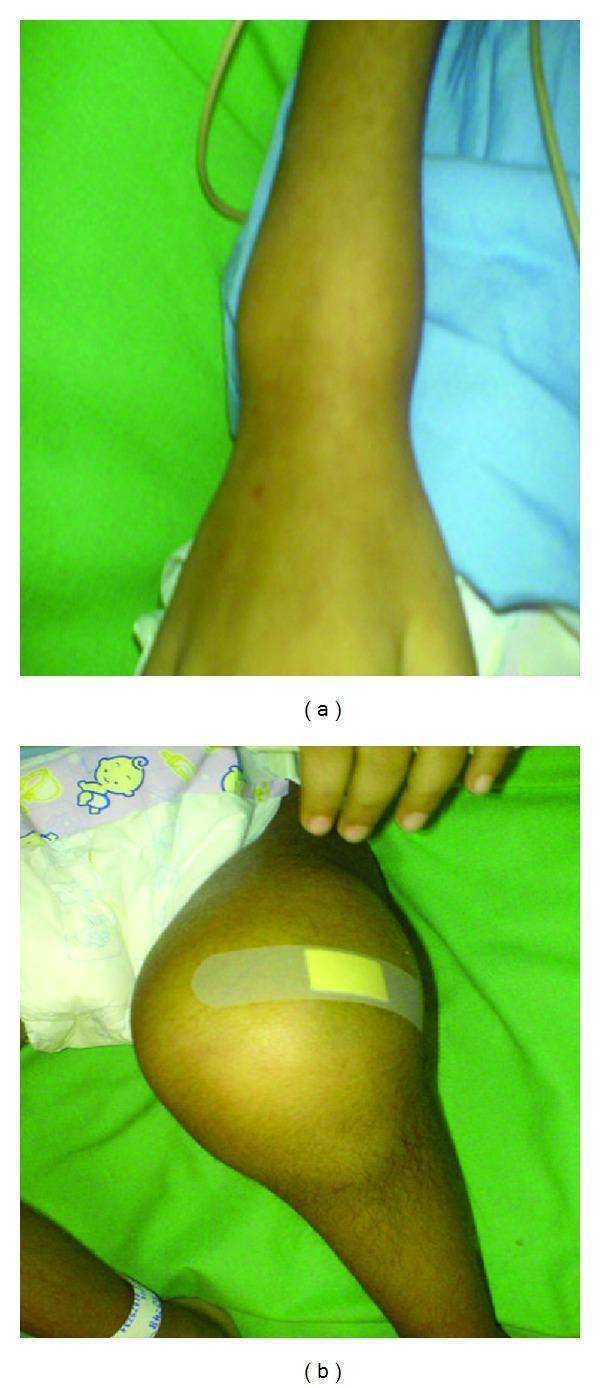
This clinical photograph consists of two panels, (a) and (b), illustrating pediatric joint manifestations associated with a nutritional deficiency, likely scurvy (vitamin C deficiency). Panel (a) shows a superior view of the right wrist, demonstrating circumferential, diffuse soft tissue swelling that obscures normal anatomical bony landmarks of the distal radius and ulna. Panel (b) shows the left knee of a child, revealing significant, tense articular swelling and joint effusion that creates a bulbous, rounded appearance of the knee joint. A small adhesive bandage is present on the anterior surface of the knee. In both images, the overlying skin remains intact without obvious discoloration, erythema, or ulceration. These findings are clinically relevant as they represent musculoskeletal manifestations of scurvy, where subperiosteal hemorrhage and metaphyseal fragility lead to painful joint swelling and pseudoparalysis in pediatric patients.
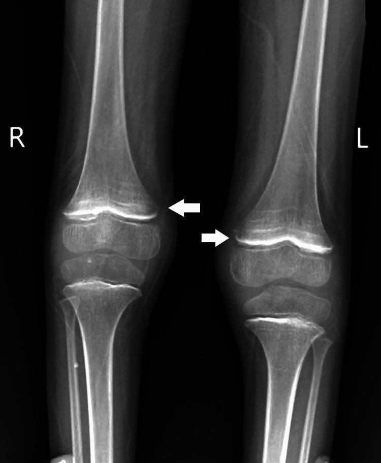
This diagnostic image is an anteroposterior (AP) X-ray of the pediatric lower limbs, specifically focusing on the bilateral knee joints. The radiograph demonstrates findings consistent with scurvy (vitamin C deficiency). Key features include dense, sclerotic bands at the metaphyses, known as the 'white line of Frankel,' and a subjacent radiolucent zone (the 'Trummerfeld zone' or scurvy zone). White arrows indicate bilateral periosteal thinning and elevation, often associated with subperiosteal hemorrhage. The epiphyses show a characteristic 'Wimberger ring sign' with a dense peripheral rim and central lucency. The overall bone density appears slightly decreased, but the architecture of the distal femur, proximal tibia, and fibula remains intact without obvious lytic or sclerotic focal lesions. This visual information is critical for pediatric radiology and nutritional deficiency diagnosis, illustrating musculoskeletal manifestations of scurvy in a growing child.
vitamin B12 deficiency child infant megaloblastic anemia neural tube defect folate
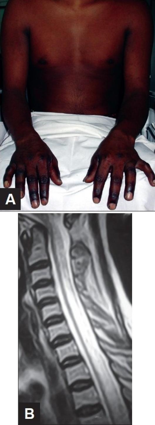
This composite figure demonstrates clinical and diagnostic findings associated with Vitamin B12 deficiency (Subacute Combined Degeneration of the spinal cord). Image A is a clinical photograph of an 18-year-old male showing significant cutaneous hyperpigmentation of the distal upper extremities. The darkening is most pronounced on the dorsal surfaces of the hands and fingers, contrasting with the lighter skin tone of the proximal arms and chest. This is a common dermatologic manifestation of megaloblastic anemia. Image B is a sagittal T2-weighted MRI of the cervical and upper thoracic spine. It reveals a long-segment, linear intramedullary hyperintense signal within the posterior aspect of the spinal cord. This finding represents edema and demyelination characteristic of Subacute Combined Degeneration, which typically affects the posterior and lateral columns while sparing the anterior column. The combination of these visual findings—distal hyperpigmentation and longitudinal posterior cord hyperintensity—is highly suggestive of severe Vitamin B12 deficiency in the context of progressive paraplegia.
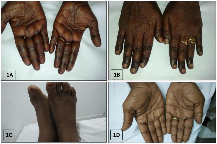
This composite of clinical photographs illustrates various patterns of cutaneous hyperpigmentation associated with megaloblastic anemia due to Vitamin B12 deficiency. Image 1A and 1D display the palmar surfaces of the hands, showing diffuse, brownish-black hyperpigmentation with prominent accentuation along the palmar creases. Image 1B focuses on the dorsal aspect of the hands, highlighting localized brownish-black pigmentation specifically over the knuckle pads and phalangeal joints. Image 1C depicts the dorsal aspect of the feet, exhibiting a similar diffuse, dusky, brownish-black discoloration across the skin surface. These dermatological findings are classic external markers of severe B12 deficiency and often present alongside systemic symptoms like pancytopenia and macrocytosis. The collection serves as an educational tool for recognizing the cutaneous manifestations of nutritional deficiencies and metabolic disorders in hematology and dermatology.
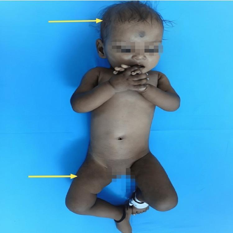
Clinical photograph of a pediatric patient exhibiting characteristic dermatological and hair changes associated with nutritional deficiencies, specifically Vitamin B12 deficiency. The image shows an infant with sparse, coarse, and hypopigmented scalp hair (indicated by the upper arrow). Significant cutaneous findings include hyperpigmentation of the knuckles on both hands and a diffuse reticular (net-like) pigmentation pattern across the lower extremities (indicated by the lower arrow). The patient also appears mildly edematous or 'plump' despite malnutrition markers. These clinical signs—knuckle hyperpigmentation and reticular skin changes—are classic cutaneous markers of megaloblastic anemia in infants, often secondary to maternal vegetarian diets or malabsorption. The visual serves as an educational tool for identifying systemic metabolic disorders through external physical examination findings in a pediatric clinical context.
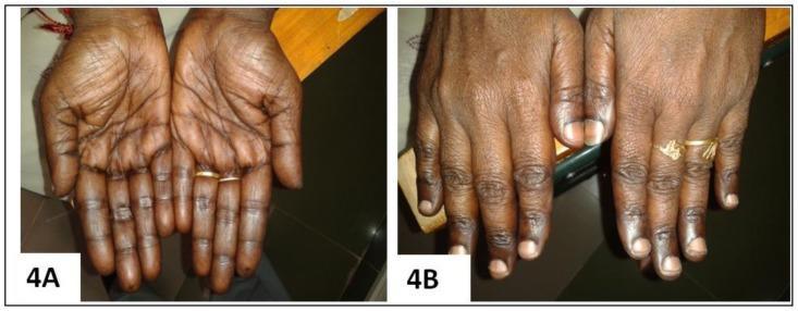
Clinical photograph comparison (Figure 4A and 4B) demonstrating the cutaneous manifestations of Vitamin B12 deficiency and its resolution following treatment. Image 4A (Pre-treatment): Palmar view showing diffuse, brownish-black hyperpigmentation with prominent accentuation along the palmar creases and longitudinal hyperpigmentation of the fingers. The knuckles exhibit characteristic darkening, a classic sign of megaloblastic anemia-associated pigmentary changes. Image 4B (Post-treatment): Dorsal view of the same patient's hands 12 weeks after initiating parenteral cyanocobalamin (Vitamin B12) therapy. There is a marked reversal of the hyperpigmentation, with the skin returning to a more uniform, lighter tone. The previously dusky knuckles and digits show significant clearing, and the nail beds appear healthy. This comparison serves as an educational tool for identifying dermatological markers of nutritional deficiencies and monitoring therapeutic response to Vitamin B12 supplementation.
Water soluble vitamins in children ppt 40slides in detail
pediatric scurvy child swollen limbs bleeding gums subperiosteal hemorrhage vitamin C deficiency
This composite of clinical photographs illustrates the classic mucocutaneous and musculoskeletal manifestations of pediatric scurvy (Vitamin C deficiency). Image A displays the lower extremities of a child, showing significant swelling and edema of the left thigh and knee area, with the limb held in a flexed, antalgic posture due to subperiosteal hemorrhage and bone pain. Image B consists of two intraoral views showing severe gingival hyperplasia, spongy swelling, intense erythema, and active friable bleeding along the gingival margins, particularly around the erupting primary teeth. Image C shows a close-up of an extremity featuring a diffuse distribution of pinpoint, non-blanching petechial hemorrhages across the skin surface. Collectively, these findings represent the clinical triad of hemorrhagic diathesis (petechiae and bleeding gums), skeletal changes (painful limb swelling), and nutritional deficiency. The images are highly relevant for pediatrics and hematology, demonstrating the physical exam signs of defective collagen synthesis and impaired capillary integrity.
This diagnostic image is an anteroposterior (AP) X-ray of the pediatric lower limbs, specifically focusing on the bilateral knee joints. The radiograph demonstrates findings consistent with scurvy (vitamin C deficiency). Key features include dense, sclerotic bands at the metaphyses, known as the 'white line of Frankel,' and a subjacent radiolucent zone (the 'Trummerfeld zone' or scurvy zone). White arrows indicate bilateral periosteal thinning and elevation, often associated with subperiosteal hemorrhage. The epiphyses show a characteristic 'Wimberger ring sign' with a dense peripheral rim and central lucency. The overall bone density appears slightly decreased, but the architecture of the distal femur, proximal tibia, and fibula remains intact without obvious lytic or sclerotic focal lesions. This visual information is critical for pediatric radiology and nutritional deficiency diagnosis, illustrating musculoskeletal manifestations of scurvy in a growing child.
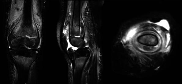
This diagnostic image set consists of three magnetic resonance imaging (MRI) views—coronal, sagittal, and axial—of a pediatric knee joint, illustrating advanced findings of scurvy (vitamin C deficiency). The T2-weighted or fluid-sensitive sequences reveal prominent, circumferential high-signal intensity fluid collections located beneath the periosteum of the distal femur and proximal tibia. This subperiosteal hemorrhage appears as a thick layer of fluid that elevates the periosteum away from the cortical bone. The coronal and sagittal views highlight the longitudinal extent of the hemorrhage along the metaphyses, while the axial view demonstrates the circumferential nature of the fluid collection around the femoral shaft. Additionally, there is evidence of diffuse bone marrow edema and soft tissue swelling. These findings are characteristic musculoskeletal manifestations of scurvy in children, resulting from impaired collagen synthesis and subsequent capillary fragility. The image serves as an educational tool for distinguishing nutritional deficiencies from other marrow infiltrative or inflammatory processes using advanced imaging.
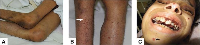
A composite of three clinical photographs demonstrating the systemic manifestations of Vitamin C deficiency (Scurvy). Panel A shows the lower extremities with prominent knee flexion contractures and diffuse ecchymoses around the joints. Panel B provides a close-up of the skin on the legs, highlighting perifollicular purpura and follicular hyperkeratosis; a white arrow points to characteristic 'corkscrew' hairs. Panel C displays the facial and oral features, including hemorrhagic gingivitis with swollen, friable, and bleeding gums. A black arrow in Panel C indicates a neurotic excoriation on the chin. The clinical findings represent the classic triad of scurvy: follicular hyperkeratosis with perifollicular hemorrhage, gingival bleeding, and musculoskeletal involvement. This content is intended for medical education regarding nutritional deficiencies and dermatologic manifestations of systemic disease.
infant thiamine beriberi wernicke child niacin pellagra dermatitis skin rash child
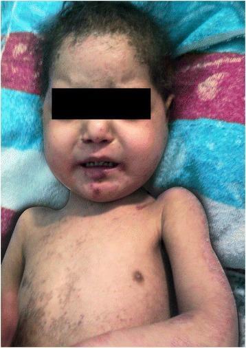
This clinical photograph displays the initial presentation of a pediatric patient with systemic dermatological manifestations of pellagra (niacin deficiency). The image shows a severe, scaly, desquamating skin rash with a widespread distribution across the face, neck, torso, and upper extremities. Key visual features include significant pigmentary alterations, characterized by areas of both hyperpigmentation and hypopigmentation, particularly prominent on the chest and arms. The facial rash exhibits erythema and scaling in a photosensitive distribution. Additional findings include thin, sparse scalp hair and visible scaliness on the scalp, potentially reflecting underlying nutritional compromise. The clinical presentation is consistent with the 'dermatitis' component of the pellagra triad, often associated with systemic conditions such as Hartnup disease or chronic malabsorption leading to secondary niacin deficiency. The educational focus is on recognizing the characteristic morphology and distribution of photosensitive nutritional dermatoses in a clinical setting.
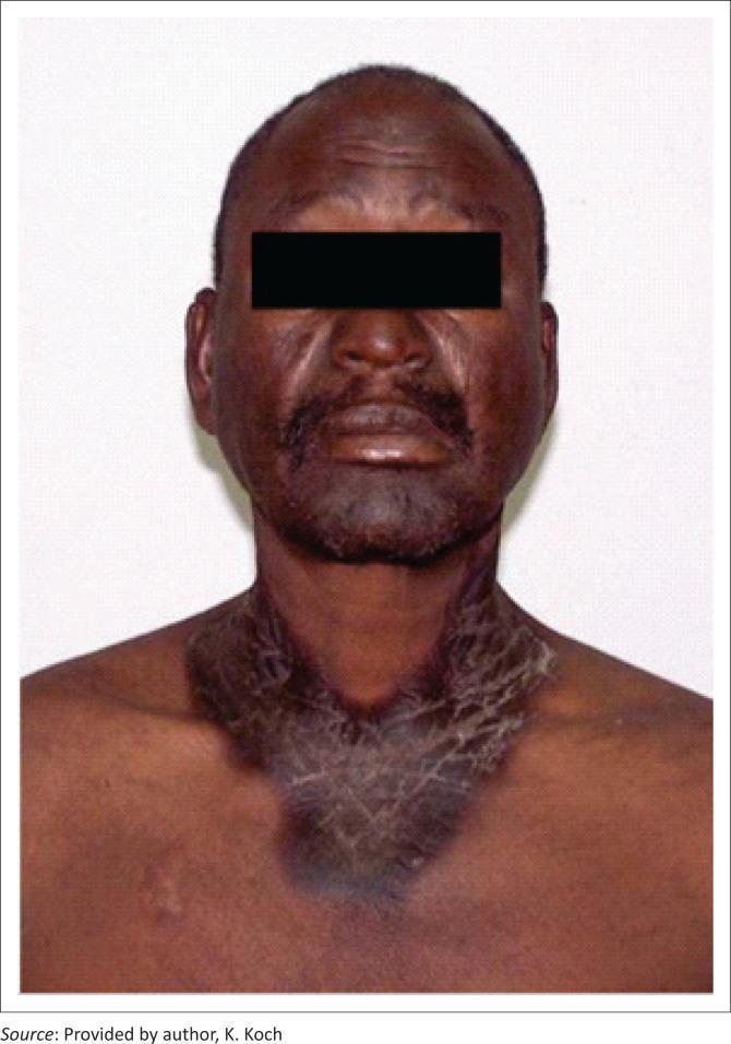
A clinical photograph of an adult male exhibiting classic dermatological manifestations of pellagra, caused by niacin (vitamin B3) deficiency. The image shows a well-defined, hyperpigmented, and xerotic (dry) skin rash with a characteristic photodistributed pattern. On the neck and upper anterior chest, the lesion forms a prominent circumferential band that transitions into a V-shaped distribution, clinically known as Casal's necklace. The affected skin appears thickened with visible scaling, fissuring, and a 'crazy-pavement' texture. Similar hyperpigmentation and skin changes are visible across the face, particularly on the forehead and perioral regions. These findings represent the 'dermatitis' component of the classic triad of pellagra (dermatitis, diarrhea, and dementia) and are frequently associated with advanced HIV or certain medications like isoniazid that interfere with niacin metabolism. This visual material is used in dermatology and nutrition education to demonstrate systemic nutritional deficiencies and photosensitive eruptions.
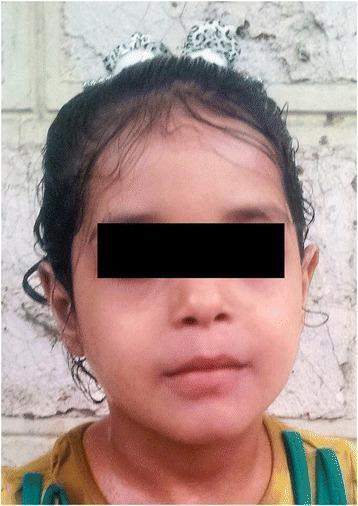
This clinical photograph shows the frontal view of a pediatric patient following treatment for niacin deficiency (pellagra). The image demonstrates a complete resolution of previously severe dermatological symptoms. The facial skin appears smooth and healthy, with an even tone and a total absence of the scaly, desquamating rash and hyperpigmentation characteristic of pellagra. The forehead and cheeks show no signs of inflammation or photosensitivity-induced lesions. The scalp and hairline appear normal with no evidence of alopecia or dermatitis. This visual serves as an educational comparison to show therapeutic success in managing Hartnup disease or secondary nutritional deficiencies through oral niacin supplementation. The patient's eyes are masked for privacy, while the remaining facial features highlight the restoration of normal skin integrity and texture in a clinical dermatology context.
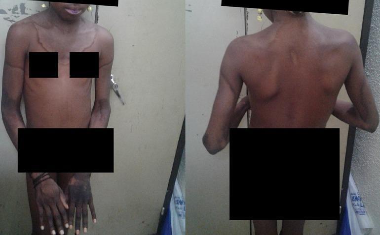
Clinical photograph of a pediatric patient presenting with classical dermatological manifestations of pellagra (niacin deficiency). The image shows well-demarcated, symmetrical, hyperpigmented, and hyperkeratotic plaques. A prominent feature is the 'Casal necklace,' a circumferential band of darkened, thickened skin extending around the neck and onto the upper thorax and back. Additional acral involvement is visible, characterized by symmetrical hyperpigmentation on the dorsal aspects of the hands, wrists, and forearms. The skin in affected areas appears dry and scaly with erythematous margins, consistent with photosensitive dermatitis. The patient exhibits a wasted body habitus, suggesting underlying malnutrition or cachexia. This presentation illustrates the 'Dermatitis' component of the pellagra triad (Dermatitis, Diarrhea, Dementia) and is key for clinical diagnosis in resource-limited settings where dietary niacin or tryptophan intake is insufficient.
vitamin B12 deficiency infant hyperpigmentation knuckles sparse hair megaloblastic anemia child
Clinical photograph of a pediatric patient exhibiting characteristic dermatological and hair changes associated with nutritional deficiencies, specifically Vitamin B12 deficiency. The image shows an infant with sparse, coarse, and hypopigmented scalp hair (indicated by the upper arrow). Significant cutaneous findings include hyperpigmentation of the knuckles on both hands and a diffuse reticular (net-like) pigmentation pattern across the lower extremities (indicated by the lower arrow). The patient also appears mildly edematous or 'plump' despite malnutrition markers. These clinical signs—knuckle hyperpigmentation and reticular skin changes—are classic cutaneous markers of megaloblastic anemia in infants, often secondary to maternal vegetarian diets or malabsorption. The visual serves as an educational tool for identifying systemic metabolic disorders through external physical examination findings in a pediatric clinical context.
Clinical photograph comparison (Figure 4A and 4B) demonstrating the cutaneous manifestations of Vitamin B12 deficiency and its resolution following treatment. Image 4A (Pre-treatment): Palmar view showing diffuse, brownish-black hyperpigmentation with prominent accentuation along the palmar creases and longitudinal hyperpigmentation of the fingers. The knuckles exhibit characteristic darkening, a classic sign of megaloblastic anemia-associated pigmentary changes. Image 4B (Post-treatment): Dorsal view of the same patient's hands 12 weeks after initiating parenteral cyanocobalamin (Vitamin B12) therapy. There is a marked reversal of the hyperpigmentation, with the skin returning to a more uniform, lighter tone. The previously dusky knuckles and digits show significant clearing, and the nail beds appear healthy. This comparison serves as an educational tool for identifying dermatological markers of nutritional deficiencies and monitoring therapeutic response to Vitamin B12 supplementation.
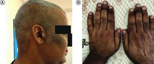
Clinical photograph in two panels (A and B) demonstrating cutaneous hyperpigmentation associated with vitamin B12 deficiency. Panel A shows a lateral view of the head and neck, highlighting patchy, irregular melanoderma on the temporal region and cheek, with a mottled distribution across the scalp. The pigmentation ranges from light tan to dark brown and appears non-elevated and non-scaly. Panel B displays the dorsal surfaces of both hands, revealing generalized darkening with accentuated intensity over the knuckles and phalangeal creases. A proximal-to-distal gradient is visible on the fingers, with the most intense hyperpigmentation occurring proximally. These findings are characteristic of the mucocutaneous manifestations of megaloblastic anemia and vitamin B12 deficiency, serving as a critical clinical sign for differential diagnosis from Addison’s disease.
scurvy X-ray child white line Frankel Wimberger ring Trummerfeld zone metaphysis
This diagnostic image is an anteroposterior (AP) X-ray of the pediatric lower limbs, specifically focusing on the bilateral knee joints. The radiograph demonstrates findings consistent with scurvy (vitamin C deficiency). Key features include dense, sclerotic bands at the metaphyses, known as the 'white line of Frankel,' and a subjacent radiolucent zone (the 'Trummerfeld zone' or scurvy zone). White arrows indicate bilateral periosteal thinning and elevation, often associated with subperiosteal hemorrhage. The epiphyses show a characteristic 'Wimberger ring sign' with a dense peripheral rim and central lucency. The overall bone density appears slightly decreased, but the architecture of the distal femur, proximal tibia, and fibula remains intact without obvious lytic or sclerotic focal lesions. This visual information is critical for pediatric radiology and nutritional deficiency diagnosis, illustrating musculoskeletal manifestations of scurvy in a growing child.
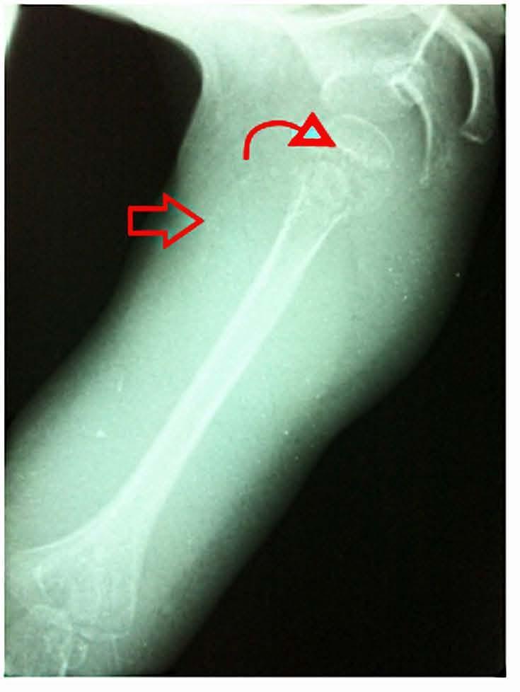
Anteroposterior diagnostic X-ray radiography of a pediatric right humerus and shoulder region, demonstrating classic skeletal manifestations of infantile scurvy (hypovitaminosis C). The image features a prominent, dense transverse radiopaque line at the distal metaphysis, known as the 'white line of Frankel' (indicated by a curved red arrow), representing a zone of provisional calcification. Along the humeral diaphysis, there is visible periosteal elevation or separation from the underlying cortex (marked by a straight red arrow), consistent with subperiosteal hemorrhage. The epiphyses appear osteopenic with a thin sclerotic rim, characteristic of Wimberger's ring sign. These findings illustrate the impact of impaired collagen synthesis on osteoblast function and capillary integrity. The educational focus is on identifying radiographic signs of nutritional deficiencies in pediatric orthopedics and radiology.
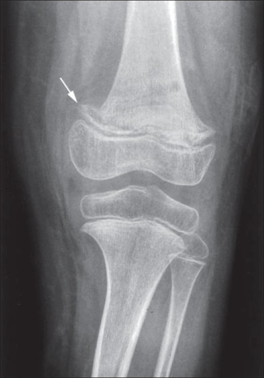
**Imaging Modality:** Conventional Radiography (X-ray). **Anatomical Region:** Pediatric knee joint, including the distal femur and proximal tibia/fibula. **Observed Pathology:** Findings consistent with infantile scurvy (hypovitaminosis C). **Characteristic Visual Features:** * **White Line of Frankel:** A dense, transverse radiopaque band is visible at the zone of provisional calcification in the distal femoral metaphysis (indicated by the white arrow). * **Pelkan Spur:** Marginal lateral bony protrusion extending from the metaphysis, resulting from the outward displacement of the zone of provisional calcification. * **Trümmerfeld Zone:** A lucent, scorbutic zone of rarefaction located immediately proximal to the dense metaphyseal line. * **Wimberger Ring Sign:** Sclerotic circumferential rimming around the epiphyseal centers of ossification, which appear osteopenic or "ground-glass" centrally. * **Generalized Osteopenia:** Diffuse reduction in bone mineral density throughout the visualized long bones. **Key Diagnostic Features:** The combination of a dense metaphyseal white line, metaphyseal spurring (Pelkan spur), and ring-shaped epiphyses is pathognomonic for skeletal scurvy in a pediatric patient.
mkdir -p /tmp/workspace/pediatric-vitamins-ppt && echo "ready"