
Causes of chest pain how do you diagnosis gastritis
Reading File
Now I'll search the medical library for both topics in parallel.
Finding Sources
Finding Sources
Searching the Web
causes of chest pain differential diagnosis gastritis diagnosis 2024
Good, I found the chest pain sections. Let me now read those sections and search for gastritis diagnosis content in parallel.
Reading File
Finding Sources
Now let me read the noncardiac/GI chest pain sections and the gastritis diagnosis sections in parallel.
Reading File
Reading File
Reading File
Now let me read the GI chest pain section and also the gastritis clinical presentation/diagnosis from Rosen's Emergency Medicine.
Reading File
Reading File
Let me read the diagnostic workup for gastritis and also the musculoskeletal/other causes of chest pain.
Reading File
Reading File
I now have excellent content from the textbooks. Let me also read a bit more about the gastritis diagnostic tests (H. pylori tests, endoscopy, etc.).
Reading File
I now have comprehensive information from the textbooks to provide a detailed answer. Let me compose the full response.
Here is a thorough breakdown of both topics.
Causes of Chest Pain
Chest pain is one of the most common presentations in medicine and can be life-threatening. It is classified broadly into cardiac and noncardiac causes.
I. Cardiac Causes
Acute Coronary Syndrome (ACS)
ACS describes a range of clinical syndromes resulting in myocardial ischemia with or without infarction. Diagnosis requires a compatible history, ECG findings, and biomarkers.
Characteristic symptoms:
- Chest, upper extremity, mandibular, or epigastric discomfort at rest or with exertion
- Usually lasts >20 minutes
- Associated with diaphoresis, nausea, or syncope
- "Angina equivalents": dyspnea or fatigue (especially in women, diabetics, post-op patients)
Subtypes:
- STEMI (ST-elevation MI) - requires immediate reperfusion (PCI or fibrinolytics)
- NSTEMI (non-ST-elevation MI)
- Unstable angina (UA)
Acute MI is defined by the rise and fall of cardiac-specific troponin plus supportive ECG changes.
Non-ACS Cardiac Causes
- Stable angina
- Pericarditis
- Myocarditis
- Aortic dissection (tearing pain radiating to the back, pulse differential between arms)
- Hypertensive emergency
II. Noncardiac Causes
Pulmonary
| Condition | Key Features |
|---|---|
| Pneumonia | Fever, purulent cough, pleuritic chest pain, dullness to percussion |
| Pulmonary embolism | Sudden pleuritic pain, dyspnea, risk factors (immobility, malignancy, DVT) |
| Pneumothorax | Sudden onset, hyperresonance over affected side |
| Pulmonary hypertension | Dyspnea on exertion, elevated JVP, right ventricular heave, loud P2 |
| Pleurisy | Inflammation of the pleura; pain worsens with deep breathing or coughing |
| Acute chest syndrome | In sickle cell patients; diffuse arthralgias + sudden chest pain |
Gastrointestinal
| Condition | Key Features |
|---|---|
| GERD | Burning, acidic taste, worsened by large meals/lying down; most common noncardiac cause |
| Peptic ulcer disease (PUD) | Epigastric/retrosternal discomfort, often linked to NSAID use |
| Biliary colic | Right upper quadrant pain radiating to chest and right scapula |
| Esophageal spasm | Painful contractions of the esophagus; can closely mimic ACS |
| Acute pancreatitis | Epigastric and retrosternal pain radiating to the back; associated with hypertriglyceridemia or alcohol |
| Boerhaave syndrome (esophageal rupture) | After violent retching; subcutaneous emphysema + left pleural effusion |
Musculoskeletal
| Condition | Key Features |
|---|---|
| Costochondritis | Chest wall tenderness reproducible on palpation; responds to NSAIDs |
| Chest wall trauma / rib fracture | History of trauma; reproducible pain on palpation |
| Cervical angina | Caused by cervical nerve root compression (C4-C8), often with neck pain |
| Chest wall tumors | Soft tissue sarcoma is most common primary malignant chest wall tumor |
Other Causes
| Condition | Key Features |
|---|---|
| Panic disorder / Anxiety | May coexist with agoraphobia, depression, substance abuse |
| Herpes zoster | Vesicular rash on erythematous base along a dermatomal distribution |
Source: Frameworks for Internal Medicine
Diagnosis of Gastritis
What Is Gastritis?
Strictly speaking, gastritis is a histologic diagnosis - it means inflammation of the gastric mucosa confirmed by endoscopy and biopsy. Clinicians commonly use the term loosely to describe dyspeptic symptoms, but without histologic confirmation, the more correct term is "gastropathy."
- Active gastritis: granulocytes predominate
- Chronic gastritis: mononuclear cells predominate
- Distribution: antral-predominant, corpus-predominant, or pangastritis
- Nonatrophic vs. atrophic: atrophic gastritis = premalignant, associated with intestinal metaplasia
Common Causes
- Helicobacter pylori infection - the most common cause; up to 95% of duodenal ulcers are associated with it
- NSAIDs / Aspirin - second most common; up to 25% of chronic NSAID users develop ulcer disease
- Alcohol, bile, cigarette smoke, glucocorticoids, stress, shock
- Suppurative (phlegmonous) gastritis - rare, bacterial (Streptococcus in ~75%), often fatal
- Zollinger-Ellison syndrome - gastrin-secreting tumor causing acid hypersecretion
Clinical Features
- Epigastric burning or bloating (burning pain is twice as common in gastritis as in PUD)
- Nausea and vomiting
- Pain typically 2-5 hours after meals or nocturnal (waking at 2-3 AM)
- Relief with antacids or food
- Key point: gastritis cannot be diagnosed based on clinical features alone
Diagnostic Approach
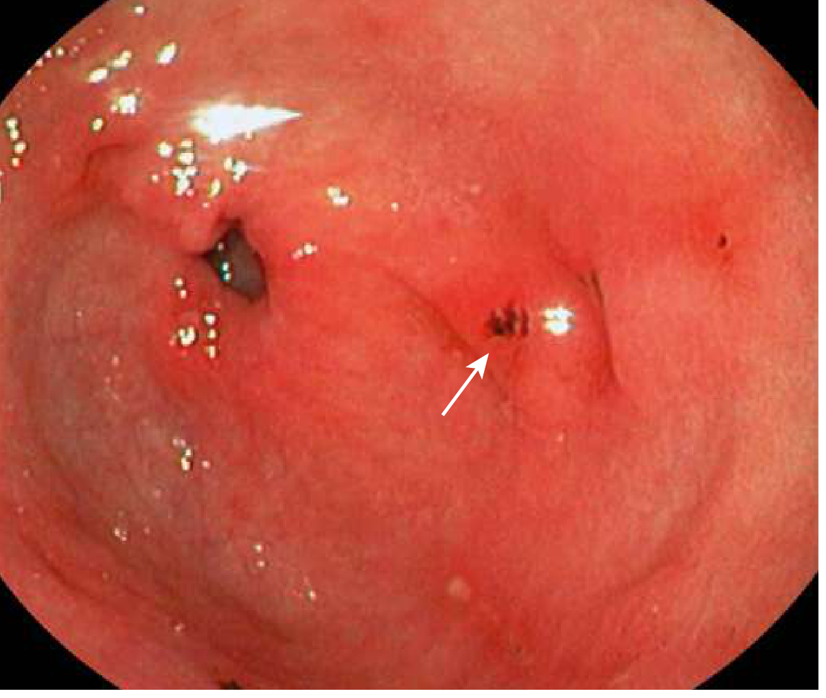
1. Upper Endoscopy (Gold Standard)
- Procedure of choice for confirming the diagnosis
- Findings: edema, erythema, mucosal friability, erosions, nodularity
- Biopsy required for histologic confirmation
- High-definition endoscopy with magnification and image enhancement allows visualization of atrophy and intestinal metaplasia (gray-white patches on white-light endoscopy; "light-blue crests" on narrow-band imaging)
- Typically not performed in the emergency department unless treating complications (e.g., acute bleeding)
2. H. pylori Testing
Testing can be done by invasive or noninvasive methods:
| Method | Type | Notes |
|---|---|---|
| Urea breath test | Noninvasive | Good for active infection and test-of-cure |
| Stool antigen test | Noninvasive | Practical for outpatient testing |
| Serum antibody (IgG) | Noninvasive | Cannot distinguish active from past infection |
| Mucosal biopsy (rapid urease test or histology) | Invasive (via endoscopy) | Most accurate; done during endoscopy |
3. Ancillary Tests (to rule out complications or other diagnoses)
- CBC: check for anemia from GI bleeding
- Stool guaiac (fecal occult blood test): evidence of slow bleeding
- Abdominal/chest X-ray: if perforation or obstruction suspected (look for pneumoperitoneum - free air under diaphragm)
- CT abdomen: can visualize deep ulcers, mural and extraluminal signs of peptic gastroduodenitis
- ECG: always consider in patients where cardiac cause cannot be excluded
- Pregnancy test: in women of childbearing age
Differential Diagnosis of Gastritis
Before diagnosing gastritis, exclude:
- Pancreatitis
- Biliary tract disease
- Small bowel obstruction
- GERD / esophagitis / esophageal spasm
- Mesenteric ischemia (especially in elderly or those with atrial fibrillation)
- Acute coronary syndrome (always consider!)
Treatment Overview (Brief)
- NSAID-related: stop the NSAID + start PPI therapy
- H. pylori-related: eradication with triple therapy (PPI + clarithromycin 500 mg BID + amoxicillin 1 g BID x 10-14 days) or quadruple therapy if penicillin-allergic or clarithromycin-resistant
Sources: Rosen's Emergency Medicine, Goldman-Cecil Medicine, Frameworks for Internal Medicine
Gcs scoring
Reading File
Finding Sources
Finding Sources
Searching the Web
Glasgow Coma Scale GCS scoring system eye verbal motor 2024
Excellent sources found. Let me read the best textbook sections in parallel.
Reading File
Reading File
Reading File
I now have comprehensive, authoritative content from multiple textbooks. Here is the complete GCS breakdown.
Glasgow Coma Scale (GCS) Scoring
The GCS was devised by Teasdale and Jennett (1974) to categorize patients with head trauma and predict outcome. It is now the most widely used tool for measuring level of consciousness in acute medical and trauma settings.
The Three Components
The total GCS score = Eye (E) + Verbal (V) + Motor (M)
Range: 3 (minimum) to 15 (maximum)
1. Eye Opening (E) - Maximum 4
| Score | Response |
|---|---|
| 4 | Opens eyes spontaneously |
| 3 | Opens eyes to verbal command / speech |
| 2 | Opens eyes to pain |
| 1 | No eye opening |
2. Verbal Response (V) - Maximum 5
| Score | Adult/Child >4 yrs | Child <4 yrs | Infant |
|---|---|---|---|
| 5 | Oriented | Oriented, social, speaks | Coos, babbles |
| 4 | Confused (disoriented conversation) | Confused, consolable | Irritable cry |
| 3 | Inappropriate words | Inappropriate words, inconsolable | Cries to pain |
| 2 | Incomprehensible sounds / moans | Agitated, incomprehensible | Moans to pain |
| 1 | No verbal response | No response | No response |
Note: If the patient is intubated, no verbal score is possible. Add "T" after the score (e.g., GCS 8T). In that case, the range is 3T to 10T.
3. Motor Response (M) - Maximum 6
| Score | Response |
|---|---|
| 6 | Obeys commands / follows commands |
| 5 | Localizes pain (purposeful movement toward stimulus) |
| 4 | Withdrawal from pain (pulls away) |
| 3 | Flexion response to pain (decorticate posturing) |
| 2 | Extension response to pain (decerebrate posturing) |
| 1 | No motor response |
The motor score independently correlates with outcome, almost as well as the full GCS score.
Interpretation / Severity Classification
| GCS Score | Severity | Clinical Meaning |
|---|---|---|
| 14-15 | Mild TBI | Alert, minor impairment |
| 9-13 | Moderate TBI | Significant impairment |
| 3-8 | Severe TBI | Coma - patient cannot obey commands, speak, or open eyes |
A GCS score of 8 or less = coma - typically the threshold for considering intubation and airway protection.
Memory Tip: "EMV 456"
- Eye: 1-4
- Motor: 1-6
- Verbal: 1-5
- Lowest possible score = 3 (not 0), as each component scores minimum 1
- Highest possible score = 15
Limitations of GCS
As noted in Tintinalli's Emergency Medicine:
- Measures behavioral responses, not the underlying pathophysiology - two patients with the same score may have very different structural injuries
- Confounded by: alcohol, drugs, paralytics, periorbital swelling (prevents eye opening), intubation, spinal or peripheral injuries
- Not designed for mild TBI - lacks granularity for minor head injury assessment
- Interrater reliability is only moderate when used by emergency physicians
- Better used as a serial measure to track trends over time rather than a single snapshot
Alternative / Related Scales
| Scale | Description |
|---|---|
| AVPU | Alert / Voice / Pain / Unresponsive - simpler, correlates well with GCS |
| ACDU | Alert / Confused / Drowsy / Unresponsive - better at detecting early deterioration |
| FOUR Score | Full Outline of Unresponsiveness - provides more neurologic detail than GCS, can assess brainstem reflexes and breathing pattern; useful in intubated patients |
| Simplified Motor Scale (sMS) | Three motor variables only; validated for trauma outcome prediction |
Quick Clinical Reference
| GCS | What it Means Clinically |
|---|---|
| 15 | Normal |
| 13-14 | Minor head injury; observe |
| 9-12 | Moderate injury; CT head, consider admission |
| ≤8 | Severe; secure airway (intubate), urgent CT, ICU |
| 3 | Deepest coma (no eye opening, no verbal, no motor) |
Sources: Plum and Posner's Diagnosis and Treatment of Stupor and Coma; Schwartz's Principles of Surgery 11th Ed.; Tintinalli's Emergency Medicine
How to do cpr for infants and children
Your credits are exhausted. You can purchase a top-up or subscribe to a plan to continue using Orris. If you're already subscribed, your daily credits will reset tomorrow.
This is a shared conversation. Sign in to Orris to start your own chat.