

Explain the reports
"esophageal squamous cell carcinoma" AND "microinvasive"
Histopathology Report Explained - Mrs. Ambika S. Gadgi
Patient Summary
| Detail | Value |
|---|---|
| Name | Mrs. Ambika S. Gadgi |
| Age / Sex | 55 years, Female |
| Referred by | Dr. Jalil Mujawar (MS, ENT) |
| Date | 24-07-2026 |
| Report No. | HPR 752/26 |
What Was Biopsied?
What the Microscope Found (Plain Language)
1. Stratified Squamous Epithelium with Severe Dysplasia to Carcinoma in Situ
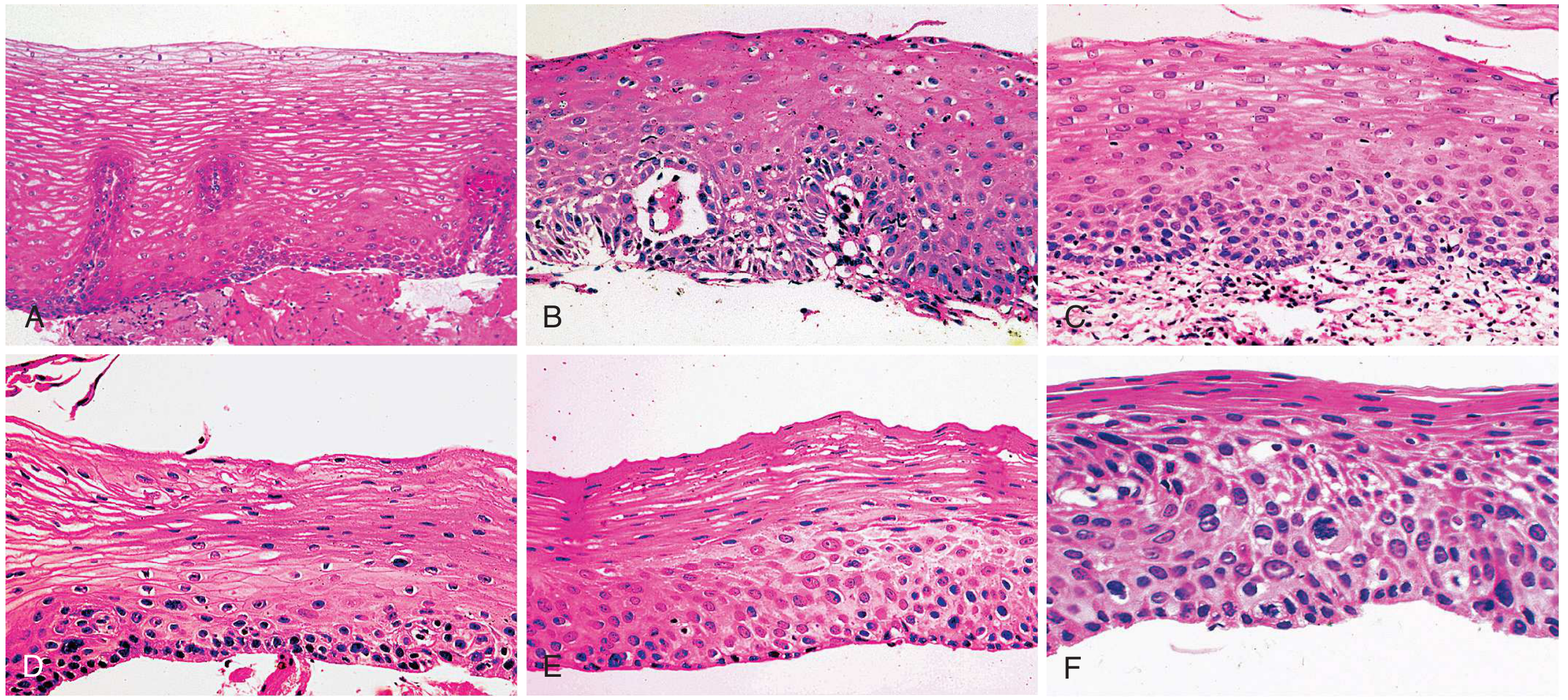
2. Abnormal Cell Features
- Pleomorphic nuclei: cells are irregular and vary in size/shape (not uniform like normal cells)
- Hyperchromatic nuclei: the cell nuclei stain very dark, indicating abnormal DNA content
- Prominent nucleoli: the "control center" within the nucleus is enlarged and visible
- Eosinophilic cytoplasm: the cell body stains pink, consistent with squamous differentiation
- High N:C ratio: the nucleus is disproportionately large relative to the cell body - a hallmark of malignancy
3. Foci of Microinvasion
4. Lymphocyte Infiltration
Final Diagnosis: Microinvasive Squamous Cell Carcinoma
Why Was This Sent to an ENT Surgeon?
What Does This Mean for the Patient?
| Aspect | Implication |
|---|---|
| Stage | Very early - microinvasive (T1a or early T1b) |
| Prognosis | Favorable if treated promptly |
| Treatment options | Endoscopic resection (EMR/ESD) is often curative at this stage; surgery may be discussed |
| Next steps | Urgent referral to a gastroenterologist/oncologist for staging (endoscopic ultrasound, CT scan) and treatment planning |
| Urgency | This is a serious diagnosis requiring prompt action, but the early detection is a significant advantage |
Important note for the patient/family: This report confirms an early cancer in the food pipe. The word "microinvasive" is actually reassuring - it means the cancer has been caught at a very early stage where treatment is most likely to succeed. The next step is to see an oncologist or gastroenterologist urgently for staging investigations (CT scan, endoscopic ultrasound) and to plan treatment. Do not delay.
How operation works give me some photos operation
endoscopic submucosal dissection ESD esophageal squamous carcinoma procedure steps
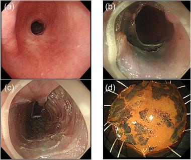
A multi-panel clinical image depicting an endoscopic submucosal dissection (ESD) procedure for metachronous early esophageal squamous cell carcinoma (SCC) in the setting of a pre-existing esophageal stricture. Panel (a) shows an endoscopic view of a severe esophageal stricture with a pinhole-sized lumen and surrounding fibrotic, erythematous mucosa resulting from a previous ESD scar. Panel (b) demonstrates the intraprocedural release of the stricture following mucosal incision and partial submucosal dissection of the lesion at the oral aspect, resulting in a widened lumen. Panel (c) shows the post-procedural ulcer base, characterized by a smooth, denuded surface without visible residual submucosal tissue. Panel (d) displays the en-bloc resected specimen, pinned for pathological examination. The specimen is roughly circular, showing orange-tinted mucosa with distinct dark-stained areas (likely from Lugol's iodine staining) identifying the squamous cell carcinoma lesion. This series illustrates the successful management of complex esophageal lesions located within fibrotic strictures using conventional endoscopic techniques.
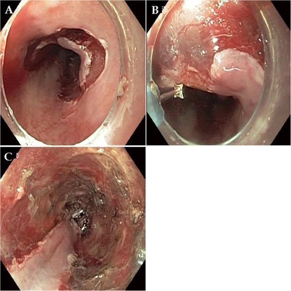
This set of three endoscopic images (A-C) illustrates the sequential stages of an Endoscopic Submucosal Dissection (ESD) procedure for esophageal squamous cell carcinoma. Image A shows the initial pre-cutting phase, where circumferential mucosal incisions are made around the outer border of the target lesion, resulting in visible tissue disruption and minor bleeding. Image B depicts the submucosal dissection phase; an endoscopic knife is visible in the field as it separates the lesion from the muscularis propria, with clear visualization of the submucosal plane. Image C captures the post-procedural state, revealing a large iatrogenic ulcer spanning approximately three-fourths of the esophageal circumference. The ulcer base is irregular, showing exposed submucosa with areas of hyperemic and dark necrotic tissue, characteristic of a comprehensive en bloc resection. This series is an educational example of advanced gastrointestinal therapeutic endoscopy, highlighting the transition from surgical planning to the final iatrogenic defect.
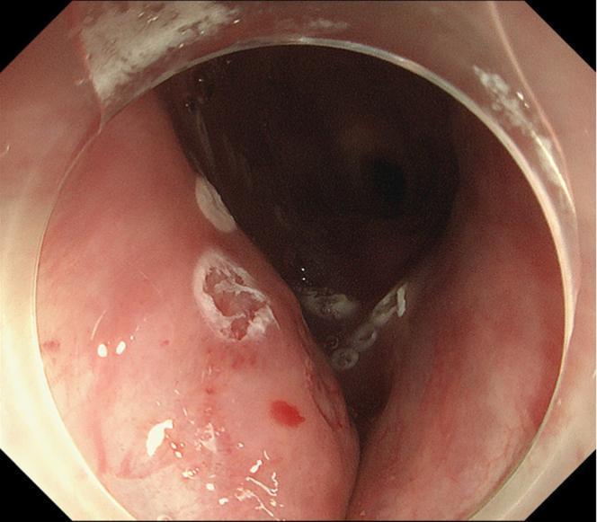
This endoscopic image displays the cervical esophagus during an Endoscopic Submucosal Dissection (ESD) procedure for squamous cell carcinoma. The primary focus is a depressed, irregular lesion within the esophageal lumen, which has been demarcated by white, ring-shaped circumferential thermal markings. These cautery spots define the resection margins. The mucosal tissue appears erythematous and edematous, consistent with inflammatory changes. At approximately the 10 o'clock position, a distinct mucosal defect or superficial ulceration is visible, characterized by a pale, granular base and slightly raised, irregular borders. Small focal areas of petechiae and subepithelial hemorrhage are noted on the surrounding pinkish mucosa. The image is captured through a transparent distal attachment (hood), facilitating better visualization of the esophageal folds and the anatomical depth of the depressed region. This visual serves as an educational example of preoperative lesion mapping and endoscopic site preparation in a patient with a complex anatomical history involving previous surgery and radiotherapy.
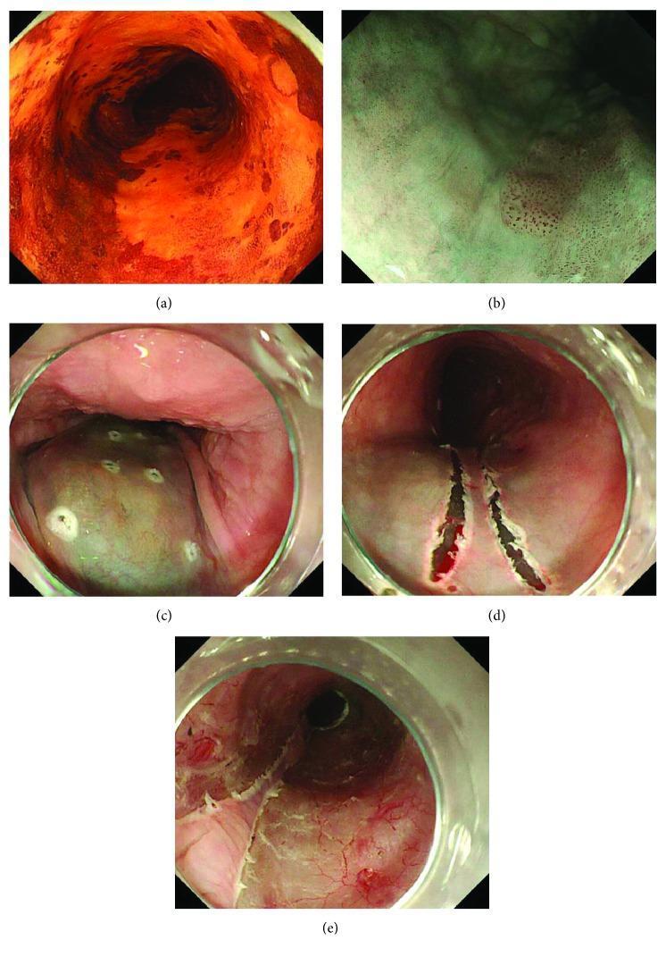
This composite of clinical endoscopic images illustrates the procedural steps for the endoscopic submucosal dissection (ESD) of subcircumferential superficial esophageal cancer. (a) Conventional endoscopy with Lugol's iodine staining reveals a large, unstained, irregular reddish-orange lesion occupying most of the esophageal circumference, characteristic of superficial squamous cell carcinoma. (b) Magnified narrow-band imaging (NBI) shows the intrapapillary capillary loop (IPCL) patterns used to evaluate depth of invasion; the absence of B2/B3 vessel patterns suggests limited mucosal involvement. (c) Pre-procedural marking of the lesion's boundaries with a Flush knife is visible, accompanied by a submucosal injection of glycerol solution, which causes the target tissue to become distended and pale/translucent. (d) Shows the initial mucosal incisions performed precisely along the marking dots on both sides of the lesion. (e) Demonstrates the final result of the 'Line-and-Branch' (LB) method, highlighting the preservation of healthy remnant mucosa to minimize the risk of post-operative esophageal stricture. This series demonstrates advanced gastroenterological techniques for organ-preserving oncological resection.
esophagectomy surgery technique Ivor Lewis diagram
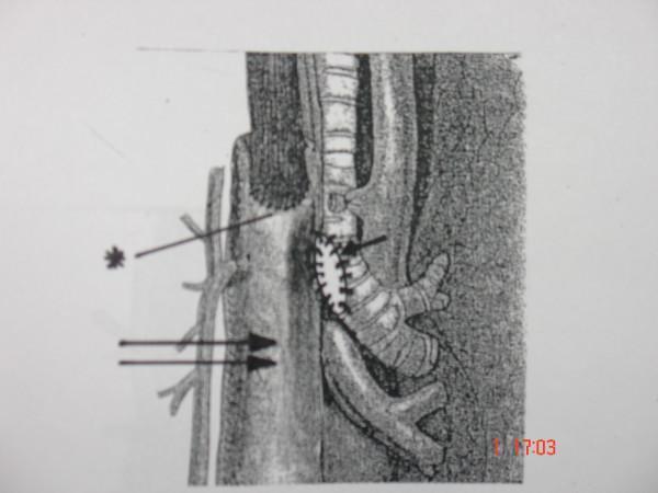
This medical illustration depicts a surgical reinforcement technique following an Ivor Lewis esophagogastrectomy. The diagram shows the intrathoracic positioning of a gastric conduit (marked by a double arrow) in relation to the tracheobronchial tree. An esophagogastric anastomosis is indicated by an asterisk (*). To reinforce a repair of the posterior membranous tracheal wall, the gastric conduit is moved into direct contact with the trachea. Single interrupted nonabsorbable sutures (indicated by a single arrow) are used to secure the gastric wall to the inferior edge of the rigid cartilaginous tracheal rings. This pedagogical image illustrates the use of a vascularized gastric pedicle as a protective flap or 'seal' for tracheal lacerations or patches, demonstrating the anatomical proximity and surgical landmarks relevant to thoracic surgery and complication management in esophagectomy cases.
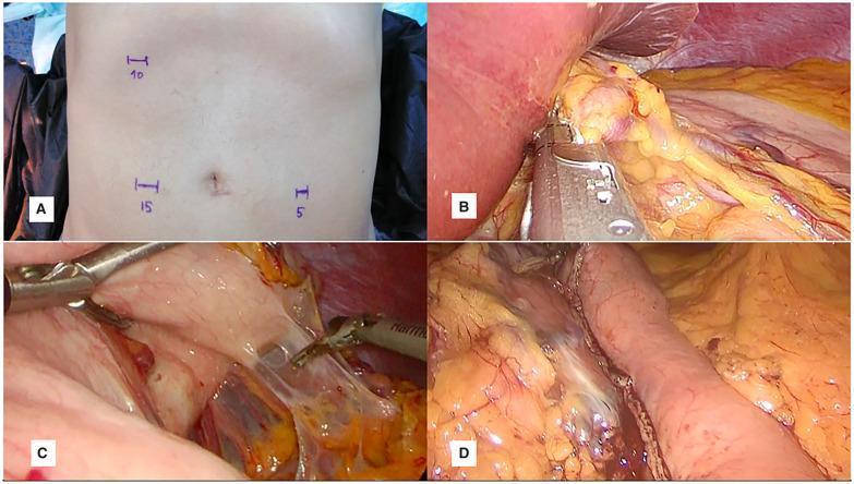
This composite of four clinical images illustrates the abdominal phase of a three-port laparoscopic esophagectomy (Ivor Lewis procedure). Image (A) is a clinical photograph of a patient's abdomen showing preoperative markings for port placement: a 10-mm port at the right costal arch, a 15-mm port right lateral to the umbilicus, and a 5-mm port left lateral to the umbilicus. Images (B, C, D) are intraoperative laparoscopic views. Image (B) demonstrates the division of the left gastric vessels using an endoscopic linear vascular stapler, with the liver retracted to expose the celiac axis area. Image (C) illustrates a non-grasping technique using open-jaw retraction with a Babcock clamp to manipulate the stomach and visualize the gastrocolic ligament without tissue trauma. Image (D) shows the final view of the tubularized gastric conduit, characterized by longitudinal staple lines and shiny serosal tissue, prepared for intrathoracic pull-up. The series highlights surgical techniques for vascular control, atraumatic gastric mobilization, and conduit formation in minimally invasive esophageal surgery.
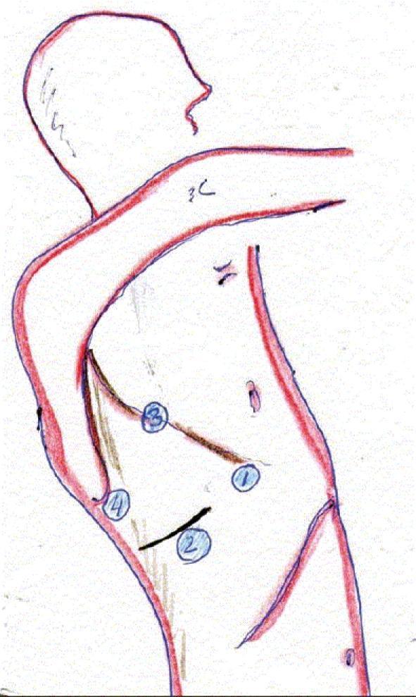
A medical illustration depicting the surgical setup for a right thoracoscopic approach in an Ivor Lewis esophagectomy. The diagram shows a patient in the left lateral decubitus position with the right arm extended forward. Four surgical trocar port sites are numerically labeled (1-4) on the lateral thoracic wall. Port 1 is positioned anteriorly near the midaxillary line. Port 2 is located inferiorly to port 1. Port 3 is placed superiorly and posteriorly, near the posterior axillary line. Port 4 is situated posteriorly and inferiorly, in proximity to the scapula tip. A brown line connects ports 3 and 1, likely indicating the surgical trajectory or line of esophageal mobilization. This diagram serves as a clinical guide for port placement during minimally invasive esophagectomy, emphasizing the relationship between thoracic landmarks and instrument access points for mediastinal lymphadenectomy and esophageal resection.
How the Operations Work for Esophageal Microinvasive Squamous Cell Carcinoma
🔬 OPTION 1: Endoscopic Resection (ESD / EMR) — Preferred for Early/Microinvasive Stage
Step-by-Step: How ESD Works
Recovery: Patient typically stays 1-3 days in hospital. The raw ulcer left behind heals on its own over 4-6 weeks. Prognosis is excellent if margins are clear.
🏥 OPTION 2: Esophagectomy (Surgical Removal) — for Deeper Invasion or Failed Endoscopy
How Ivor Lewis Esophagectomy Works
- The surgeon enters the abdomen and mobilizes the stomach, freeing it from its attachments
- The stomach is shaped into a long tube ("gastric conduit") using surgical staplers
- The left gastric blood vessels are divided
- The surgeon enters the right chest
- The diseased esophagus is removed
- The stomach tube is pulled up into the chest and joined (anastomosed) to the remaining esophagus above the tumor

According to Goldman-Cecil Medicine, surgery alone can be curative in over 60% of cases for early T1-T2 disease without lymph node involvement.
Which Treatment for Mrs. Ambika?
| Treatment | Used When | How Done |
|---|---|---|
| ESD (endoscopic) | Microinvasive, T1a - PREFERRED at this stage | Scope through mouth, no incision |
| Esophagectomy | Deeper invasion, failed ESD, T1b or higher | Major surgery, 2-phase operation |
| Chemoradiation | Can't do surgery, cervical location, or advanced stage | Radiation + chemotherapy tablets/infusion |
Bottom line: Mrs. Ambika's cancer was caught at the ideal stage for endoscopic treatment. The next step is urgent referral to a gastroenterologist or surgical oncologist for endoscopic ultrasound (EUS) staging and ESD planning. The earlier the treatment begins, the better the outcome.
What is the best step what can can I do next step next or chemotherapy or surgery or enscopt
"esophageal squamous cell carcinoma" AND "early stage" AND treatment
PMID: 37498266
What Is the Best Next Step for Mrs. Ambika?
Short answer: Do NOT start chemotherapy or surgery yet. The immediate next step is proper staging scans - then endoscopic treatment (ESD) is most likely the best option for this early cancer.
The Recommended Pathway - Step by Step
✅ STEP 1 (Do This First) — Staging Investigations
| Test | What It Shows | Why It Matters |
|---|---|---|
| Endoscopic Ultrasound (EUS) | Depth of invasion (T stage) and nearby lymph nodes | Most important test to decide ESD vs surgery |
| CT Scan (chest + abdomen) | Whether cancer has spread to distant organs or lymph nodes | Rules out metastasis |
| PET Scan (optional) | Any hidden spread elsewhere in the body | Confirms no distant disease |
✅ STEP 2 — Endoscopic Treatment (ESD) — Most Likely Best Option
"For early-stage esophageal squamous cell carcinoma, the ASGE recommends ESD over surgery, whenever possible."
- The cancer is still very early (microinvasive)
- No cuts on the body - scope goes through the mouth
- Very high cure rate at this stage
- Faster recovery (1-3 days in hospital vs weeks for surgery)
- Surgery can still be done later if ESD margins are not clear
Simple Decision Tree
Biopsy confirms microinvasive SCC
            ↓
   STAGING FIRST (EUS + CT scan)
            ↓
   ┌─────────────────┬────────────────────┐
   ↓                 ↓                    ↓
T1a (surface)    T1b (deeper but    T2-T4 or lymph
no lymph nodes   no lymph nodes)    nodes involved
   ↓                 ↓                    ↓
  ESD            ESD or Surgery     Surgery +
(Preferred)      (Discuss with      Chemoradiation
                  oncologist)       (Before/After)
Why NOT Chemotherapy First?
- Patients who cannot tolerate surgery
- More advanced cancers (T3/T4 or lymph node positive)
- Cervical esophagus location
Summary - What To Do Right Now
| Priority | Action | Where to Go |
|---|---|---|
| Urgent (this week) | See a gastroenterologist + oncologist | Referral from Dr. Jalil Mujawar |
| Within 1-2 weeks | Get EUS + CT scan for staging | GI endoscopy centre |
| After staging | Most likely ESD procedure | Tertiary hospital with advanced endoscopy |
| Do NOT do yet | Surgery or chemotherapy | Wait for staging results first |
Important: This cancer was caught at the best possible stage. The earlier ESD is done, the higher the chance of complete cure. Please consult a gastroenterologist at a major hospital as soon as possible - ideally within this week.
What is the best step what can can I do next step next or chemotherapy or surgery or enscopt
Best Next Steps for Mrs. Ambika (Simple Version)
🚫 Do NOT start chemotherapy or surgery yet
✅ Do This in Order:
- Go back to Dr. Jalil Mujawar and ask for urgent referral to a cancer specialist (oncologist) and gastroenterologist at a major hospital
- Endoscopic Ultrasound (EUS) - checks exact depth of cancer
- CT Scan (chest + abdomen) - checks if cancer has spread anywhere
- These tests decide which treatment is best
- Since the cancer is caught very early, the most likely treatment is ESD (Endoscopic Submucosal Dissection) - done through the mouth, no cuts, 1-3 days hospital stay, very high cure rate
- The 2023 ASGE guideline specifically recommends ESD over surgery for early esophageal squamous cell carcinoma
When Is Each Treatment Used?
| Treatment | Used When |
|---|---|
| ESD (Endoscopy) | Early cancer (most likely for Mrs. Ambika) ✅ |
| Surgery | Cancer is deeper or ESD failed |
| Chemo + Radiation | Advanced cancer or unfit for surgery |
⚠️ The most important thing right now is to NOT delay. Book an appointment at a major hospital cancer centre this week. The cancer is early and highly treatable - but waiting makes it worse. Time matters.
Esd operation photos
ESD endoscopic submucosal dissection esophageal cancer step by step procedure photos
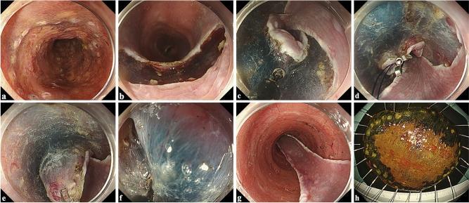
This eight-panel clinical image sequence illustrates a traction-assisted Endoscopic Submucosal Dissection (ESD) procedure for large esophageal cancer. (a) Displays Lugol's iodine chromoendoscopy revealing an unstained neoplastic area with circumferential marking dots. (b) Shows the initial distal (anal) mucosal incision. (c) Demonstrates the completion of circumferential mucosal incisions with trimming of the submucosal edges. (d-f) Illustrate the traction phase, where a metallic clip-with-line is anchored to the oral mucosal edge; pulling the thread provides counter-traction, enhancing visualization of the blue-stained submucosal plane for precise dissection. (g) Visualizes the post-resection esophageal ulcer bed showing clear muscularis propria. (h) Presents the final en bloc resected specimen after ex vivo iodine staining, highlighting the irregular tumor margins for pathological assessment. The sequence serves as a gastrointestinal endoscopy educational resource for advanced therapeutic techniques in managing early-stage esophageal squamous cell carcinoma.
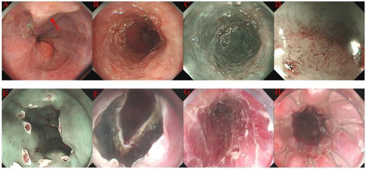
A composite of eight endoscopic images (A-H) demonstrating the stepwise procedure of Endoscopic Submucosal Dissection (ESD) for superficial esophageal cancer. (A) White light imaging shows distal esophageal varices marked with a red arrow. (B-C) Visualization of a superficial 0-IIb esophageal lesion using white light and Narrow Band Imaging (NBI), highlighting mucosal irregularity. (D) Magnifying endoscopy with NBI reveals abnormal intraepithelial papillary capillary loops (IPCL), categorized as type B1 and B2, indicative of squamous cell carcinoma. (E) Circumferential electrocautery marking of the lesion's margins for resection guidance. (F-G) Intra-procedural views showing mucosal/submucosal incision and the final post-resection wound surface (ESD ulcer). (H) Placement of a fully covered self-expanding metal stent (SEMS) within the esophageal lumen following the procedure. This series illustrates advanced therapeutic endoscopy techniques for managing early-stage gastrointestinal malignancies in the context of portal hypertension.
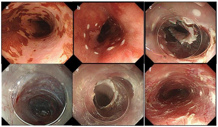
A series of six endoscopic clinical photographs (a-f) documenting the step-by-step procedure of Endoscopic Submucosal Tunnel Dissection (ESTD) for a circumferential superficial esophageal neoplastic lesion. (a) Chromoendoscopy using iodine staining highlights patchy, non-staining neoplastic areas against brown-stained healthy mucosa. (b) White circumferential coagulation markings delineate the lesion's boundaries. (c) A complete circumferential mucosal incision is shown, performed using an endoscopic knife at the distal margin. (d) Initiation of a submucosal tunnel, revealing the submucosal layer with visible vasculature. (e) Extended submucosal tunnel creation from an oral-to-anal approach, maintaining a stable dissection plane between the mucosa and muscularis propria. (f) Final post-resection view showing a large artificial ulcer after en bloc removal of the lesion. The series demonstrates advanced interventional gastroenterology techniques used to treat extensive esophageal neoplasia while preserving luminal integrity and minimizing procedural time compared to conventional endoscopic submucosal dissection (ESD).
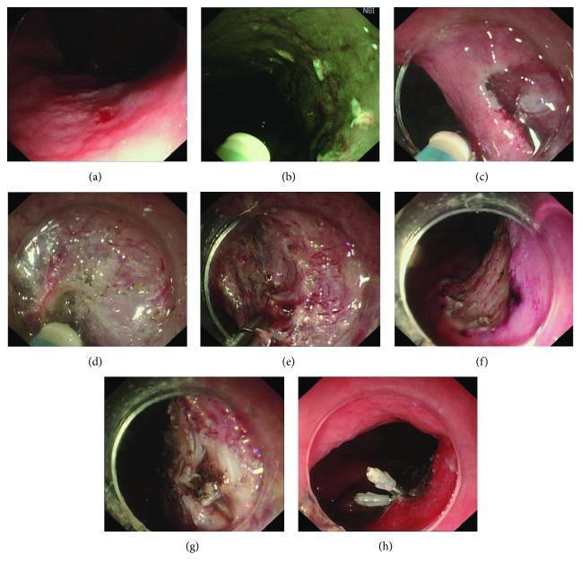
A series of eight endoscopic clinical photographs (a-h) illustrating the step-by-step Endoscopic Submucosal Dissection (ESD) procedure in the gastrointestinal tract. (a) Displays the initial gastric or esophageal lesion under white light endoscopy, characterized by mucosal irregularity and erythema. (b) Shows Narrow Band Imaging (NBI) used to identify and mark the lateral margins of the lesion. (c) Visualizes the circumferential mucosal incision. (d) Demonstrates active submucosal dissection using an electrosurgical knife to separate the lesion from the deeper muscular layer. (e) Shows intraprocedural hemostasis of a bleeding vessel using hot biopsy forceps. (f) Displays the post-resection artificial ulcer base after complete en bloc removal. (g) Identifies an intraprocedural complication involving visible damage to the muscularis propria layer. (h) Illustrates the therapeutic management of the complication, where mechanical endoscopic clips are used to close the muscularis propria defect to prevent luminal perforation. This series serves as an educational resource for gastroenterology trainees to understand ESD stages, instrument usage, and complication management.
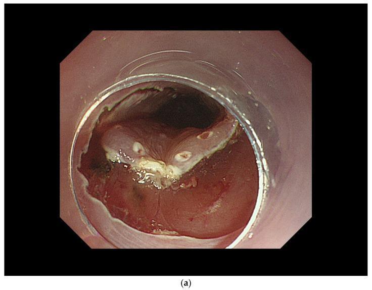
This endoscopic clinical photograph depicts a key step in Endoscopic Submucosal Dissection (ESD) of a superficial esophageal squamous cell carcinoma (SCC). The image shows the initial mucosal incision phase. Centrally, a circular transparent distal hood (cap) is attached to the endoscope to maintain a stable field of view and provide tissue tension. A circumferential mucosal incision is visible, characterized by a sharp boundary where the mucosal layer has been cut, exposing the underlying whitish-yellow submucosal layer. The surrounding esophageal mucosa appears erythematous and slightly edematous, consistent with the procedural trauma and inflammatory response. The lumen beyond the lesion is visible as a dark aperture. This stage is critical for defining the lateral margins of the en bloc resection, allowing subsequent dissection of the submucosa to separate the lesion from the muscularis propria.
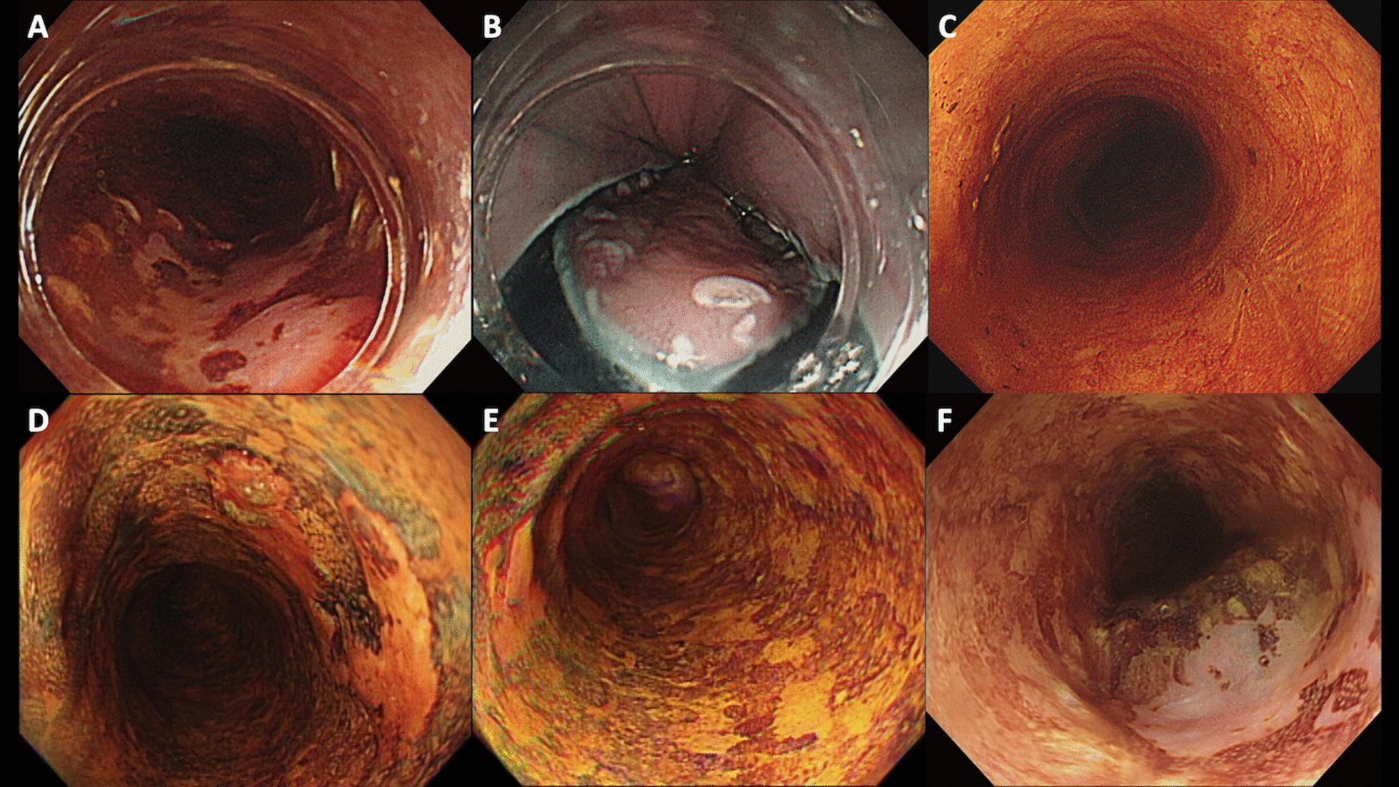
This composite of six endoscopic images (A-F) illustrates the progression and surveillance of superficial esophageal squamous cell carcinoma (SESCC) post-endoscopic submucosal dissection (ESD), categorized by Lugol staining patterns. Images A and D show primary lesions: Type A background (A) displays a uniform pinkish-red hue, while Type D background (D) exhibits numerous irregular Lugol-void lesions (LVLs) with a variegated appearance. Image B shows the intraoperative ESD procedure. Image C demonstrates a successful post-procedural outcome with a smooth, homogeneous orange-red scar indicating no recurrence. In contrast, images E and F depict high-risk surveillance findings; image E shows a background with extensive LVLs (Type D), and image F demonstrates a metachronous recurrence, characterized by a textured, heterogeneous elevation and abnormal mucosal color compared to the primary scar tissue. This series highlights the clinical significance of Lugol chromoendoscopy in identifying high-risk mucosal backgrounds prone to metachronous esophageal cancer.
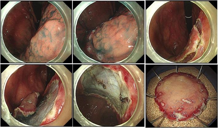
This composite of clinical endoscopic images demonstrates a step-by-step endoscopic submucosal dissection (ESD) for a gastric dysplasia lesion. The initial frames show the identification of the target lesion, where 0.1% indigo carmine dye has been applied to delineate the margins and surface morphology. The subsequent frames illustrate the active dissection phase: a circumferential mucosal incision is performed using an IT knife (electrosurgical generator), followed by the dissection of the submucosal layer to separate the lesion from the underlying muscularis propria. The visualization shows the 'lifting' of the mucosal layer away from the deeper gastric wall, exposing a clean, white submucosal base. The final frame depicts the pathological specimen after en bloc resection, pinned out on a dark board with surgical pins to maintain its orientation and size for histological examination. This procedure is critical in managing high-grade dysplasia (HGD) or early gastric cancer, allowing for complete removal with lower local recurrence rates compared to piecemeal resection techniques.
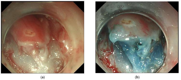
This endoscopic clinical photograph depicts the stages of endoscopic submucosal dissection (ESD) performed on an esophageal lesion following prior radiofrequency ablation (RFA). Image (a) shows the post-RFA mucosal environment, characterized by pink tissue with interspersed white, irregular patches indicating mild submucosal fibrosis and thermal effect. Image (b) demonstrates the procedural step of submucosal elevation. A glycerol solution mixed with indigo carmine dye (blue) has been injected, successfully expanding the submucosal space and creating a distinct blue-tinged cushion. This elevation facilitates the dissection of the target tissue from deeper muscular layers. The content illustrates management strategies for residual or recurrent superficial esophageal squamous cell neoplasia (SESCN), highlighting how ESD can overcome RFA-induced fibrosis through deep submucosal plane dissection. The visual is intended for intermediate to advanced gastroenterology training, focusing on therapeutic endoscopy techniques and tissue response to thermal ablation.
ESD Procedure - Step by Step with Real Photos
🔴 STEP 1 — Find the Cancer with Iodine Staining + Mark the Borders
🟡 STEP 2 — Identify the Cancer Borders Under Special Light (NBI)
🟢 STEP 3 — Inject Fluid to Lift the Lesion + Make the Cut
🔵 STEP 4 — Tunnel Under the Lesion and Remove It
🟣 STEP 5 — Full Dissection, Bleeding Control, Clip if Needed
✅ STEP 6 — The Cancer is Removed (Specimen + Healed Scar)
Summary of All 6 Steps
| Step | What Happens | Photo Above |
|---|---|---|
| 1 | Iodine staining finds the cancer, borders marked | Photo 1 |
| 2 | NBI confirms exact cancer edges | Photo 2 |
| 3 | Fluid injected to lift lesion, circular cut made | Photo 3 |
| 4 | Tunnel cut underneath the cancer | Photo 4 |
| 5 | Cancer lifted out, bleeding sealed, clips if needed | Photo 5 |
| 6 | Cancer removed, ulcer heals into clean scar | Photo 6 |
The entire procedure takes 30-90 minutes. The patient is sedated (asleep). No cuts on the skin. Hospital stay is just 1-3 days. For early-stage cancer like Mrs. Ambika's, this is the most recommended treatment worldwide.