
Shoulder joint anatomy
shoulder joint anatomy diagram glenohumeral
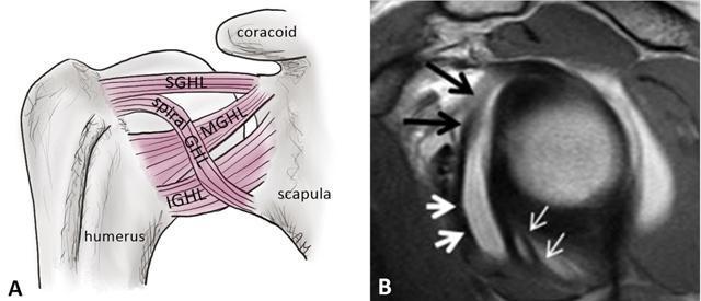
This educational image set illustrates the anatomy of the glenohumeral ligaments, focusing on the fasciculus obliquus (spiral glenohumeral ligament). Image A is an anatomical diagram of the anterior shoulder joint, labeling the Superior Glenohumeral Ligament (SGHL), Middle Glenohumeral Ligament (MGHL), Inferior Glenohumeral Ligament (IGHL), and the Spiral GHL (fasciculus obliquus) crossing between the humerus and scapula. Image B is a sagittal oblique PD-weighted MR arthrogram of the shoulder, demonstrating clinical correlation. Black arrows point to the Middle Glenohumeral Ligament (MGHL), which presents as a low-signal (dark) band. Thick white arrows indicate the fasciculus obliquus located inferiorly. Thin white arrows highlight the frenula capsulae (synovial bands) within the axillary recess. The contrast-enhanced joint fluid helps delineate these capsuloligamentous structures. This comparison is useful for musculoskeletal radiology and orthopedic surgery to understand shoulder stability and the visualization of anatomical variants during arthrography.
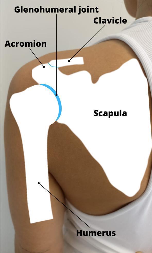
This anatomical diagram overlays a skeletal conceptualization onto a clinical photograph of a human posterior shoulder. The visual illustrates the primary components of the shoulder girdle and their spatial relationships. The scapula is depicted as a large, flat, white silhouette positioned over the upper back, featuring the acromion process superiorly. The clavicle is shown extending medially from its articulation with the acromion. The humerus is represented as a vertical long bone silhouette. The glenohumeral joint, a synovial ball-and-socket joint, is specifically highlighted with a blue semi-circular arc, indicating the interface between the humeral head and the glenoid fossa of the scapula. Labels identify the Humerus, Scapula, Clavicle, Acromion, and the Glenohumeral joint. This educational material is designed for medical students and clinicians to understand the surface anatomy and underlying skeletal structure relevant to physical examination, joint mobilization, and manual therapy techniques for shoulder pathologies such as rotator cuff injuries or impingement syndromes.
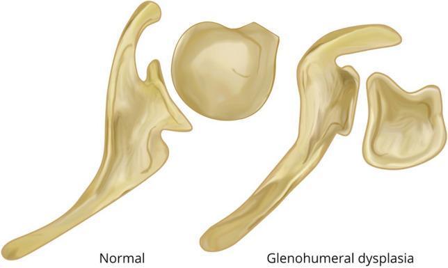
Anatomical comparison diagram of the glenohumeral joint in an axial view, illustrating the morphological differences between a normal shoulder and one with glenohumeral dysplasia secondary to brachial plexus birth injury (BPBI). The left side of the diagram depicts a 'Normal' joint, showing a spherical, well-contoured humeral head centrally seated within a single concave glenoid fossa. The right side, labeled 'Glenohumeral dysplasia,' demonstrates progressive pathological changes: the glenoid fossa is flattened and bi-concave, featuring a distinctive false posterior-inferior facet (pseudo-glenoid). The corresponding humeral head is shown as flattened and retroverted, articulating abnormally with the posterior aspect of the dysplastic glenoid. This illustration serves to teach the secondary skeletal remodeling and posterior subluxation that occurs when muscular imbalance (internal rotation contracture) persists in developing pediatric shoulders after nerve injury. It is a key educational resource for orthopedic surgery and physical medicine and rehabilitation specializing in neonatal brachial plexus palsy.
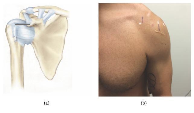
This composite educational graphic focuses on shoulder anatomy and surface landmark localization for clinical procedures. Figure (a) is an anatomical diagram of the right shoulder joint depicting the scapula, clavicle, and proximal humerus. It highlights the coracoclavicular and acromioclavicular ligaments, as well as the glenohumeral capsule. A horizontal black line illustrates the measurement distance between the coracoid process and the intertubercular (bicipital) groove. Figure (b) is a clinical photograph showing the anterior aspect of a human male's left shoulder with surface markings. Vertical skin markings labeled A, B, and C correspond to the underlying anatomy: (A) represents the coracoid process, (B) marks the intertubercular groove at 0° of external rotation, and (C) marks the same groove at 45° of external rotation. Visible ultrasound gel near markings B and C suggests that these points were localized using musculoskeletal ultrasound. This material is designed to teach clinical localization of the long head of the biceps tendon relative to fixed bony landmarks for diagnostic or therapeutic injections.
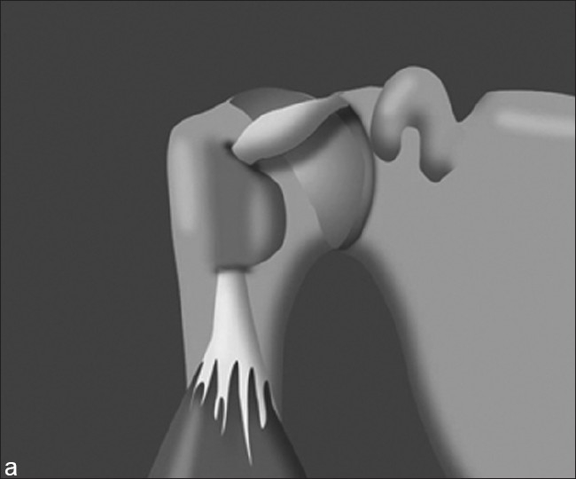
**Imaging Modality:** Medical illustration/diagram. **Anatomical Region:** Shoulder joint (glenohumeral joint), specifically the anterosuperior aspect. **Observed Pathology/Entity:** Hypertrophy of the long head of the biceps (LHB) tendon. **Characteristic Visual Features:** The illustration depicts a three-dimensional rendering of the shoulder anatomy with the humerus in a neutral position (arm at the side). The long head of the biceps tendon is shown originating from the supraglenoid tubercle and passing through the bicipital groove. The intra-articular portion of the LHB tendon exhibits significant fusiform thickening and enlargement (hypertrophy) compared to its distal portion. Key landmarks visible include the humeral head, the greater and lesser tuberosities forming the bicipital groove, the coracoid process, and the glenoid. **Clinical Context:** This finding is often associated with chronic tendinopathy or the "hourglass biceps" phenomenon, where the enlarged tendon becomes mechanically trapped or restricted within the bicipital groove during joint motion, potentially leading to pain and limited range of motion (terminal elevation). **Key Diagnostic Features:** - Pronounced thickening of the intra-articular LHB tendon segment. - Positioned within the glenohumeral space proximal to the intertubercular sulcus.
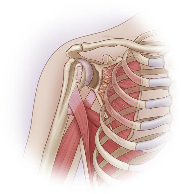
This medical anatomical diagram illustrates a right shoulder joint (glenohumeral joint) with significant orthopedic pathology. The illustration reveals an anterior-inferior bone defect of the glenoid, representing a structural loss of bone mass in the lower-front rim of the socket. Accompanying this bony lesion is a complete, irreparable tear of the subscapularis tendon, shown as a discontinuous and retracted muscle-tendon unit. A notable anatomical abnormality is the absence of the coracoid process, which typically serves as a landmark and attachment point for the conjoint tendon. The humeral head shows signs of degenerative wear, and the surrounding thoracic cage (ribs and intercostal muscles) and clavicle are depicted for orientation. The diagram serves as an educational tool for identifying combined soft-tissue and bony instability in the shoulder, often requiring complex reconstructive procedures like bone grafting or muscle transfer.
rotator cuff muscles supraspinatus infraspinatus teres minor subscapularis anatomy
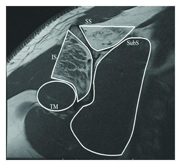
A T1-weighted MRI scan in the sagittal oblique plane demonstrating the rotator cuff musculature of the shoulder. The image highlights the anatomical arrangement and tissue characteristics of the four primary rotator cuff muscles, delineated by white outlines: the supraspinatus (SS), infraspinatus (IS), subscapularis (SubS), and teres minor (TM). Clinically significant findings include increased T1 signal intensity (hyperintensity) within the muscle bellies of the supraspinatus and infraspinatus, indicative of advanced fatty infiltration and muscle atrophy. In contrast, the subscapularis and teres minor maintain a normal, lower signal intensity (isointense to other skeletal muscles). The spatial relationship shows the supraspinatus superiorly, the infraspinatus posteriorly, the subscapularis anteriorly, and the teres minor inferior to the infraspinatus. This diagnostic image is used in orthopedics and radiology to assess chronic rotator cuff tears and associated muscle degeneration, which has implications for surgical prognosis and functional outcomes.
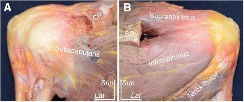
This clinical photograph displays a human cadaveric specimen dissection of a right shoulder, focusing on the rotator cuff musculature and their humeral insertions. The content is presented in two panels: (A) Anterior aspect and (B) Posterior aspect. In panel A, the subscapularis muscle is clearly labeled, demonstrating its broad muscle belly and fibers converging toward the lesser tuberosity of the humerus. The coracoid process (CP) is visible superiorly. In panel B, the posterior rotator cuff muscles are identified in relation to the scapular spine (SS). The supraspinatus is positioned superior to the spine, while the infraspinatus and teres minor are situated inferiorly, showing their tendinous progression toward the greater tuberosity. Spatial orientation is provided by superior (Sup) and lateral (Lat) directional indicators. The specimen has undergone resection of the acromion to provide an unobstructed view of the tendon footprints. This visual resource is intended for medical education regarding shoulder anatomy, surgical landmarks, and musculoskeletal pathology.
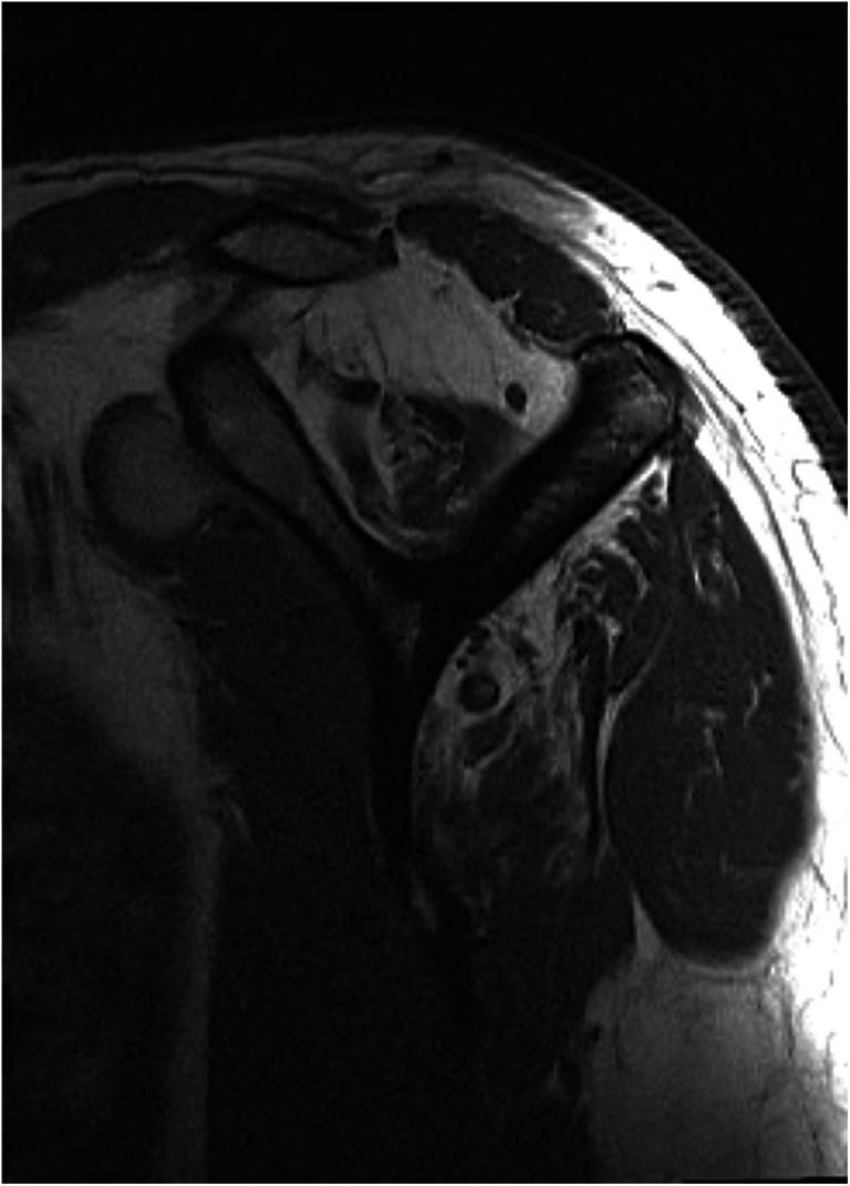
This diagnostic image is a T1-weighted parasagittal MR slice of the shoulder, specifically oriented to evaluate the rotator cuff musculature. The image demonstrates advanced fatty infiltration and muscular atrophy, critical for determining the reparability of chronic rotator cuff tears. The supraspinatus muscle shows a significant loss of muscle bulk (atrophy) and high signal intensity (hyperintensity), indicating replacement of muscle fibers by adipose tissue, consistent with a high Goutallier stage. Similarly, the infraspinatus muscle exhibits marked fatty degeneration. In contrast, the subscapularis and teres minor muscles maintain a normal, low-signal appearance (hypointense) relative to the fat, indicating preserved muscle quality without significant degeneration. This comparison serves as a key clinical indicator in orthopedic radiology for surgical planning and prognosis in patients with tendon pathology.
shoulder joint ligaments capsule bursa bursae anatomy
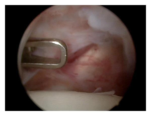
This diagnostic image is an intraoperative clinical photograph showing an arthroscopic view of an anterior shoulder capsule release. The visual field displays the internal shoulder joint environment, characterized by fibrous, pinkish-red vascularized tissue representing the joint capsule. A metallic, silver-colored surgical instrument with a U-shaped distal end is positioned on the left side of the frame, currently interacting with the capsular tissue. A linear incision or 'release' is visible in the anterior capsule, appearing as a dark, longitudinal disruption in the tissue fibers with slightly retracted edges. The procedure aims to increase joint mobility, typically in cases of adhesive capsulitis, by surgically dividing tightened ligaments. The image demonstrates the surgical technique of interval capsule incision while avoiding damage to the adjacent subscapularis tendon. This content is suitable for orthopedic surgical education, illustrating arthroscopic anatomy and procedural landmarks.
This composite educational graphic focuses on shoulder anatomy and surface landmark localization for clinical procedures. Figure (a) is an anatomical diagram of the right shoulder joint depicting the scapula, clavicle, and proximal humerus. It highlights the coracoclavicular and acromioclavicular ligaments, as well as the glenohumeral capsule. A horizontal black line illustrates the measurement distance between the coracoid process and the intertubercular (bicipital) groove. Figure (b) is a clinical photograph showing the anterior aspect of a human male's left shoulder with surface markings. Vertical skin markings labeled A, B, and C correspond to the underlying anatomy: (A) represents the coracoid process, (B) marks the intertubercular groove at 0° of external rotation, and (C) marks the same groove at 45° of external rotation. Visible ultrasound gel near markings B and C suggests that these points were localized using musculoskeletal ultrasound. This material is designed to teach clinical localization of the long head of the biceps tendon relative to fixed bony landmarks for diagnostic or therapeutic injections.
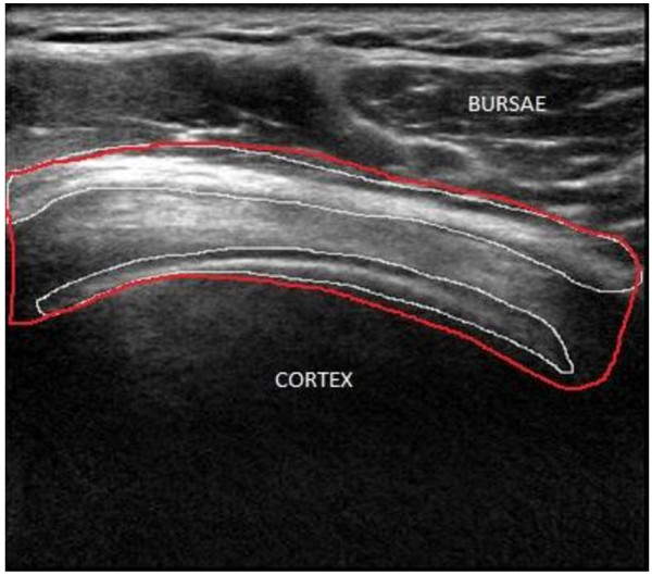
**Imaging Modality:** Musculoskeletal Ultrasound (B-mode). **Anatomical Region:** Shoulder joint, specifically focusing on the subacromial-subdeltoid region and the underlying humeral head. **Key Landmarks and Observed Structures:** - **Bursae:** The subacromial-subdeltoid bursa is visible as a hypoechoic plane superior to the rotator cuff tendons. - **Cortex:** The hyperechoic, curvilinear bony cortex of the humeral head is visible at the inferior aspect of the image, characterized by strong acoustic shadowing deep to the interface. - **Rotator Cuff:** The supraspinatus tendon is positioned between the bursa and the bony cortex, exhibiting a fibrillar echotexture. **Image Annotations:** - **Manual Segmentation:** Red and white contour lines delineate the boundaries of the anatomical layers. - **Labels:** Text overlays identify the "BURSAE" (superiorly) and "CORTEX" (inferiorly). **Diagnostic/Visual Features:** The image demonstrates standard anatomical relationships used in the assessment of subacromial impingement or tendinopathy. The clear differentiation between the superficial deltoid muscle, the intermediate bursa/tendon complex, and the deep bony cortex provides a baseline for evaluating inflammatory changes or structural tears.
Shoulder Joint Anatomy
1. Bones of the Shoulder Complex
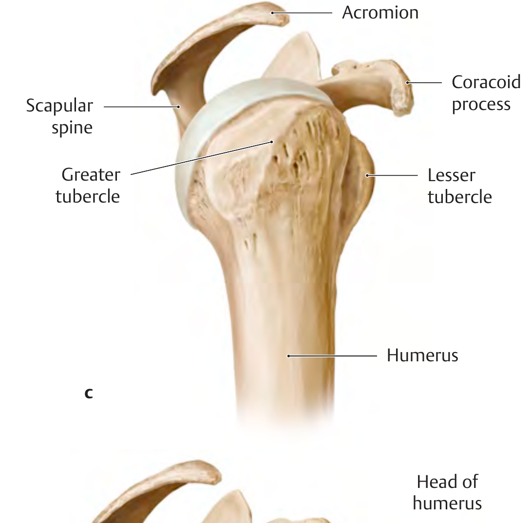
- Humerus - the proximal end articulates at the glenohumeral joint. Key landmarks include the head, anatomical neck, surgical neck, greater tubercle (insertions for supraspinatus, infraspinatus, teres minor), and lesser tubercle (insertion for subscapularis). The intertubercular (bicipital) groove between the tubercles carries the long head of biceps tendon.
- Scapula - a flat triangular bone bearing three processes: the glenoid (articulating fossa), coracoid (anterior projection, site of muscle/ligament attachments), and acromion (a lateral extension of the scapular spine that roofs the subacromial space).
- Clavicle - connects the upper limb to the axial skeleton via the sternoclavicular joint.
2. The Four Joints
| Joint | Type | Function |
|---|---|---|
| Glenohumeral (GHJ) | Ball-and-socket (spheroidal) | Primary axis of arm motion - flexion, extension, abduction, adduction, rotation |
| Acromioclavicular (ACJ) | Plane synovial | Rotation; helps suspend shoulder girdle |
| Sternoclavicular (SCJ) | Saddle-type | Elevation of clavicle; only true articulation of upper limb with axial skeleton |
| Scapulothoracic | Functional (not a true synovial joint) | Scapular protraction, retraction, rotation - every 1° of scapulothoracic motion permits 2° of GHJ motion |
3. The Glenohumeral Joint in Detail
- The glenoid labrum (increases depth of glenoid concavity)
- Glenohumeral ligaments (reinforce the capsule)
- The rotator cuff muscles (primary dynamic stabilizers)
4. Joint Capsule and Ligaments
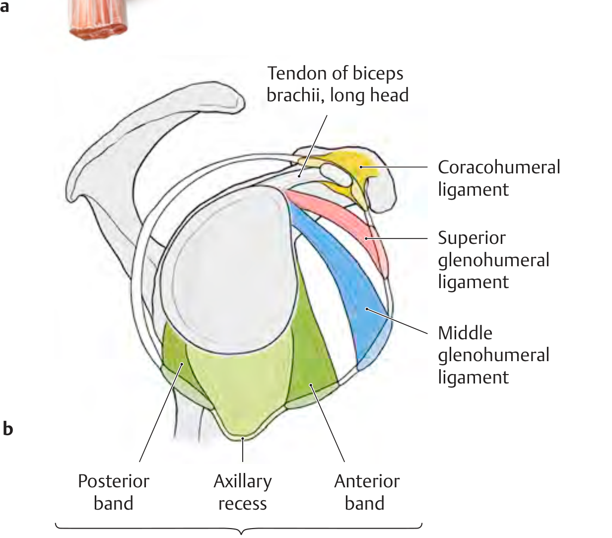
Glenohumeral Ligaments (GHLs)
| Ligament | Origin | Insertion | Function |
|---|---|---|---|
| Superior GHL (SGHL) | Upper margin of glenoid | Intertubercular groove + lesser tubercle | Limits inferior translation with arm adducted; forms rotator interval with coracohumeral ligament |
| Middle GHL (MGHL) | Upper glenoid margin | Anatomical neck of humerus | Resists anterior translation at 45-60° abduction |
| Inferior GHL (IGHL) | Inferior glenoid margin (anterior + posterior bands + axillary recess) | Middle of anatomical neck / surgical neck | Primary anterior-inferior stabilizer at 90° abduction; acts as a hammock during abduction |
Coracohumeral Ligament (CHL)
Rotator Interval
5. The Rotator Cuff
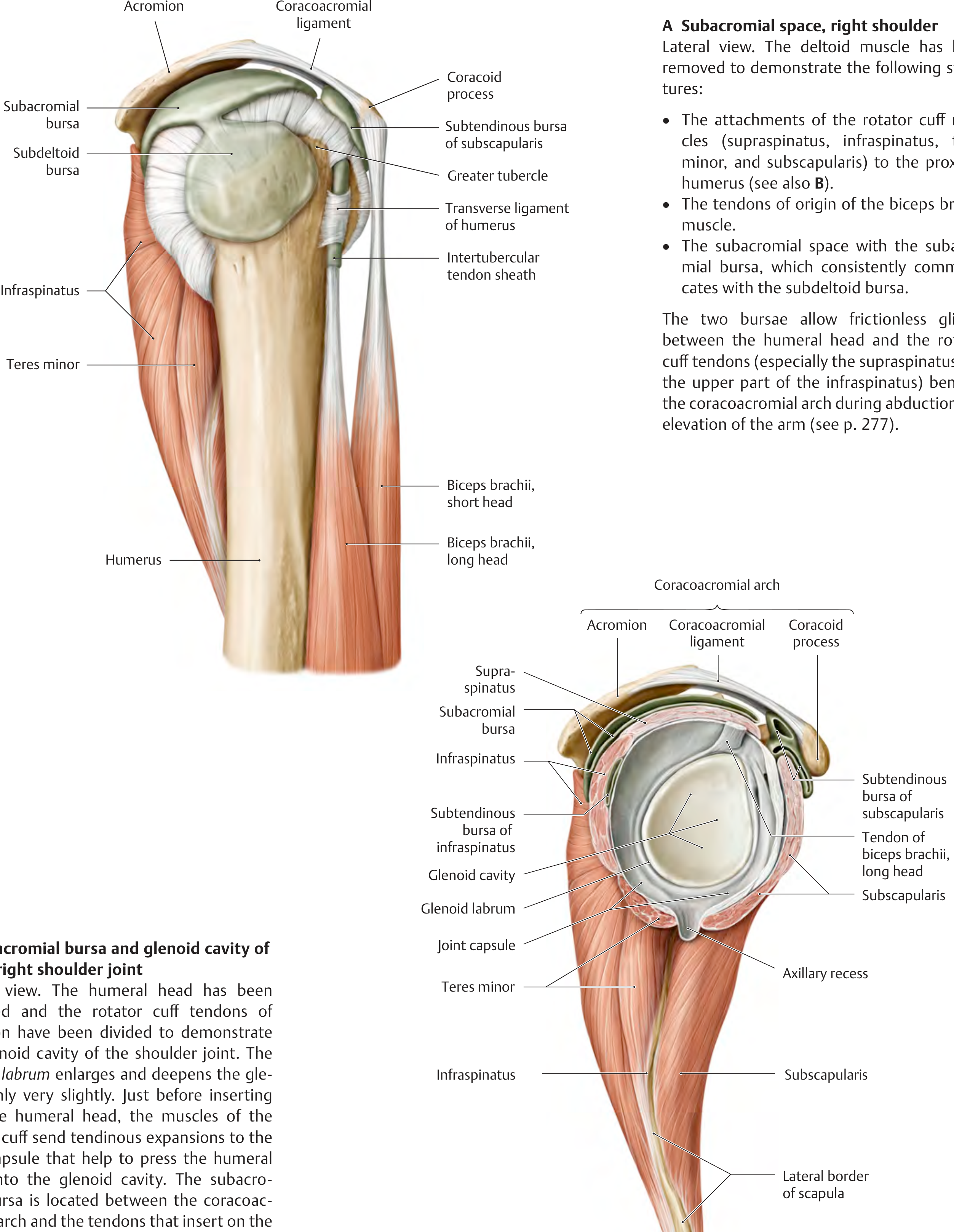
| Muscle | Origin | Insertion | Action |
|---|---|---|---|
| Supraspinatus | Supraspinous fossa (posterior/superior scapula) | Greater tubercle (superior facet) | Initiates abduction (0-15°); balances deltoid to keep humerus centered in glenoid |
| Infraspinatus | Infraspinous fossa (posterior scapula, below spine) | Greater tubercle (middle facet) | Primary external rotator |
| Teres Minor | Lateral border of scapula, inferior to infraspinatus | Greater tubercle (inferior facet) / posterior humerus | External rotation (with infraspinatus) |
| Subscapularis | Subscapular fossa (anterior scapula) | Lesser tubercle | Internal rotation; anterior stabilizer |
6. Subacromial Space and Bursae
- Subacromial bursa - communicates consistently with the subdeltoid bursa. These two bursae allow frictionless gliding of the humeral head and rotator cuff tendons (especially supraspinatus and upper infraspinatus) beneath the arch during abduction and elevation.
- Subtendinous bursa of subscapularis - lies between subscapularis and the glenoid neck; often communicates with the joint cavity.
- Subtendinous bursa of infraspinatus - situated at the infraspinatus tendon near insertion.
7. Long Head of the Biceps Tendon
8. Neurovascular Supply
- Arterial supply: Primarily the anterior and posterior circumflex humeral arteries (from axillary artery), with contributions from the suprascapular and subscapular arteries.
- Nerve supply: Axillary nerve (C5, C6) - supplies the deltoid and teres minor, and the sensory "regimental badge" area over the lateral deltoid; the suprascapular nerve (C5, C6) - supplies supraspinatus and infraspinatus; subscapular nerves supply subscapularis.
The axillary nerve is at particular risk in shoulder dislocations and proximal humeral fractures - always test sensation over the lateral deltoid after such injuries.
9. Arthroscopic Anatomy (Key Landmarks)
10. Clinical Correlations
- Shoulder dislocations: 45% of all joint dislocations. Typically anterior-inferior (from forcible external rotation of the raised arm). Complications include Bankart lesion (avulsion of anteroinferior labrum), Hill-Sachs lesion (posterolateral humeral head impression fracture), axillary nerve injury, and rotator cuff tear.
- Subacromial impingement: Compression of the supraspinatus tendon between the humeral head and the coracoacromial arch during arm elevation.
- Rotator cuff tears: Supraspinatus is most commonly torn; chronic tears lead to fatty infiltration of the muscle belly visible on MRI.
- SLAP lesion: Superior labrum tear from anterior to posterior at the biceps anchor.