
Antigen antibody reaction essay
Antigen-Antibody Reaction
Introduction
Definition of Key Terms
Structural Basis of the Reaction
Forces Involved in Antigen-Antibody Binding
-
Hydrophobic interactions - The most important force. When nonpolar groups on the antigen and antibody are in close proximity, they exclude water molecules, gaining thermodynamic stability. This accounts for the bulk of binding energy.
-
Hydrogen bonds - Form between hydrogen donor and acceptor groups on the antigen and antibody surface (e.g., -OH, -NH, -C=O groups).
-
Van der Waals-London dispersion forces - Weak electrostatic interactions between temporary induced dipoles in adjacent atoms. Effective only at very short range (< 3-4 Å).
-
Ionic (electrostatic) interactions - Attractions between oppositely charged groups (e.g., carboxylate and amino groups).
-
Steric complementarity - Not a force per se, but close spatial fit allows all the above forces to act simultaneously, greatly amplifying the total binding energy.
Kinetics of the Reaction
-
The affinity constant (Ka) from the slope of the line
-
The total antibody-binding site concentration from the X-intercept
-
Henry's Clinical Diagnosis and Management by Laboratory Methods
Affinity vs. Avidity
-
Affinity reflects the tightness of fit of a single antigenic determinant to a single antigen-binding site. It is independent of the number of binding sites.
-
Avidity is the total binding strength of all binding sites of an antibody molecule for a multivalent antigen. A typical IgG molecule binds at least 10,000 times more strongly to a multivalent antigen when both its antigen-binding sites are engaged compared to only one.
Factors Affecting the Reaction
| Factor | Effect |
|---|---|
| pH | Optimal pH is usually 6.5-8.5. Extremes denature proteins and disrupt ionic bonds |
| Temperature | Higher temperature increases reaction rate (K₁) but may also increase dissociation (K₂). Optimal is often 37°C |
| Ionic strength (salt concentration) | Moderate NaCl (0.15 mol/L, normal saline) is standard. Higher concentrations inhibit binding. Fluoride ion (F⁻) offers modest improvement over Cl⁻ in immunochemical buffers |
| Ion species | Cationic salts inhibit binding in order: Cs⁺ > Rb⁺ > NH₄⁺ > K⁺ > Na⁺ > Li⁺. For anionic salts: SCN⁻ > NO₃⁻ > I⁻ > Br⁻ > Cl⁻ > F⁻ |
| Polymers (e.g., PEG) | High-MW polymers such as polyethylene glycol (PEG 6000, 3-5 g/dL) promote immune complex formation through steric exclusion, effectively increasing local protein concentration without altering complex composition |
- Tietz Textbook of Laboratory Medicine, 7th Edition
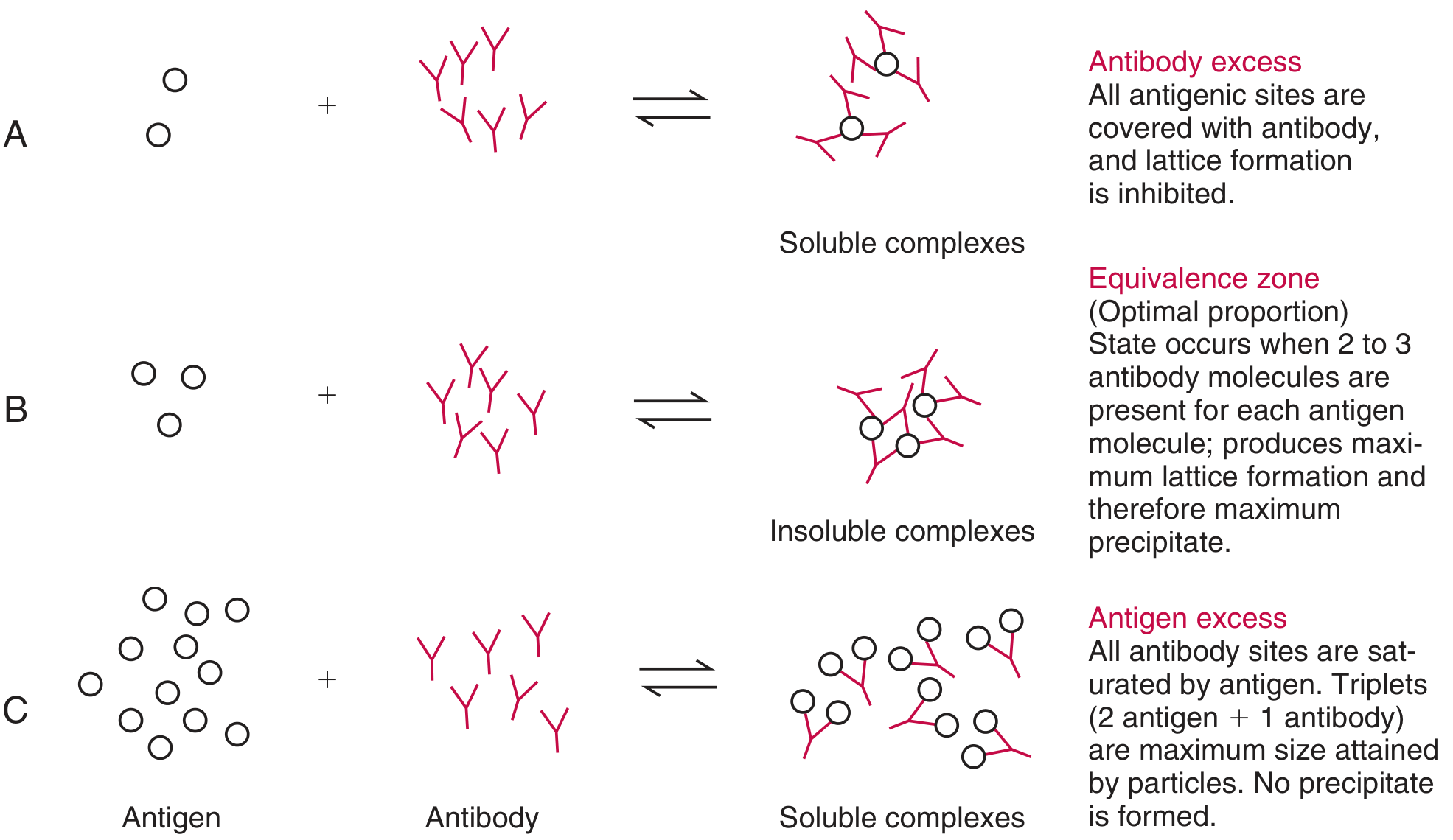
The Precipitin Reaction
-
Zone of antibody excess (prozone): [Ab] >> [Ag]. All antigenic sites are rapidly saturated by antibody before cross-linking can occur. Small soluble complexes form. No precipitate. Free antibody is detectable in solution.
-
Zone of equivalence: Optimal Ag:Ab ratio (approximately 2-3 antibody molecules per antigen molecule). Maximal cross-linking and lattice formation. Maximum precipitate. Neither free antigen nor free antibody is detectable.
-
Zone of antigen excess (postzone): [Ag] >> [Ab]. All antibody sites are saturated. Triplets (2 Ag + 1 Ab) are the maximum size complex formed. Small soluble complexes result. No precipitate. Free antigen is detectable.
- Tietz Textbook of Laboratory Medicine, 7th Edition
Types of Antigen-Antibody Reactions
1. Precipitation
2. Agglutination
3. Complement Fixation
4. Neutralization
5. Opsonization
6. Cytotoxicity (ADCC)
Polyclonal vs. Monoclonal Antibodies
Biological Consequences and Clinical Significance
- Protective immunity: Neutralization of viruses, bacteria, and toxins
- Complement activation: IgG- or IgM-Ag complexes activate the classical complement pathway, promoting inflammation, opsonization, and microbial lysis
- Hypersensitivity reactions: Inappropriate or excessive Ag-Ab reactions underlie all four types of hypersensitivity:
- Type I (anaphylaxis): IgE binds allergen on mast cell surface → degranulation
- Type II (cytotoxic): IgG/IgM bind cell-surface antigens → complement/ADCC-mediated cell lysis
- Type III (immune complex): Excess Ag-Ab complexes deposit in tissues → complement activation and inflammation. Low complement levels and "lumpy" immunoglobulin/C3 deposits along glomerular basement membranes are characteristic findings. - Jawetz, Melnick & Adelberg's Medical Microbiology
- Type IV: Cell-mediated, not involving Ag-Ab directly
Applications in Laboratory Diagnostics
| Method | Principle |
|---|---|
| ELISA | Enzyme-labeled Ab or Ag; colorimetric detection |
| Radioimmunoassay (RIA) | Radiolabeled Ab or Ag; competitive binding |
| Fluorescent immunoassay (FIA) | Fluorophore-labeled Ab; fluorescence detection |
| Chemiluminescent immunoassay (CLIA) | Acridinium ester or electrochemiluminescent labels |
| Nephelometry/Turbidimetry | Precipitation complexes scatter/absorb light |
| Agglutination assays | Particle clumping (latex, erythrocytes) |
| Western blot | Electrophoresis + Ab probing |
| Immunofluorescence | Direct/indirect tissue staining |
Summary
- Henry's Clinical Diagnosis and Management by Laboratory Methods (pp. 901-902, 1026-1028, 1087-1088)
- Tietz Textbook of Laboratory Medicine, 7th Edition (pp. 793-795)
- Sherris & Ryan's Medical Microbiology, 8th Edition (p. 73)
- Jawetz, Melnick & Adelberg's Medical Microbiology, 28th Edition
Generate this in a word document
ls -la /tmp/workspace/ 2>/dev/null || mkdir -p /tmp/workspace && ls -la /tmp/workspace/
mkdir -p /tmp/workspace/antigen-antibody && python3 << 'EOF' from docx import Document from docx.shared import Pt, Inches, RGBColor from docx.enum.text import WD_ALIGN_PARAGRAPH from docx.oxml.ns import qn from docx.oxml import OxmlElement doc = Document() # --- Page margins --- from docx.shared import Inches section = doc.sections[0] section.top_margin = Inches(1) section.bottom_margin = Inches(1) section.left_margin = Inches(1.2) section.right_margin = Inches(1.2) # --- Default font --- style = doc.styles['Normal'] style.font.name = 'Calibri' style.font.size = Pt(11) # --- Heading styles --- h1_style = doc.styles['Heading 1'] h1_style.font.name = 'Calibri' h1_style.font.size = Pt(16) h1_style.font.bold = True h1_style.font.color.rgb = RGBColor(0x1F, 0x49, 0x7D) h2_style = doc.styles['Heading 2'] h2_style.font.name = 'Calibri' h2_style.font.size = Pt(13) h2_style.font.bold = True h2_style.font.color.rgb = RGBColor(0x2E, 0x74, 0xB5) h3_style = doc.styles['Heading 3'] h3_style.font.name = 'Calibri' h3_style.font.size = Pt(11.5) h3_style.font.bold = True h3_style.font.color.rgb = RGBColor(0x2E, 0x74, 0xB5) # ===== TITLE PAGE ===== title_para = doc.add_paragraph() title_para.alignment = WD_ALIGN_PARAGRAPH.CENTER run = title_para.add_run('Antigen-Antibody Reaction') run.font.name = 'Calibri' run.font.size = Pt(24) run.font.bold = True run.font.color.rgb = RGBColor(0x1F, 0x49, 0x7D) doc.add_paragraph() # spacer subtitle = doc.add_paragraph() subtitle.alignment = WD_ALIGN_PARAGRAPH.CENTER run2 = subtitle.add_run('A Comprehensive Essay on Immunological Binding, Kinetics, and Clinical Applications') run2.font.name = 'Calibri' run2.font.size = Pt(12) run2.font.italic = True run2.font.color.rgb = RGBColor(0x44, 0x72, 0xC4) doc.add_paragraph() doc.add_paragraph() # horizontal rule via bottom border on a paragraph hr_para = doc.add_paragraph() pPr = hr_para._p.get_or_add_pPr() pBdr = OxmlElement('w:pBdr') bottom = OxmlElement('w:bottom') bottom.set(qn('w:val'), 'single') bottom.set(qn('w:sz'), '6') bottom.set(qn('w:space'), '1') bottom.set(qn('w:color'), '2E74B5') pBdr.append(bottom) pPr.append(pBdr) doc.add_paragraph() # ===== SECTION 1: INTRODUCTION ===== doc.add_heading('1. Introduction', level=1) intro = ( 'The antigen-antibody (Ag-Ab) reaction, also called an immunological or immune reaction, ' 'is the specific binding between an antigen (a foreign substance capable of inducing an ' 'immune response) and the antibody produced against it. This interaction is the cornerstone ' 'of both humoral immunity and diagnostic immunology. It underlies neutralization of pathogens, ' 'complement activation, opsonization, agglutination, precipitation, and a wide range of ' 'laboratory assays used in clinical diagnosis. The reaction is characterized by high ' 'specificity, reversibility, and dependence on noncovalent intermolecular forces.' ) p = doc.add_paragraph(intro) p.paragraph_format.space_after = Pt(8) # ===== SECTION 2: KEY TERMS ===== doc.add_heading('2. Definition of Key Terms', level=1) doc.add_heading('Antigen (Ag)', level=2) ag_text = ( 'An antigen is any substance that can present antigenic sites (epitopes) to produce ' 'corresponding antibodies. Antigens range from small molecules such as haptens and hormones ' 'to macromolecules such as proteins, glycoproteins, glycolipids, and microbial structural ' 'components. An antigen must possess at least one epitope - a specific molecular region ' 'recognized by the antibody. Artificial chemical compounds can also act as antigens, ' 'particularly as haptens (small molecules that become immunogenic when coupled to a carrier ' 'protein). (Henry\'s Clinical Diagnosis and Management by Laboratory Methods)' ) doc.add_paragraph(ag_text).paragraph_format.space_after = Pt(6) doc.add_heading('Antibody (Ab)', level=2) ab_text = ( 'Also called immunoglobulin (Ig), an antibody is a plasma protein produced by B lymphocytes ' 'in response to antigenic stimulation. Five classes of immunoglobulin exist: IgG, IgA, IgM, ' 'IgD, and IgE. Each antibody molecule is composed of variable regions (which bind antigens) ' 'and constant regions (which mediate effector functions). The hypervariable domain ' '(complementarity-determining regions, CDRs) assembles to interact with a wide variety of ' 'epitopes. (Henry\'s Clinical Diagnosis and Management by Laboratory Methods)' ) doc.add_paragraph(ab_text).paragraph_format.space_after = Pt(6) doc.add_heading('Epitope (Antigenic Determinant)', level=2) epi_text = ( 'The specific molecular site on an antigen recognized by the antibody\'s antigen-binding site ' '(paratope). Most antigenic macromolecules carry multiple different epitopes; if two or more ' 'are identical, the antigen is said to be multivalent.' ) doc.add_paragraph(epi_text).paragraph_format.space_after = Pt(8) # ===== SECTION 3: STRUCTURAL BASIS ===== doc.add_heading('3. Structural Basis of the Reaction', level=1) struct_text = ( 'The antigen-binding site (paratope) is located in the variable (V) domains of both the heavy ' 'and light chains of the antibody (VH/VL regions). This region is highly specific due to the ' 'unique three-dimensional configuration of the CDRs, which form a pocket or groove ' 'complementary to the shape of the epitope. Binding is therefore a lock-and-key type of ' 'recognition - the antigen molecule must be close enough to allow some of its atoms to fit into ' 'complementary recesses on the antibody surface. (Henry\'s Clinical Diagnosis and Management ' 'by Laboratory Methods)' ) doc.add_paragraph(struct_text).paragraph_format.space_after = Pt(8) # ===== SECTION 4: FORCES ===== doc.add_heading('4. Forces Involved in Antigen-Antibody Binding', level=1) forces_intro = ( 'The Ag-Ab reaction does not involve covalent bonds. Instead, it is the cumulative effect of ' 'several weak noncovalent intermolecular forces that collectively produce a stable interaction ' 'when there is close structural complementarity:' ) doc.add_paragraph(forces_intro).paragraph_format.space_after = Pt(4) forces = [ ('Hydrophobic interactions', 'The most important force. When nonpolar groups on the antigen ' 'and antibody are in close proximity, they exclude water molecules, gaining thermodynamic ' 'stability. This accounts for the bulk of binding energy.'), ('Hydrogen bonds', 'Form between hydrogen donor and acceptor groups on the antigen and antibody ' 'surface (e.g., -OH, -NH, -C=O groups).'), ('Van der Waals-London dispersion forces', 'Weak electrostatic interactions between temporary ' 'induced dipoles in adjacent atoms. Effective only at very short range (< 3-4 A).'), ('Ionic (electrostatic) interactions', 'Attractions between oppositely charged groups ' '(e.g., carboxylate and amino groups).'), ('Steric complementarity', 'Not a force per se, but close spatial fit allows all the above ' 'forces to act simultaneously, greatly amplifying the total binding energy.'), ] for title, desc in forces: p = doc.add_paragraph(style='List Bullet') run_bold = p.add_run(title + ': ') run_bold.bold = True p.add_run(desc) p.paragraph_format.space_after = Pt(3) note = doc.add_paragraph( 'Because each individual force is weak, the reaction is reversible - it can be disrupted ' 'by extremes of pH, high salt concentration, or chaotropic agents such as urea. ' '(Henry\'s Clinical Diagnosis and Management by Laboratory Methods)' ) note.paragraph_format.space_before = Pt(6) note.paragraph_format.space_after = Pt(8) # ===== SECTION 5: KINETICS ===== doc.add_heading('5. Kinetics of the Reaction', level=1) kinetics_text = ( 'The Ag-Ab reaction follows the law of mass action and can be represented as a reversible ' 'equilibrium:' ) doc.add_paragraph(kinetics_text) eq1 = doc.add_paragraph() eq1.alignment = WD_ALIGN_PARAGRAPH.CENTER run_eq = eq1.add_run('Ag + Ab ⇌ AgAb') run_eq.font.bold = True run_eq.font.size = Pt(12) run_eq.font.color.rgb = RGBColor(0x1F, 0x49, 0x7D) doc.add_paragraph( 'The rate of formation of the Ag-Ab complex is represented as:' ) eq2 = doc.add_paragraph() eq2.alignment = WD_ALIGN_PARAGRAPH.CENTER run_eq2 = eq2.add_run('d[AgAb]/dt = K\u2081[Ag][Ab] - K\u2082[AgAb]') run_eq2.font.italic = True run_eq2.font.size = Pt(11) doc.add_paragraph( 'where K\u2081 is the association rate constant and K\u2082 is the dissociation rate constant. ' 'At equilibrium, the net rate is zero, giving the affinity constant (Ka):' ) eq3 = doc.add_paragraph() eq3.alignment = WD_ALIGN_PARAGRAPH.CENTER run_eq3 = eq3.add_run('Ka = K\u2081/K\u2082 = [AgAb] / ([Ag][Ab])') run_eq3.font.italic = True run_eq3.font.size = Pt(11) doc.add_paragraph( 'The Scatchard plot - plotting bound antigen (B) on the x-axis vs. the B/F ratio of bound ' 'to free antigen on the y-axis - allows determination of the affinity constant (Ka) from ' 'the slope and the total antibody-binding site concentration from the X-intercept. ' '(Henry\'s Clinical Diagnosis and Management by Laboratory Methods)' ).paragraph_format.space_after = Pt(8) # ===== SECTION 6: AFFINITY vs AVIDITY ===== doc.add_heading('6. Affinity vs. Avidity', level=1) doc.add_heading('Affinity', level=2) doc.add_paragraph( 'Affinity reflects the tightness of fit of a single antigenic determinant to a single ' 'antigen-binding site. It is independent of the number of binding sites on the antibody.' ).paragraph_format.space_after = Pt(4) doc.add_heading('Avidity', level=2) doc.add_paragraph( 'Avidity is the total binding strength of all binding sites of an antibody molecule for ' 'a multivalent antigen. A typical IgG molecule binds at least 10,000 times more strongly ' 'to a multivalent antigen when both its antigen-binding sites are engaged compared to ' 'only one site. IgM (a pentamer with 10 binding sites) therefore has far greater avidity ' 'than IgG (2 binding sites) even when individual site affinity is equal. This is clinically ' 'significant because early immune responses produce IgM with low individual affinity, but ' 'high overall avidity allows effective pathogen clearance. As the immune response matures, ' 'affinity maturation occurs - somatic hypermutation and selection produce B cell clones ' 'with progressively higher-affinity antibodies. ' '(Henry\'s Clinical Diagnosis and Management by Laboratory Methods)' ).paragraph_format.space_after = Pt(8) # ===== SECTION 7: FACTORS ===== doc.add_heading('7. Factors Affecting the Reaction', level=1) doc.add_paragraph( 'Several physicochemical factors modulate the rate and efficiency of Ag-Ab binding:' ).paragraph_format.space_after = Pt(4) # Table table = doc.add_table(rows=1, cols=2) table.style = 'Table Grid' hdr_cells = table.rows[0].cells hdr_cells[0].text = 'Factor' hdr_cells[1].text = 'Effect' for cell in hdr_cells: for para in cell.paragraphs: for run in para.runs: run.bold = True run.font.color.rgb = RGBColor(0xFF, 0xFF, 0xFF) cell._tc.get_or_add_tcPr() shading = OxmlElement('w:shd') shading.set(qn('w:val'), 'clear') shading.set(qn('w:color'), 'auto') shading.set(qn('w:fill'), '2E74B5') cell._tc.tcPr.append(shading) rows_data = [ ('pH', 'Optimal range 6.5-8.5. Extremes denature proteins and disrupt ionic bonds.'), ('Temperature', 'Higher temperature increases association rate (K1) but may also increase dissociation. Optimal is often 37 C.'), ('Ionic strength', 'Moderate NaCl (0.15 mol/L, normal saline) is standard. Higher concentrations inhibit binding.'), ('Ion species', 'Inhibition by cations in order: Cs+ > Rb+ > NH4+ > K+ > Na+ > Li+. For anions: SCN- > NO3- > I- > Br- > Cl- > F-.'), ('Polymers (e.g., PEG)', 'High-MW polymers like PEG 6000 (3-5 g/dL) promote immune complex formation via steric exclusion, without altering complex composition.'), ] for factor, effect in rows_data: row_cells = table.add_row().cells row_cells[0].text = factor row_cells[1].text = effect for cell in row_cells: for para in cell.paragraphs: para.runs[0].font.size = Pt(10.5) if para.runs else None doc.add_paragraph().paragraph_format.space_after = Pt(4) doc.add_paragraph( '(Tietz Textbook of Laboratory Medicine, 7th Edition)' ).runs[0].font.italic = True # ===== SECTION 8: PRECIPITIN ===== doc.add_heading('8. The Precipitin Reaction', level=1) doc.add_paragraph( 'When soluble antigen and antibody react in the correct proportions in solution, they form ' 'large insoluble lattice complexes that precipitate out of solution. This is called the ' 'precipitin reaction. The size and solubility of the complex depends critically on the ' 'antigen-to-antibody (Ag:Ab) ratio. Three zones are defined:' ).paragraph_format.space_after = Pt(4) zones = [ ('Zone of Antibody Excess (Prozone)', '[Ab] >> [Ag]. All antigenic sites are rapidly saturated ' 'by antibody before cross-linking can occur. Small, soluble complexes form. No precipitate. ' 'Free antibody is detectable in solution.'), ('Zone of Equivalence', 'Optimal Ag:Ab ratio (approximately 2-3 antibody molecules per antigen ' 'molecule). Maximal cross-linking and lattice formation. Maximum precipitate. Neither free ' 'antigen nor free antibody is detectable in solution.'), ('Zone of Antigen Excess (Postzone)', '[Ag] >> [Ab]. All antibody sites are saturated. Triplets ' '(2 Ag + 1 Ab) are the maximum size complex formed. Small, soluble complexes result. No ' 'precipitate. Free antigen is detectable in solution.'), ] for i, (title, desc) in enumerate(zones, 1): p = doc.add_paragraph(style='List Number') run_b = p.add_run(title + ': ') run_b.bold = True p.add_run(desc) p.paragraph_format.space_after = Pt(3) doc.add_paragraph( 'This zonal behavior explains the prozone (hook effect) phenomenon seen in immunoassays - ' 'a falsely low or negative result at very high antigen concentrations, because massive ' 'antigen excess prevents lattice formation. (Tietz Textbook of Laboratory Medicine, 7th Edition)' ).paragraph_format.space_before = Pt(6) # ===== SECTION 9: TYPES ===== doc.add_heading('9. Types of Antigen-Antibody Reactions', level=1) types = [ ('Precipitation', 'Soluble antigens + soluble antibodies form insoluble lattice complexes. ' 'Basis of double immunodiffusion (Ouchterlony), radial immunodiffusion (RID), ' 'immunoelectrophoresis, turbidimetry, and nephelometry.'), ('Agglutination', 'Particulate antigens (cells, bacteria, latex particles) + antibodies form ' 'visible clumping. IgM is the most efficient agglutinating antibody due to its pentameric ' 'structure and high avidity. Used in blood grouping (ABO, Rh), Widal test, VDRL, and latex ' 'agglutination assays.'), ('Complement Fixation', 'IgG and IgM antibodies bound to antigen expose sites on their Fc ' 'regions that bind and activate C1q, initiating the classical complement pathway. This leads ' 'to opsonization (via C3b), inflammation (via C3a, C5a), and membrane attack complex (MAC) ' 'formation with lysis of target cells. (Sherris & Ryan\'s Medical Microbiology, 8th Edition)'), ('Neutralization', 'Antibodies bind to and block the active site or receptor-binding domain of ' 'a toxin or pathogen, preventing its harmful effects. IgG, IgM, and secretory IgA are the ' 'main neutralizing antibodies.'), ('Opsonization', 'Antibodies (particularly IgG) coat pathogens; their Fc regions are then ' 'recognized by Fc receptors on macrophages and neutrophils, promoting phagocytosis.'), ('Antibody-Dependent Cellular Cytotoxicity (ADCC)', 'Antibody-coated target cells are killed ' 'by NK cells, macrophages, neutrophils, and eosinophils through Fc receptor engagement.'), ] for title, desc in types: p = doc.add_paragraph(style='List Bullet') r = p.add_run(title + ': ') r.bold = True p.add_run(desc) p.paragraph_format.space_after = Pt(4) # ===== SECTION 10: POLYCLONAL vs MONOCLONAL ===== doc.add_heading('10. Polyclonal vs. Monoclonal Antibodies', level=1) doc.add_heading('Polyclonal Antibodies', level=2) doc.add_paragraph( 'Arise from immunization with complex antigens presenting multiple epitopes. The resulting ' 'mixture of antibodies targets different epitopes on the same antigen, giving broader coverage ' 'but less specificity. Their avidity to complex antigens is usually stronger than a single ' 'monoclonal antibody.' ).paragraph_format.space_after = Pt(4) doc.add_heading('Monoclonal Antibodies', level=2) doc.add_paragraph( 'Produced by hybridoma technology (Kohler & Milstein, 1975) - somatic cell fusion of a single ' 'antibody-producing B cell with a myeloma cell, followed by clonal selection. They are ' 'homogeneous antibodies directed against a single epitope, giving extreme specificity. ' 'They enable analysis on an epitope-by-epitope basis and are the backbone of modern ' 'diagnostic immunoassays and therapeutic biologics. ' '(Henry\'s Clinical Diagnosis and Management by Laboratory Methods)' ).paragraph_format.space_after = Pt(8) # ===== SECTION 11: BIOLOGICAL CONSEQUENCES ===== doc.add_heading('11. Biological Consequences and Clinical Significance', level=1) doc.add_paragraph( 'The Ag-Ab reaction mediates critical biological outcomes in vivo:' ).paragraph_format.space_after = Pt(4) consequences = [ 'Protective immunity: neutralization of viruses, bacteria, and toxins', 'Complement activation: IgG- or IgM-Ag complexes activate the classical complement pathway, ' 'promoting inflammation, opsonization, and microbial lysis', 'Hypersensitivity reactions:', ] for c in consequences: p = doc.add_paragraph(style='List Bullet') p.add_run(c) p.paragraph_format.space_after = Pt(3) # Hypersensitivity sub-list hyper = [ ('Type I (Anaphylaxis)', 'IgE binds allergen on mast cell surface → degranulation → histamine release'), ('Type II (Cytotoxic)', 'IgG/IgM bind cell-surface antigens → complement/ADCC-mediated cell lysis'), ('Type III (Immune Complex)', 'Excess Ag-Ab complexes deposit in tissues → complement activation and inflammation. ' 'Characteristic findings include low complement levels and "lumpy" immunoglobulin/C3 deposits along ' 'glomerular basement membranes. (Jawetz, Melnick & Adelberg\'s Medical Microbiology)'), ('Type IV (Delayed)', 'Cell-mediated, not involving Ag-Ab directly'), ] for title, desc in hyper: p = doc.add_paragraph(style='List Bullet 2') r = p.add_run(title + ': ') r.bold = True p.add_run(desc) p.paragraph_format.space_after = Pt(3) # ===== SECTION 12: APPLICATIONS ===== doc.add_heading('12. Applications in Laboratory Diagnostics', level=1) doc.add_paragraph( 'The specificity of Ag-Ab reactions has made them the basis of numerous clinical laboratory methods:' ).paragraph_format.space_after = Pt(4) # Diagnostics table diag_table = doc.add_table(rows=1, cols=2) diag_table.style = 'Table Grid' dh = diag_table.rows[0].cells dh[0].text = 'Method' dh[1].text = 'Principle' for cell in dh: for para in cell.paragraphs: for run in para.runs: run.bold = True run.font.color.rgb = RGBColor(0xFF, 0xFF, 0xFF) shading2 = OxmlElement('w:shd') shading2.set(qn('w:val'), 'clear') shading2.set(qn('w:color'), 'auto') shading2.set(qn('w:fill'), '1F497D') cell._tc.get_or_add_tcPr().append(shading2) diag_rows = [ ('ELISA', 'Enzyme-labeled Ab or Ag; colorimetric detection'), ('Radioimmunoassay (RIA)', 'Radiolabeled Ab or Ag; competitive binding'), ('Fluorescent Immunoassay (FIA)', 'Fluorophore-labeled Ab; fluorescence detection'), ('Chemiluminescent Immunoassay (CLIA)', 'Acridinium ester or electrochemiluminescent labels'), ('Nephelometry / Turbidimetry', 'Precipitation complexes scatter or absorb light'), ('Agglutination assays', 'Particle clumping (latex, erythrocytes)'), ('Western blot', 'Electrophoresis + antibody probing'), ('Immunofluorescence', 'Direct/indirect tissue staining'), ] for method, principle in diag_rows: row = diag_table.add_row().cells row[0].text = method row[1].text = principle doc.add_paragraph() doc.add_paragraph( 'Modern immunoassays can detect analytes at concentrations of less than 0.1 pg/mL, and ' 'highly sensitive single-molecule immunoassays (digital ELISA) hold promise for detecting ' 'cancer recurrence through monitoring of individual protein molecules. ' '(Henry\'s Clinical Diagnosis and Management by Laboratory Methods)' ).paragraph_format.space_after = Pt(8) # ===== SECTION 13: SUMMARY ===== doc.add_heading('13. Summary', level=1) summary = ( 'The antigen-antibody reaction is a highly specific, reversible, noncovalent interaction ' 'between an epitope on an antigen and the paratope on an antibody. Its driving forces include ' 'hydrophobic interactions, hydrogen bonds, van der Waals forces, and ionic interactions - all ' 'dependent on close structural complementarity. The reaction follows mass action kinetics, ' 'characterized by the affinity constant (Ka) and measured by the Scatchard plot. Avidity ' '(total multi-site binding strength) is distinct from affinity and is especially important ' 'for pentameric IgM and early immune responses. The proportion of antigen to antibody ' 'determines whether precipitation, soluble complexes, or the prozone/postzone phenomena occur. ' 'In vivo, Ag-Ab reactions drive protective immunity through neutralization, opsonization, ' 'complement activation, and ADCC, but also underlie pathological hypersensitivity states. ' 'In the laboratory, these interactions underpin virtually every immunoassay used in modern ' 'clinical diagnosis.' ) doc.add_paragraph(summary) # ===== REFERENCES ===== doc.add_page_break() doc.add_heading('References', level=1) refs = [ 'Henry, J.B. et al. Henry\'s Clinical Diagnosis and Management by Laboratory Methods. ' 'Chapter 45: Immunoassays and Immunochemistry (pp. 901-902, 1026-1028, 1087-1088).', 'Burtis, C.A., Bruns, D.E. Tietz Textbook of Laboratory Medicine, 7th Edition. ' 'Chapter 26: Immunochemical Techniques (pp. 793-795).', 'Carroll, K.C. et al. Sherris & Ryan\'s Medical Microbiology, 8th Edition. ' 'Chapter 2: Innate Host Defenses (p. 73).', 'Brooks, G.F. et al. Jawetz, Melnick & Adelberg\'s Medical Microbiology, 28th Edition. ' 'Chapter on Immunopathology: Type III Hypersensitivity.', ] for ref in refs: p = doc.add_paragraph(style='List Bullet') p.add_run(ref) p.paragraph_format.space_after = Pt(4) # Save out_path = '/tmp/workspace/antigen-antibody/Antigen_Antibody_Reaction.docx' doc.save(out_path) print(f'Saved: {out_path}') EOF
Antigen Antibody Reaction
Word Document · DOCX
- Title page with a styled heading and subtitle
- 13 numbered sections covering the full essay: Introduction, Key Terms, Structural Basis, Binding Forces, Kinetics (with equations), Affinity vs. Avidity, Factors Affecting the Reaction, Precipitin Reaction (with zones), Types of Reactions, Polyclonal vs. Monoclonal Antibodies, Biological Consequences, Laboratory Applications, and Summary
- Two formatted tables - one for factors affecting the reaction and one for diagnostic methods
- Bulleted and numbered lists for key concepts
- Color-coded headings in a professional blue scheme (Calibri font throughout)
- References page citing Henry's, Tietz, Sherris & Ryan, and Jawetz textbooks