
Skin anatomy physiology function and skin appendages
Reading File
Now let me search the medical library for comprehensive skin anatomy content:
Finding Sources
Finding Sources
Excellent finds. Now let me read the most comprehensive sources in parallel:
Reading File
Reading File
Reading File
Good content - let me now read deeper into the histology textbook for skin appendages and more detail on the epidermis/dermis:
Reading File
Reading File
I have rich content. Let me fetch key images and read a bit more on sweat glands and nails:
Loading Image
Reading File
Excellent. I now have comprehensive content from the textbooks. Let me compile the full, detailed response:
Skin: Anatomy, Physiology, Functions, and Appendages
1. Overview of the Integumentary System
The skin (cutis/integument) is the largest organ of the body, covering an external surface area of approximately 1.8 m² and constituting 15-20% of total body mass. Together with its derivatives (hair, nails, sweat glands, sebaceous glands), it forms the integumentary system.
Skin is broadly categorized as:
- Thick skin - palms of hands and soles of feet; hairless, thicker epidermis, greater abrasion resistance
- Thin skin - everywhere else; thinner epidermis, contains hair follicles
2. Structure: Layers of the Skin
Skin is divided into two primary layers (plus a deeper hypodermis):
A. Epidermis (5% of skin)
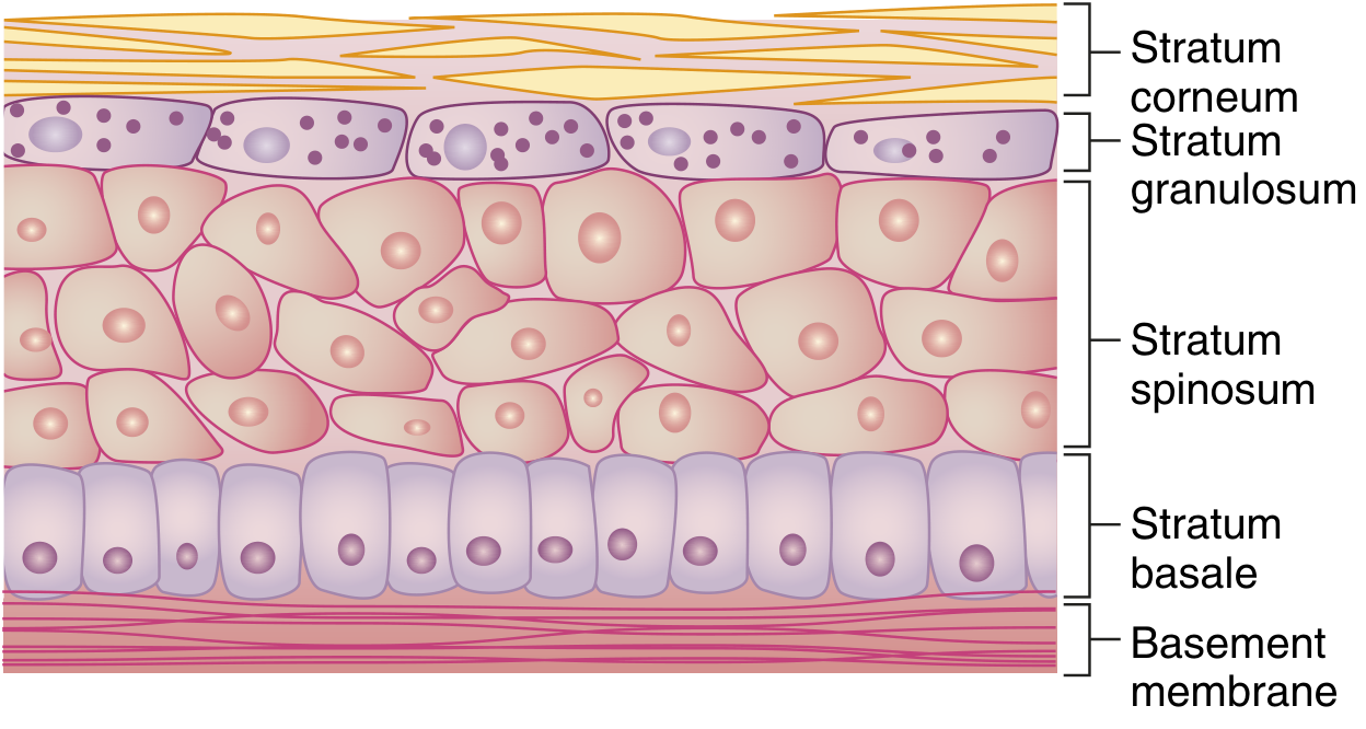
Composed of keratinized stratified squamous epithelium derived from ectoderm. It grows continuously but maintains normal thickness through desquamation. There are 5 layers (strata) from deep to superficial:
| Layer | Features |
|---|---|
| Stratum Basale (germinativum) | Deepest layer; single row of columnar/cuboidal cells; site of mitosis; contains melanocytes |
| Stratum Spinosum | "Prickle cell layer"; cells connected by desmosomes; contains Langerhans cells (APCs) |
| Stratum Granulosum | Cells contain keratohyalin granules; lamellar bodies released here form the water barrier |
| Stratum Lucidum | Present only in thick skin (palms, soles); translucent dead cells |
| Stratum Corneum | Most superficial; flat, anucleate, fully keratinized dead cells; protective barrier |
Keratinocytes are classified according to their depth and degree of differentiation - they proliferate in the basal layer, ascend, lose nuclei and organelles, and ultimately form the stratum corneum. - Bailey and Love's Short Practice of Surgery, 28th ed.
Epidermal cell replacement time: ~30 days (cells progress from basal layer to corneum in approximately 30 days, then shed).
Cells of the Epidermis
- Keratinocytes - predominant cell type (~90%); produce keratin
- Melanocytes - neural crest-derived dendritic cells in the basal layer; synthesize melanin (brown-black pigment) transferred to keratinocytes via membrane processes; protect against UV radiation. Ethnic differences in skin colour are determined by variations in melanin amount, combination, and distribution, not by differences in melanocyte number. - Bailey and Love's
- Langerhans cells - dendritic antigen-presenting cells in the stratum spinosum; part of the skin immune system
- Merkel cells - mechanoreceptors in the basal layer; associated with nerve endings; function in light touch discrimination
Epidermal Water Barrier
The lipid envelope (5 nm) - ceramides, cholesterol, free fatty acids - and the cell envelope (15 nm) - cross-linked structural proteins including loricrin (the major protein, ~80% of CE mass), involucrin, filaggrin, and cystatins - together form a "Teflon-like" barrier preventing transcutaneous water loss. Destruction over large areas (e.g., severe burns) can cause life-threatening fluid loss. - Histology: A Text and Atlas, 9e
B. Dermis (95% of skin)
Composed of dense irregular connective tissue derived from mesoderm. It is structurally divided into two layers:
| Layer | Description |
|---|---|
| Papillary dermis (superficial) | Delicate collagen and elastin fibres in ground substance; contains capillary and lymphatic networks; forms dermal papillae projecting into the epidermis |
| Reticular dermis (deep) | Coarse branching collagen fibres layered parallel to the skin surface; thicker, provides tensile strength |
The epidermis and dermis meet at the dermoepidermal junction (DEJ) - a basement membrane zone. In thick skin this creates an undulating surface with dermal papillae and rete ridges (epidermal ridges), which increases surface area and forms fingerprints.
Dermis contains:
- Collagen (type I - reticular; type III - papillary)
- Elastic fibres (elastin + fibrillin microfibrils)
- Ground substance (glycosaminoglycans, proteoglycans)
- Blood vessels, lymphatics
- Nerve fibres and sensory receptors
- Fibroblasts, mast cells, macrophages
- Skin appendages (hair follicles, sweat glands, sebaceous glands)
C. Hypodermis (Subcutaneous Layer)
- Deep to the dermis; equivalent to subcutaneous fascia
- Contains variable amounts of adipose tissue arranged in lobules separated by connective tissue septa
- Functions: energy storage, thermal insulation, mechanical cushioning, attachment of skin to underlying structures - Histology: A Text and Atlas, 9e
3. Functions of the Skin
The skin performs multiple vital functions:
1. Barrier / Protection
- Mechanical barrier - keratin and stratified epithelium resist abrasion and injury
- Permeability barrier - lipid envelope prevents transcutaneous water loss (TEWL); also prevents absorption of harmful external substances
- UV barrier - melanin absorbs and dissipates UV radiation, protecting DNA
- Chemical barrier - acid surface pH (~4.5-5.5) protects against pathogenic microorganisms
- Infection barrier - intact skin prevents microbial invasion; skin microbiome contributes to protection
2. Thermoregulation
- Sweat glands - eccrine sweating dissipates heat through evaporation
- Cutaneous blood vessels - vasodilation cools the body; vasoconstriction conserves heat
- Subcutaneous fat - acts as thermal insulator
- Hair erection (piloerection) via arrector pili muscles traps warm air in cold conditions
3. Sensory Function
Skin contains an array of sensory receptors conveying touch, pressure, vibration, pain, temperature, and itch to the CNS:
| Receptor | Location | Modality |
|---|---|---|
| Meissner's corpuscle | Dermal papillae; fingertips, lips | Fine/discriminative touch; rapidly adapting |
| Pacinian corpuscle | Deep dermis/hypodermis | Pressure and vibration; rapidly adapting |
| Merkel's disc | Stratum basale | Fine touch, texture; slowly adapting |
| Ruffini corpuscle | Reticular dermis | Stretch, torque, sustained pressure; rapidly adapting |
| Free nerve endings | Epidermis and dermis | Pain, temperature, itch |
| Krause end bulbs | Mucous membranes | Temperature (cold) |
4. Immune Function
- Langerhans cells process antigens and present to T-lymphocytes
- Skin-associated lymphoid tissue (SALT) participates in immune surveillance
- Provides immunologic information to effector cells in lymphatic tissue
5. Endocrine / Metabolic Functions
- Vitamin D synthesis - UV-B converts 7-dehydrocholesterol in the skin to cholecalciferol (vitamin D3)
- Secretes hormones, cytokines, and growth factors
- Converts precursor molecules into hormonally active substances
6. Excretion
- Eccrine sweat glands excrete water, electrolytes (Na⁺, Cl⁻, K⁺), urea, lactate
- Sebaceous glands secrete sebum
- Apocrine glands contribute to excretion
7. Absorption
- Lipid-soluble substances can be absorbed transdermally (basis of transdermal drug delivery systems - nicotine patches, steroid hormones, antiemetics)
4. Skin Appendages (Epidermal Derivatives)
Skin appendages are downgrowths of epidermal epithelium during development (ectodermal origin). They extend into the dermis and hypodermis.
A. Hair Follicles and Hair
Hair follicles are invaginations of the epidermis into the dermis in which a hair is formed. Present over almost the entire body except: palmar surfaces of hands, plantar surfaces of feet, lips, labia minora, glans penis, and nipples.
Hair distribution is influenced by sex hormones: facial hair appears at puberty in males; pubic and axillary hair at puberty in both sexes. Scalp hair thins with age due to reduced estrogen.
Structure of the Hair Follicle:
- Hair bulb - base of follicle; contains the dermal papilla (vascular connective tissue) and matrix cells (rapidly dividing stem cells that produce hair)
- Inner root sheath - surrounds and shapes the hair
- Outer root sheath - continuity with the epidermis
- Bulge region - in the isthmus; contains hair follicle stem cells important for hair cycling and wound repair
- Arrector pili muscle - smooth muscle; causes piloerection ("goose bumps")
Hair Growth Cycle:
- Anagen (growth phase) - matrix cells actively divide and differentiate
- Catagen (transition) - regression and apoptosis of lower follicle
- Telogen (resting/shedding) - hair shed; follicle rests before new anagen
Hair structure: keratinized thread with medulla (centre), cortex, and cuticle. Hair colour depends on melanin type - eumelanin (brown-black), phaeomelanin (red-yellow).
B. Nails
Nails are keratinized plates on the dorsal surface of the distal phalanges of fingers and toes.
Structure:
- Nail plate - hard, translucent keratinized structure
- Nail bed - epithelium under the nail plate
- Nail matrix (root) - beneath the proximal nail fold; site of nail formation (keratinisation of matrix cells)
- Lunula - whitish crescent-shaped area at the proximal nail; visible part of the matrix
- Hyponychium - junction of nail bed and fingertip skin
- Eponychium (cuticle) - proximal fold of skin overlying the nail matrix
- Paronychium - lateral nail folds
Functions: protection of fingertips, manipulation of small objects, scratching, cosmetic.
Growth rate: fingernails ~3-4 mm/month; toenails ~1-2 mm/month.
C. Sebaceous Glands
- Holocrine glands - entire cell disintegrates to release secretion (sebum)
- Found almost everywhere on the skin except palms and soles
- Open into hair follicle canal (pilosebaceous unit); independent sebaceous glands open directly on certain surfaces (lips, eyelids - Meibomian glands, areola - Montgomery glands)
- Secretion: Sebum - mixture of triglycerides, wax esters, squalene, cholesterol, free fatty acids
- Functions of sebum: lubricates and waterproofs skin and hair, antimicrobial properties (acidic pH), may have roles in vitamin E delivery and pheromone communication
- Stimulated by androgens (especially testosterone/DHT); active at puberty; overactivity leads to acne
- Inactive in childhood, become active at puberty
D. Sweat Glands (Sudoriferous Glands)
Two types:
1. Eccrine (Merocrine) Sweat Glands
- Most numerous sweat glands (~3-4 million on the body)
- Found over most of the body surface; greatest density on palms, soles, and forehead
- Not associated with hair follicles - open directly onto the skin surface
- Structure: simple coiled tubular gland; coiled secretory portion in deep dermis/hypodermis; straight/coiled duct rising to the surface
- Secretion: watery sweat - water, NaCl, KCl, urea, lactic acid, ammonia, IgA
- Functions:
- Thermoregulation (primary function) - evaporative cooling
- Minor excretory role
- Helps maintain acidic skin pH
- Innervated by cholinergic sympathetic nerve fibres (unusual - sympathetic but uses ACh)
2. Apocrine Sweat Glands
- Larger than eccrine glands
- Found in limited locations: axillae, areola of nipple, perianal region, external ear canal (ceruminous glands), eyelids (Moll's glands)
- Associated with hair follicles - drain into the upper portion of the follicle above the sebaceous gland
- Become active at puberty (androgen-dependent)
- Secretion: viscous, oily, protein-rich fluid containing carbohydrates, lipids, proteins, and pheromone-like compounds; odourless when secreted but odour develops when broken down by skin bacteria
- Function: pheromone secretion (sex attractant function - well-established in animals, possible in humans); emotional sweating (anxiety, fear, sexual excitement)
- Innervated by adrenergic sympathetic nerve fibres
| Feature | Eccrine | Apocrine |
|---|---|---|
| Distribution | Whole body | Axilla, areola, anogenital |
| Hair follicle | Independent | Associated |
| Onset | Birth | Puberty |
| Secretion | Watery (electrolytes, urea) | Viscous (lipids, proteins) |
| Function | Thermoregulation | Pheromones, emotional |
| Innervation | Cholinergic sympathetic | Adrenergic sympathetic |
E. Mammary Glands
Mammary glands are modified apocrine glands. They are epidermal appendages specialized for milk production (lactation) and are considered part of the integumentary system.
5. Sensory Receptors of the Dermis
The dermis contains encapsulated and unencapsulated nerve endings:
- Encapsulated mechanoreceptors: Meissner's corpuscles, Pacinian corpuscles, Ruffini corpuscles, Krause end bulbs
- Unencapsulated receptors: Merkel's discs (associated with Merkel cells in the epidermis), free nerve endings
Ruffini corpuscles are elongated fusiform encapsulated receptors (1-2 µm length) that respond to sustained mechanical stress, stretch, and torque. They are considered rapidly adapting (phasic) receptors that generate action potentials at the beginning and end of a stimulus. - Histology: A Text and Atlas, 9e
6. Summary Diagram
SKIN
├── Epidermis (5%)
│   ├── Stratum Corneum      ← dead, keratinized cells
│   ├── Stratum Lucidum      ← only in thick skin
│   ├── Stratum Granulosum   ← lamellar bodies, water barrier
│   ├── Stratum Spinosum     ← Langerhans cells, desmosomes
│   └── Stratum Basale       ← stem cells, melanocytes, Merkel cells
│
├── Dermis (95%)
│   ├── Papillary layer      ← delicate collagen, capillaries, receptors
│   └── Reticular layer      ← coarse collagen, elastic fibres
│
├── Hypodermis
│   └── Adipose tissue, loose connective tissue
│
└── Appendages (epidermal origin)
    ├── Hair follicles + arrector pili
    ├── Nails
    ├── Sebaceous glands      (holocrine; sebum; androgen-driven)
    ├── Eccrine sweat glands  (thermoregulation; cholinergic)
    └── Apocrine sweat glands (pheromones; adrenergic; puberty onset)
Sources:
- Histology: A Text and Atlas with Correlated Cell and Molecular Biology, 9e (Pawlina), pp. 1278-1342
- Bailey and Love's Short Practice of Surgery, 28th ed. - Functional Anatomy and Physiology of Skin; Surgical Anatomy of the Skin
- Rosen's Emergency Medicine, Surface Anatomy and Skin
Give me some pathology like bulla papule and so on
Finding Sources
Finding Sources
Finding Sources
Finding Sources
Fitzpatrick's is the gold standard for dermatology. Let me read that plus the Harriet Lane and Goldman-Cecil sections in parallel:
Finding Sources
Reading File
Reading File
Excellent - I have comprehensive data. Let me also fetch Fitzpatrick's table and the Goldman-Cecil morphology section, plus grab some images:
Finding Sources
Reading File
Loading Image
Now let me get Harrison's table on primary lesions and the Symptom to Diagnosis section for secondary lesions and special lesions:
Reading File
Reading File
I now have comprehensive, authoritative content from Harrison's, Goldman-Cecil, Harriet Lane, and Symptom to Diagnosis. Here is the full response:
Skin Pathology: Morphology of Skin Lesions
Dermatology has its own vocabulary. The correct approach to any rash is to identify the primary lesion first - the typical element of the eruption - and then note secondary changes, configuration, distribution, and colour. The differential diagnosis of a rash should be based on the morphology of the lesion, which serves as the first pivotal point. - Goldman-Cecil Medicine
PRIMARY SKIN LESIONS
These arise directly from the disease process - they have not been modified by scratching, treatment, or time.
1. Macule
- Definition: Flat, circumscribed area of colour change, < 1-2 cm in diameter; NOT elevated or depressed; not palpable
- Pathology: Change in melanin, blood (haemoglobin), or exogenous pigment in the epidermis or superficial dermis; no structural change in skin surface
- Examples: Freckle (ephelid), cafe-au-lait spot, vitiligo, early measles rash, petechia (small)
- Key point: You can SEE it but cannot FEEL it
2. Patch
- Definition: A large macule, > 1-2 cm (some sources say > 0.5 cm); flat, non-palpable area of colour change
- Examples: Vitiligo patches, port-wine stain (nevus flammeus), Mongolian spot, large areas of tinea versicolor
- Key point: Differs from macule only in SIZE
3. Papule
- Definition: Small, solid, elevated, well-circumscribed lesion < 0.5-1 cm in diameter; palpable; raised above surrounding skin
- Pathology: May result from epidermal hyperplasia, dermal infiltration, or accumulation of metabolic products
- Examples: Warts (verruca vulgaris), molluscum contagiosum, closed comedone (whitehead), lichen planus (flat-topped violaceous papule), acne lesions, insect bites
- Key point: You can SEE it AND FEEL it; it is SOLID (no fluid)
4. Plaque
- Definition: Large (> 1 cm), flat-topped, raised lesion - essentially a large papule or confluence of papules
- Pathology: Same processes as papule but larger; edges may be distinct (psoriasis) or gradually blend (eczema)
- Examples: Psoriasis (well-demarcated silvery-scaled plaques), eczematous dermatitis, mycosis fungoides, discoid lupus erythematosus
- Key point: Think of it as a "raised plateau" - large surface area, flat top
5. Nodule
- Definition: Solid, firm lesion raised above the skin surface, 0.5-5 cm in diameter; extends deeper into the dermis or subcutaneous tissue than a papule
- Pathology: Dermal or subcutaneous infiltration (inflammatory cells, neoplastic cells, deposits)
- Examples: Lipoma, rheumatoid nodule, erythema nodosum, dermatofibroma, large melanocytic nevus, lymphoma cutis
- Key point: DEEPER than a papule; can be felt rolling under the fingers
6. Tumor / Mass
- Definition: Solid, raised growth > 5 cm in diameter (some sources: > 2 cm); extends into the dermis/subcutis
- Examples: Large cutaneous malignancies, large lipoma, pilomatrixoma
- Key point: Distinguished from nodule by SIZE only
7. Vesicle
- Definition: Small, fluid-filled blister < 0.5-1 cm in diameter; raised above surrounding skin; fluid often visible and translucent
- Pathology: Intraepidermal oedema (spongiosis), acantholysis, or subepidermal separation; roof = epidermis; content = clear serous fluid
- Examples:
- Herpes simplex / herpes zoster (varicella) - grouped vesicles on erythematous base
- Allergic contact dermatitis (poison ivy/oak)
- Dyshidrotic eczema (pompholyx) - vesicles on palms and soles
- Early chickenpox
- Key point: Fluid-filled; SMALL (< 1 cm); translucent
8. Bulla
- Definition: A large vesicle (fluid-filled blister) > 0.5-1 cm in diameter; elevated, often tense or flaccid
- Pathology: Subepidermal (tense bullae - BP, EBA) or intraepidermal (flaccid bullae - pemphigus vulgaris) blister formation
- Sub-types by location:
- Intraepidermal - flaccid, easily ruptured (e.g., pemphigus vulgaris - acantholysis above the basal layer)
- Subepidermal - tense, more durable (e.g., bullous pemphigoid - IgG + complement at BMZ)
- Examples:
- Bullous pemphigoid - tense bullae, elderly, autoimmune (anti-BP180/BP230)
- Pemphigus vulgaris - flaccid bullae, mucosal involvement, Nikolsky sign positive
- Epidermolysis bullosa - inherited fragility
- Impetigo (bullous type) - honey-coloured rupturing bullae, Staph. aureus
- Burns, severe contact dermatitis
- Key point: Fluid-filled; LARGE (> 1 cm); may be tense or flaccid
| Vesicle | Bulla | |
|---|---|---|
| Size | < 1 cm | > 1 cm |
| Content | Clear fluid | Clear/serosanguineous |
| Examples | Herpes, contact derm | Pemphigoid, pemphigus |
9. Pustule
- Definition: Well-circumscribed, elevated lesion filled with purulent material (pus = dead neutrophils + bacteria/debris); any size
- Pathology: Intraepidermal or subepidermal collection of neutrophils
- Key point: Presence of pustules does NOT necessarily indicate infection - sterile pustules occur in many inflammatory conditions
- Examples:
- Infected: Folliculitis, impetigo, furuncle
- Sterile: Psoriasis (pustular psoriasis), acne vulgaris, rosacea, palmoplantar pustulosis
10. Wheal (Urtica / Hive)
- Definition: Transient (typically < 24 hours), raised, well-circumscribed lesion with an erythematous periphery and central pallor; an inflamed oedematous papule or plaque formed by superficial local oedema
- Pathology: Mast cell degranulation → histamine release → transient vasodilation and increased vascular permeability in the dermis → localised oedema
- Examples: Urticaria (hives), allergic reactions, dermatographism
- Key point: TRANSIENT - comes and goes within hours; oedematous; itchy
11. Cyst
- Definition: Encapsulated, fluctuant lesion filled with soft semi-solid or liquid material (not just fluid); has an epithelial lining
- Examples: Epidermal inclusion cyst, sebaceous cyst, pilar cyst, dermoid cyst, milia
- Key point: Has a WALL (cyst capsule) - distinguishes it from a simple vesicle/bulla
12. Comedone
- Definition: A plug of keratinous material and skin oils retained in a hair follicle
- Open comedone (blackhead): Open to the air; black colour from oxidised melanin (not dirt)
- Closed comedone (whitehead): Covered by thin layer of skin; flesh-coloured or pinkish
- Pathology: Sebaceous duct obstruction; precursor of acne vulgaris
- Symptom to Diagnosis, 4e
13. Telangiectasia
- Definition: Dilated, superficial blood vessel visible on the skin surface; does not blanch with pressure (unlike erythema)
- Examples: Rosacea, hereditary haemorrhagic telangiectasia (HHT/Osler-Weber-Rendu), CREST syndrome, spider naevi, ataxia-telangiectasia
SECONDARY SKIN LESIONS
These develop from primary lesions due to scratching, infection, healing, or time.
| Lesion | Definition | Key Features |
|---|---|---|
| Scale | Excessive accumulation / shedding of stratum corneum cells | Seen in psoriasis (silvery), tinea (fine), pityriasis rosea, ichthyosis |
| Crust | Dried exudate (serum, pus, blood) on the skin surface | Yellow = serous/infected; red = haemorrhagic; "honey-coloured" = impetigo |
| Erosion | Superficial loss of epidermis ONLY; does NOT reach the dermis | Heals WITHOUT scarring; moist, shallow |
| Ulcer | Full-thickness loss of epidermis AND at least part of the dermis | Heals WITH scarring; may show necrosis, base exposed |
| Fissure | Linear or wedge-shaped epidermal crack/tear extending into the dermis | Painful; at sites of repeated movement - heels, fingers, lips |
| Excoriation | Linear, angular erosion caused by scratching; may be crusted | Sign of pruritus; seen in atopic dermatitis, scabies, neurotic excoriation |
| Lichenification | Thickened, leathery epidermis with accentuated skin markings | Chronic scratching/rubbing; typical of chronic atopic dermatitis |
| Scar | New connective tissue replacing damaged dermis after full-thickness injury | May be hypo/hyperpigmented; hypertrophic scar = stays within wound; keloid = grows beyond wound |
| Atrophy | Loss of skin substance | Epidermal atrophy = shiny, thin, wrinkled; dermal atrophy = depression; caused by topical steroids, lichen sclerosus |
SPECIAL / VASCULAR LESIONS
| Lesion | Definition | Examples |
|---|---|---|
| Petechiae | Pinpoint (< 2 mm) red/purple non-blanching spots from RBC extravasation | Thrombocytopenia, vasculitis, meningococcaemia |
| Purpura | Larger red-purple non-blanching areas from RBC extravasation | Palpable = leukocytoclastic vasculitis; non-palpable = coagulopathy |
| Ecchymosis | Large area of purpura ("bruise") from haemorrhage into skin | Trauma, coagulopathy |
| Retiform purpura | Net/lace-like purpura with concave borders | Occlusive vasculopathy (thrombosis, emboli); potentially life-threatening |
Purpura is caused by extravasation of red blood cells into the skin, resulting in a reddish to dark purple to black colour. Palpable purpura suggests small vessel leukocytoclastic vasculitis. Retiform purpura - with concave, net-like, scalloped borders - can signal a life-threatening disorder. - Goldman-Cecil Medicine
CONFIGURATION PATTERNS (Arrangement of Lesions)
The arrangement of lesions is a key diagnostic clue:
| Pattern | Description | Example |
|---|---|---|
| Linear | Along a line | Contact dermatitis (linear), lichen striatus |
| Dermatomal | Following a dermatome | Herpes zoster |
| Annular | Ring-like configuration | Tinea corporis (ringworm), erythema migrans (Lyme) |
| Serpiginous | Wavy/coiled/snake-like | Cutaneous larva migrans |
| Targetoid | Bull's-eye (central dark zone, pale ring, outer red ring) | Erythema multiforme |
| Nummular/Discoid | Coin-shaped | Nummular eczema |
| Grouped/Herpetiform | Clustered on an erythematous base | Herpes simplex, herpes zoster |
| Filiform | Thread-like | Filiform warts |
QUICK REFERENCE SUMMARY TABLE
| Lesion | Flat/Raised | Fluid? | Size | Key Example |
|---|---|---|---|---|
| Macule | Flat | No | < 1-2 cm | Freckle, vitiligo |
| Patch | Flat | No | > 1-2 cm | Port-wine stain |
| Papule | Raised | No (solid) | < 1 cm | Wart, comedone |
| Plaque | Raised (flat-top) | No (solid) | > 1 cm | Psoriasis |
| Nodule | Raised (deep) | No (solid) | 0.5-5 cm | Lipoma, rheumatoid nodule |
| Tumor | Raised (deep) | No (solid) | > 5 cm | Large malignancy |
| Vesicle | Raised | Yes (clear) | < 1 cm | Herpes, contact derm |
| Bulla | Raised | Yes (clear) | > 1 cm | Pemphigoid, burns |
| Pustule | Raised | Yes (pus) | Any size | Folliculitis, pustular psoriasis |
| Wheal | Raised (transient) | Oedema | Variable | Urticaria |
| Cyst | Raised | Semi-solid | Variable | Epidermal cyst |
Sources:
- Harrison's Principles of Internal Medicine, 22e (2025) - Tables 39-1 and 39-2
- Goldman-Cecil Medicine, International Edition - Chapter 403: Morphology, Configuration, Palpation
- The Harriet Lane Handbook, 23e (Johns Hopkins) - Evaluation and Clinical Descriptions of Skin Findings
- Symptom to Diagnosis: An Evidence-Based Guide, 4e - Constructing a Differential Diagnosis
Everything about olfactory
Reading File
This is a broad anatomical/physiological topic. Let me search across headings and paragraphs simultaneously:
Finding Sources
Finding Sources
Excellent sources found. Now read all the key sections in parallel:
Reading File
Reading File
Reading File
Excellent content. Now fetch images and read Ganong + Cummings on olfactory dysfunction:
Loading Image
Loading Image
Reading File
Reading File
Now read the olfactory pathways, dysfunction, and clinical correlates:
Reading File
Reading File
Reading File
I now have comprehensive, authoritative content from Guyton & Hall, Costanzo, Ganong, Gray's Anatomy, and Cummings Otolaryngology. Let me compile the full response:
Everything About the Olfactory System
1. Overview
Olfaction is the sense of smell - the detection and perception of airborne chemical stimuli (odorants). It plays roles in pleasure, food intake, memory, emotional responses, and survival (detecting hazardous odours, spoiled food). Uniquely among special senses, the olfactory pathway does NOT relay through the thalamus before reaching primary olfactory cortex - it projects directly to the cortex. - Gray's Anatomy for Students
2. Anatomy of the Olfactory Region
Location: The Olfactory Membrane / Epithelium
- Located in the superior part of the nasal cavity - roof near the septum, folds over the superior turbinate and upper surface of the middle turbinate
- Total surface area: ~5 cm² (Guyton & Hall) to ~10 cm² (Ganong)
- Appears yellowish in colour (due to pigmentation) - macroscopically indistinguishable from respiratory epithelium
- Bounded by the olfactory cleft (cribriform plate superiorly, superior turbinate laterally, superior septum medially)
3. Histology of the Olfactory Epithelium
The olfactory epithelium (OE) is a pseudostratified columnar epithelium containing three main cell types:
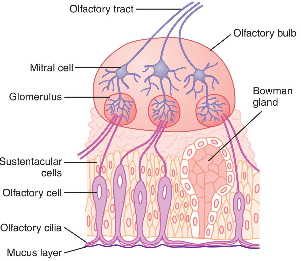
A. Olfactory Receptor Neurons (ORNs)
- ~100 million bipolar neurons in the olfactory epithelium (Guyton) / 6-30 million in young adults (Cummings)
- Bipolar neurons - unique because they are both primary sensory neurons AND CNS-derived (originally from the CNS/neural placode, not peripheral nervous system)
- Dendrite - projects apically toward the epithelial surface, ending in an olfactory vesicle (knob)
- Olfactory cilia - 4 to 25 nonmotile cilia per cell (0.3 µm diameter, up to 200 µm long) project from the knob into the overlying mucus layer; contain the odorant receptor proteins; greatly increase surface area for odorant binding
- Axon - single, unmyelinated axon at the basal pole; among the smallest and slowest fibres in the nervous system; forms bundles (fila olfactoria) surrounded by olfactory ensheathing cells (specialised glia)
- Axons pass through foramina of the cribriform plate of the ethmoid bone to reach the olfactory bulb
ORNs are unique first-order sensory neurons in that they simultaneously contact the outside environment (cilia in nasal mucus) AND the central nervous system. - Cummings Otolaryngology
B. Supporting (Sustentacular) Cells
- Columnar epithelial cells interspersed among ORNs; lined with microvilli at their mucosal surface
- Secrete mucus that provides the appropriate molecular and ionic environment for odour detection
- Contain secretory granules
- Also release neurotrophic factors stimulating genesis of new olfactory sensory neurons
C. Basal Cells (Stem Cells)
- Located at the base of the olfactory epithelium
- Undifferentiated stem cells - undergo mitosis to replace ORNs that are continuously lost due to environmental damage
- Source of adult neurogenesis in the olfactory system - one of the few sites in the body where new neurons are generated throughout life
- ORNs survive only 1-2 months (Ganong) before being replaced
- In animals, the global ORN population undergoes complete regeneration every 4-6 weeks; human turnover rate is less certain
D. Other Cell Types
- Bowman glands - submucosal seromucous glands in the lamina propria beneath the basement membrane; secrete mucus onto the olfactory surface; the mucus dissolves odorant molecules and facilitates their diffusion to receptor cilia
- Olfactory ensheathing cells - specialised glial cells in the lamina propria AND olfactory bulb; surround axon bundles; guide axonal regeneration after injury; being investigated for therapeutic transplantation in nerve repair
- Microvillar cells and duct cells - present in smaller numbers
4. Odorant Receptors
- There are approximately 1000 olfactory receptor genes in humans, accounting for ~3% of the human genome; approximately 400 functional odorant receptors are encoded (the rest are pseudogenes)
- All odorant receptors are G-protein-coupled receptors (GPCRs) - 7-transmembrane proteins
- The olfactory system can discriminate perhaps more than 1 million distinct odours
- Each ORN expresses only ONE type of odorant receptor gene
- Each receptor type is expressed in approximately 5,000 neurons confined to one spatial zone of the epithelium
- The epithelium is divided into spatial zones, each expressing different receptor types - Principles of Neural Science (Kandel)
5. Signal Transduction (How We Detect Smell)
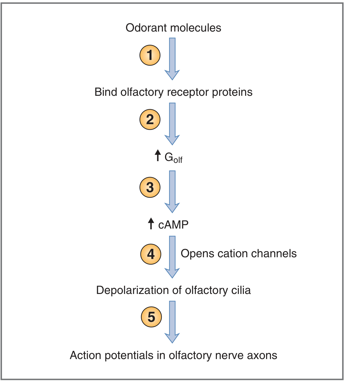
The transduction cascade converts a chemical signal into an action potential:
Step 1: Odorant molecules dissolve in the nasal mucus (aided by odorant-binding proteins) and bind to odorant receptor proteins on the olfactory cilia
Step 2: Receptor activation causes the G-protein (Golf) α-subunit to dissociate and activate adenylyl cyclase
Step 3: Adenylyl cyclase catalyses conversion of ATP → cAMP (second messenger); intracellular cAMP levels rise
Step 4: cAMP opens cation channels in the ciliary membrane, increasing permeability to Na⁺, K⁺, and Ca²⁺; net inward Ca²⁺ current produces a graded receptor potential (depolarisation)
Step 5: Ca²⁺ then opens Ca²⁺-activated Cl⁻ channels, causing further depolarisation (due to high intracellular Cl⁻ in ORNs)
Step 6: If the receptor potential exceeds threshold → action potential generated and propagated along the olfactory nerve (CN I) toward the olfactory bulb
This cAMP amplification cascade greatly multiplies the excitatory effect of even a small number of odorant molecules binding to the receptors. - Guyton & Hall
6. Olfactory Encoding - How Do We Recognise Smells?
- Olfactory receptor proteins are NOT dedicated to a single odorant; each responds to a variety of odorants
- Different receptor proteins have different response profiles to the same odorant
- Each odorant produces a unique across-fiber pattern of activity across a population of receptors
- This pattern is projected onto targeted glomeruli in the olfactory bulb, creating an "odour map"
- The CNS interprets these odour maps to identify the specific smell - Costanzo Physiology
7. The Olfactory Bulb
The olfactory bulb (OB) is a small ovoid structure resting on the cribriform plate of the ethmoid bone.
Cells of the olfactory bulb:
| Cell | Role |
|---|---|
| Mitral cells | Large 2nd-order neurons; receive ORN axons in glomeruli; project to olfactory cortex |
| Tufted cells | Similar to mitral cells; also 2nd-order projection neurons |
| Periglomerular cells | Inhibitory interneurons connecting adjacent glomeruli; sharpen olfactory signals |
| Granule cells | Inhibitory interneurons (no axons); make reciprocal dendrodendritic synapses with mitral/tufted cells; release GABA; provide lateral inhibition |
Convergence in glomeruli: Approximately 1,000 ORN axons synapse onto 1 mitral cell via glomeruli. Each olfactory sensory neuron projects to only 1-2 glomeruli. This convergence creates a precise two-dimensional odour map. - Ganong's Review of Medical Physiology
Lateral inhibition - periglomerular and granule cells inhibit neighbouring mitral cells (analogous to horizontal cells in the retina), sharpening and focusing olfactory signals.
8. The Olfactory Pathway
Odorant → Olfactory Receptor Neuron (ORN) in nasal epithelium
                    ↓ (unmyelinated axons through cribriform plate)
           Olfactory Bulb (CN I = Olfactory Nerve)
           Synapse in Glomeruli (ORN → Mitral cells)
                    ↓
           Olfactory Tract
           Divides into:
           ├── LATERAL olfactory stria
           │     → Primary Olfactory Cortex:
           │       • Piriform (prepiriform) cortex ← main; conscious discrimination
           │       • Olfactory tubercle
           │       • Amygdala ← emotional responses to smell
           │       • Entorhinal cortex ← olfactory memory
           │       • Anterior olfactory nucleus
           │
           └── MEDIAL olfactory stria
                 → Anterior commissure → contralateral olfactory bulb
                    (inhibition of mitral cells - enhances localization)
                 → Septal area / diagonal band

From Piriform cortex → Orbitofrontal cortex (via thalamus) ← conscious odour identification
From Piriform cortex → Hypothalamus / Brainstem (via MFB) ← autonomic responses
                                                              (salivation, gastric contraction)
Key unique feature: The olfactory pathway is the only special sense that does NOT relay through the thalamus to reach the primary cortex - it projects directly. - Gray's Anatomy for Students
From the olfactory cortex:
- → Orbitofrontal cortex - conscious discrimination of odours
- → Amygdala - emotional and fear responses to odours (e.g., smell of smoke triggers alarm)
- → Entorhinal cortex - olfactory memory (explains why smells trigger vivid memories - Proust phenomenon)
- → Hypothalamus/brainstem (via medial forebrain bundle, MFB) - autonomic responses: salivation, gastric contraction, arousal via reticular formation
9. Olfactory Adaptation
- With prolonged exposure to an odour, adaptation occurs rapidly - sensitivity to that particular odorant decreases
- Adaptation is specific - sensitivity to other odours is not affected
- Mechanisms include receptor desensitisation and central adaptation
- The olfactory system is extremely sensitive: some odorants detected at <1 part per trillion (e.g., mercaptans)
- Odorants typically contain 1-30 carbon atoms with functional groups (ketones, aldehydes, esters, thiols, etc.)
10. The Vomeronasal Organ (Accessory Olfactory System)
- A separate patch of olfactory epithelium along the nasal septum (more developed in other mammals)
- Concerned with perception of pheromones
- Contains ~100 GPCRs structurally different from main olfactory receptors
- Projects to the accessory olfactory bulb → amygdala and hypothalamus → regulation of reproductive and ingestive behaviour
- Rudimentary in adult humans (debated whether functional) - Ganong's
11. Olfactory Dysfunction - Definitions
Cummings Otolaryngology defines:
| Term | Definition |
|---|---|
| Normosmia | Normal olfactory function |
| Hyposmia (microsmia) | Quantitatively reduced olfactory function |
| Functional anosmia | Reduced to the extent of having no useful function in daily life |
| Anosmia | Complete absence of olfactory function |
| Specific anosmia | Inability to smell ONE specific odorant despite intact general olfaction; considered a normal physiological trait |
| Hyperosmia (superosmia) | Abnormally heightened smell; rare; associated with migraine |
| Parosmia (dysosmia, cacosmia) | Distorted quality of smell IN THE PRESENCE of an odorant |
| Phantosmia | Smell perceived WITHOUT any odorant present - an "olfactory hallucination" |
| Orthonasal olfaction | Smell via sniffing (anterograde airflow through nostrils) |
| Retronasal olfaction | Smell during eating/drinking (airflow from pharynx back to olfactory cleft) - contributes to flavour perception |
12. Causes of Olfactory Dysfunction
| Category | Causes |
|---|---|
| Conductive/Transport | Nasal polyps, rhinosinusitis, mucosal oedema (most common), deviated septum - block odour access to olfactory cleft |
| Sensorineural | Post-viral (most common after infection), head trauma (cribriform plate fracture severs ORN axons), neurodegenerative disease |
| Post-viral | URTI, COVID-19 (parosmia and anosmia); direct epithelial damage + supporting cell destruction |
| Traumatic | Cribriform plate fracture → anosmia (axons physically severed as they pass through cribriform foramina) |
| Neurodegenerative | Parkinson's disease, Alzheimer's disease - olfactory loss is often an early sign; olfactory bulb affected early |
| Congenital | Kallmann syndrome - anosmia + hypogonadotrophic hypogonadism (olfactory neurons fail to migrate); olfactory bulb aplasia |
| Toxic/Drug | Intranasal zinc (topical), cocaine, heavy metals |
| Aging | OE undergoes progressive metaplasia; regenerative capacity exhausted; cumulative damage leads to inhomogeneous (checkerboard) OE pattern |
| Tumour | Olfactory groove meningioma, olfactory neuroblastoma (esthesioneuroblastoma) |
13. Olfactory Neuroblastoma (Esthesioneuroblastoma)
A malignant tumour arising from the olfactory neuroepithelium of the upper nasal cavity. It is relevant to the olfactory system as a direct pathological derivative of ORNs. Presents with nasal obstruction, epistaxis, and anosmia. - Robbins & Cotran Pathologic Basis of Disease
14. Olfactory Nerve - CN I Summary
| Feature | Detail |
|---|---|
| Type | Special sensory (smell) |
| Cell bodies | Olfactory receptor neurons in nasal epithelium |
| Axons | Unmyelinated; fila olfactoria through cribriform plate |
| 1st synapse | Olfactory bulb (in glomeruli, with mitral cells) |
| Primary cortex | Piriform cortex (no thalamic relay!) |
| Testing | Asking patient to identify familiar odours (coffee, vanilla) with one nostril at a time |
| Clinical lesion | Anosmia from head trauma (cribriform plate fracture), tumour, or infection |
15. Clinical Correlates
- Cribriform plate fractures are the most common traumatic cause of anosmia - ORN axons are severed as they pass through the tiny foramina
- COVID-19 causes anosmia and parosmia by damaging supporting cells and sustentacular cells in the OE (not primarily the ORNs themselves), disrupting the microenvironment for receptor function
- Kallmann syndrome - congenital anosmia + hypogonadism; GnRH neurons fail to migrate from the olfactory placode; associated with aplasia/hypoplasia of the olfactory bulbs
- Early Parkinson's / Alzheimer's - hyposmia often precedes motor or cognitive symptoms; olfactory testing is being explored as a biomarker
- Olfactory training - repeated exposure to strong odorants (rose, lemon, eucalyptus, clove) may facilitate ORN regeneration and improve post-viral and post-COVID anosmia/parosmia - Cummings Otolaryngology
- Foster-Kennedy syndrome - anosmia + ipsilateral optic atrophy + contralateral papilloedema from a frontal lobe tumour compressing the olfactory tract
Sources:
- Guyton and Hall Textbook of Medical Physiology, 14e - Olfactory Membrane, Stimulation of Olfactory Cells
- Ganong's Review of Medical Physiology, 26e - Olfactory Epithelium, Odorant Receptors, Signal Transduction, Olfactory Sensory Pathway
- Costanzo Physiology, 7e - Olfactory Transduction, Pathways, Encoding
- Gray's Anatomy for Students - Olfactory System (no thalamic relay, limbic connections)
- Cummings Otolaryngology Head and Neck Surgery - Olfactory Neuroepithelium, Olfactory Dysfunction, Olfactory Training
- Principles of Neural Science (Kandel), 6e - Odorant encoding, spatial zones, receptor targeting
This is a shared conversation. Sign in to Orris to start your own chat.