I now have comprehensive textbook content from both Cummings Otolaryngology and KJ Lee's Essential Otolaryngology. Let me compile the full detailed response.
Infratemporal Fossa: Detailed Anatomy, Communications, Clinical Importance, and the Fisch Approach
(Based on Cummings Otolaryngology Head and Neck Surgery, 7th Ed.; K.J. Lee's Essential Otolaryngology, 11th Ed.)
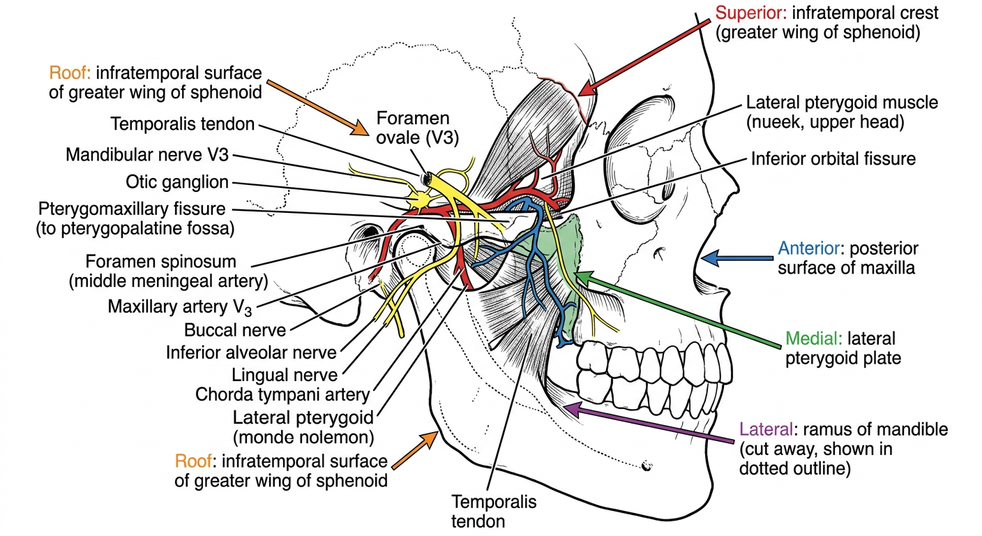
1. BOUNDARIES (Walls) of the Infratemporal Fossa
The infratemporal fossa is an irregular, wedge-shaped space deep to the zygomatic arch and posterior to the maxilla. It has no true floor.
| Wall | Boundary |
|---|
| Anterior | Posterior surface of the maxilla; inferior orbital fissure |
| Posterior | Mastoid and tympanic portions of the temporal bone |
| Superior (roof) | Inferior surface of the greater wing of the sphenoid + squamous portion of temporal bone (infratemporal crest) |
| Medial | Lateral pterygoid plate, lateral portion of the clivus, C1 vertebra, inferior surface of the petrous temporal bone; tensor and levator palatini muscles, superior constrictor |
| Lateral | Zygomatic arch and ascending ramus/coronoid process of the mandible |
| Inferior | No bony floor; bounded by the superior limit of the posterior belly of the digastric muscle and the angle of the mandible |
The fossa is continuous superiorly with the temporal fossa (through the space deep to the zygomatic arch).
- Cummings Otolaryngology, p. 3349-3350
- KJ Lee's Essential Otolaryngology, p. 703
2. CONTENTS of the Infratemporal Fossa
Muscular
- Lateral pterygoid muscle - two heads:
- Superior head: arises from the roof of the infratemporal fossa (infratemporal surface of greater wing of sphenoid)
- Inferior head: arises from the lateral aspect of the lateral pterygoid plate
- Both insert onto the TMJ (articular disc + neck of condyle)
- Medial pterygoid muscle: originates from the medial aspect of the lateral pterygoid plate; inserts on the medial surface of the angle of the mandible and ascending ramus
- Temporalis tendon: insertion on the coronoid process
Neural
- Mandibular nerve (V3) - the dominant nerve of the ITF; exits the skull through the foramen ovale and immediately divides into:
- Anterior division (mainly motor): masseteric nerve, deep temporal nerves, nerve to lateral pterygoid, buccal nerve (sensory to buccal mucosa)
- Posterior division (mainly sensory): auriculotemporal nerve, lingual nerve, inferior alveolar nerve
- Motor supply: temporalis, masseter, medial and lateral pterygoids (muscles of mastication)
- Chorda tympani (branch of CN VII): exits petrotympanic fissure, joins the lingual nerve in the ITF; carries taste from anterior 2/3 tongue and preganglionic parasympathetics to submandibular ganglion
- Otic ganglion: lies medial to V3, just below foramen ovale; receives lesser petrosal nerve (preganglionic parasympathetics from CN IX); postganglionic fibres ride on the auriculotemporal nerve to the parotid gland
- Lesser petrosal nerve: carries parasympathetics from CN IX (Jacobson's nerve) to the otic ganglion
Vascular
Maxillary artery (terminal branch of ECA):
- Enters the ITF posterior to the ramus of the mandible, passes lateral to the lateral pterygoid muscle
- Divided into 3 parts:
- Mandibular part (1st part): branches include the middle meningeal artery (exits via foramen spinosum), inferior alveolar artery, anterior tympanic artery, deep auricular artery
- Pterygoid part (2nd part): deep temporal arteries, masseteric artery, pterygoid branches, buccal artery
- Pterygopalatine part (3rd part): enters pterygopalatine fossa via pterygomaxillary fissure
Pterygoid venous plexus: lies around the pterygoid muscles; drains posteriorly into the maxillary vein (to retromandibular vein) and anteriorly into the facial vein. Has important anastomoses with:
- Cavernous sinus (via emissary veins through foramen ovale/spinosum)
- Ophthalmic veins (via inferior orbital fissure)
- Pharyngeal venous plexus
Clinical significance: Infection in the ITF can spread hematogenously to the cavernous sinus (cavernous sinus thrombosis), the orbit, and even intracranially.
- Cummings Otolaryngology, p. 3349-3351
3. COMMUNICATIONS of the Infratemporal Fossa
| Adjacent Structure | Pathway / Route |
|---|
| Orbit | Inferior orbital fissure |
| Middle cranial fossa | Foramen ovale (V3) and foramen spinosum (middle meningeal artery) |
| Pterygopalatine fossa | Pterygomaxillary fissure |
| Temporal fossa | Between zygomatic arch and cranium (deep to arch) |
| Cavernous sinus | Via pterygoid plexus emissary connections |
| Parapharyngeal space | Medially, without a distinct bony boundary |
| Nasopharynx / peritubal space | Along eustachian tube (surgical access relevant) |
Anterior to V3 and the middle meningeal artery, following the floor of the middle cranial fossa and horizontal ICA, the ITF gives access to the cavernous sinus, sphenoid sinus, and nasopharynx.
- Cummings Otolaryngology, p. 3350
4. CLINICAL IMPORTANCE
A. Spread of Infection
The ITF's communications make it a conduit for deep space neck infections:
- Dental infections (molar teeth) can spread to the parapharyngeal, masticator, and ITF spaces
- The pterygoid venous plexus can carry septic emboli to the cavernous sinus
B. Trismus
Infection or tumor in the ITF compresses/infiltrates the pterygoid muscles and TMJ - a classic presenting sign of ITF pathology
C. Tumors
The ITF is a pathway for perineural spread and for extension of:
- Nasopharyngeal carcinoma - classic spread into ITF via eustachian tube and lateral wall
- Juvenile nasopharyngeal angiofibroma (JNA) - grows into the ITF via the pterygomaxillary fissure
- Parotid malignancies - extend posteriorly
- Glomus jugulare tumors - extend into posterior ITF
- Salivary gland cancers, squamous cell carcinoma, cholesteatoma of the temporal bone
- Chordoma, chondrosarcoma - from clivus/petrous apex into ITF
D. Perineural Spread
V3 in the ITF is a major route for perineural invasion of head and neck cancers (especially adenoid cystic carcinoma), tracking intracranially to the Gasserian ganglion
E. Anesthetic Relevance
- Inferior alveolar nerve block (dental anesthesia): targets the mandibular foramen inside the ITF
- V3 block at foramen ovale and infratemporal pterygopalatine fossa blocks are performed for trigeminal neuralgia and pain management
5. THE FISCH INFRATEMPORAL FOSSA APPROACH
Ugo Fisch was the first to develop a structured series of lateral skull base approaches to the infratemporal fossa. He divided these into three basic types (A, B, C), each progressively more anterior in access.
Overview of the Three Types
| Type | Principal Access Target | Key Step |
|---|
| A | Jugular bulb, vertical petrous ICA, posterior ITF | Radical mastoidectomy + anterior transposition of facial nerve + cervical dissection |
| B | Petrous apex, clivus, superior ITF | Type A steps (usually without FN transposition) + temporalis reflection |
| C | Rostral clivus, cavernous sinus, sphenoid sinus, pterygopalatine fossa, nasopharynx, anterosuperior ITF | Type B + pterygoid base removal |
FISCH TYPE A APPROACH
Indications:
- Glomus jugulare/tympanicum tumors (paragangliomas)
- Salivary gland cancers
- Squamous cell carcinoma of the temporal bone
- Cholesteatoma
- Neurinoma (schwannoma of lower cranial nerves)
- Meningioma, rhabdomyosarcoma, teratoma, myxoma
Step-by-Step Technique:
-
Incision: Large C-shaped postauricular incision extending anterosuperiorly into the temporal scalp and anteroinferiorly into the neck (depending on the required cervical/anterior exposure needed)
-
Flap elevation: Flap raised superficial to temporalis, SCM, and postauricular musculature
-
EAC transection: Cartilaginous external auditory canal (EAC) transected at the bony-cartilaginous junction; a periosteal flap pedicled on the EAC anteriorly is created
-
EAC closure: The lateral EAC is everted and closed primarily (meatal skin everted through meatus). The periosteal flap is rotated anteriorly and sutured to the closed EAC to reinforce the closure medially
-
Middle ear entry: Osseous EAC skin elevated circumferentially to the tympanic annulus; middle ear entered. Incudostapedial joint separated, incus removed, tensor tympani tendon sectioned, EAC skin/TM with malleus removed
-
Radical mastoidectomy: Removal of osseous EAC and all air cell tracts lateral and adjacent to the otic capsule. Stapes suprastructure removed. Eustachian tube obliterated
-
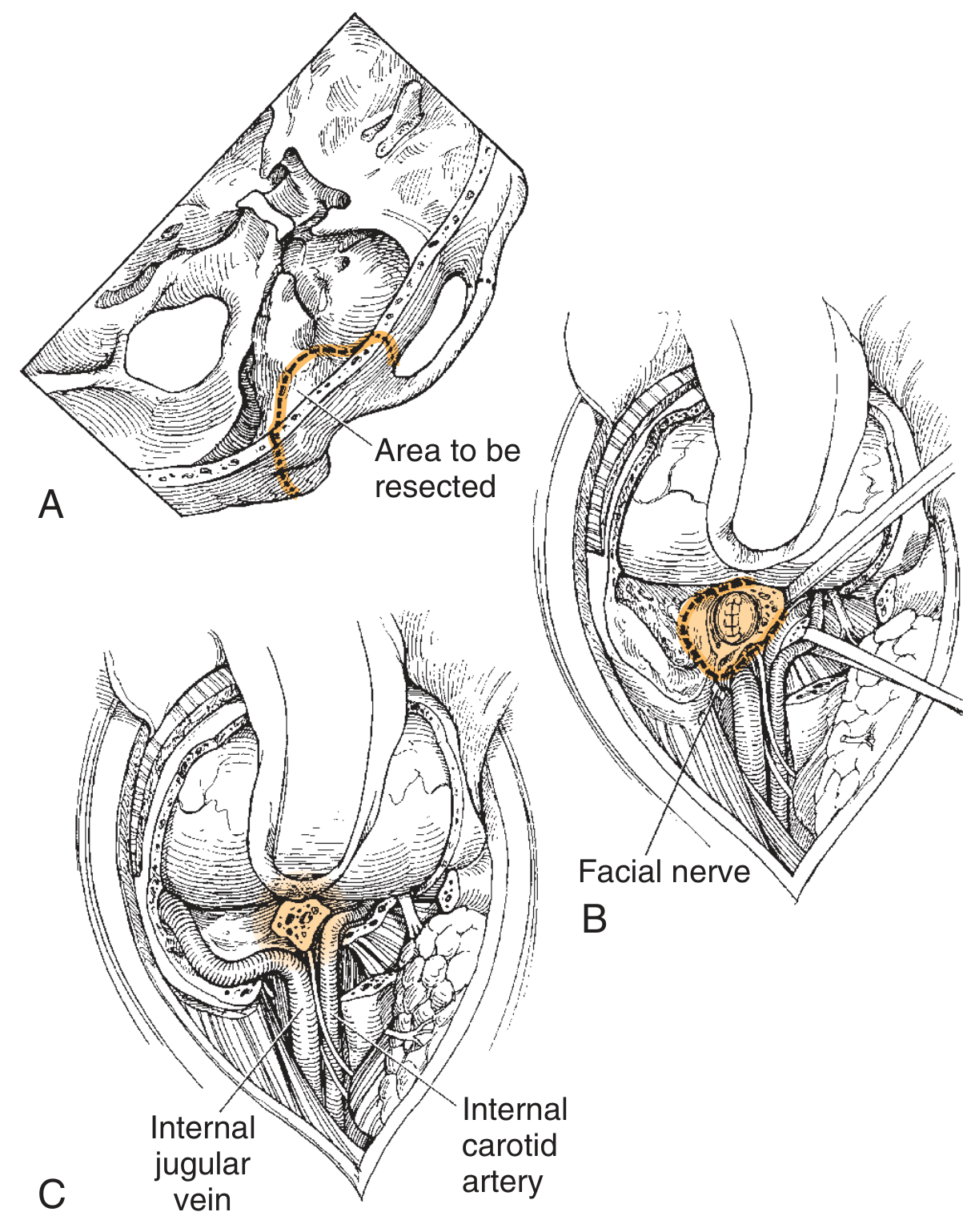
Facial nerve skeletonization: CN VII skeletonized from the geniculate ganglion to the stylomastoid foramen in preparation for transposition
-
Mastoid tip removal: SCM released from mastoid tip; tip itself skeletonized and removed; bone removed over the posterior belly of digastric
-
Cervical dissection: CN IX, X, XI, XII, ICA, and internal jugular vein (IJV) exposed in the neck
-
Facial nerve anterior transposition: The facial nerve is mobilized from stylomastoid foramen, translocated anteriorly (cutting GSPN if needed for type B/extensive type A)
-
ICA dissection: Internal carotid artery dissected from the neck up to the skull base, to its position below the cochlea; bone removed over carotid canal to expose the vertical (petrous) ICA
-
Jugular bulb and sigmoid sinus exposure: Sigmoid sinus ligated below the mastoid emissary vein; jugular bulb exposed
-
Tumor removal: With vascular control achieved, the paraganglioma or tumor is resected (see surgical illustration below)
Reconstruction: Recontouring with allogenic tissue matrix, temporalis muscle-fascia flap, or microvascular free flap; layered closure; pressure dressing for 24 hours.
Result: Full exposure of the jugular bulb, vertical petrous ICA, and posterior ITF.
Fig. - Fisch Type A infratemporal fossa approach: (B) Facial nerve identification, (C) Internal jugular vein and internal carotid artery exposed - Cummings Otolaryngology, Fig. 178.15/178.17
FISCH TYPE B APPROACH
Indications:
- Chordoma (clivus)
- Chondroma / low-grade chondrosarcoma
- Squamous cell carcinoma
- Dermoid/epidermoid cysts of petrous apex
- Meningioma, craniopharyngioma, plasmacytoma, arachnoid cyst
Key differences from Type A:
- Steps identical to Type A up to facial nerve transposition, which is usually not required
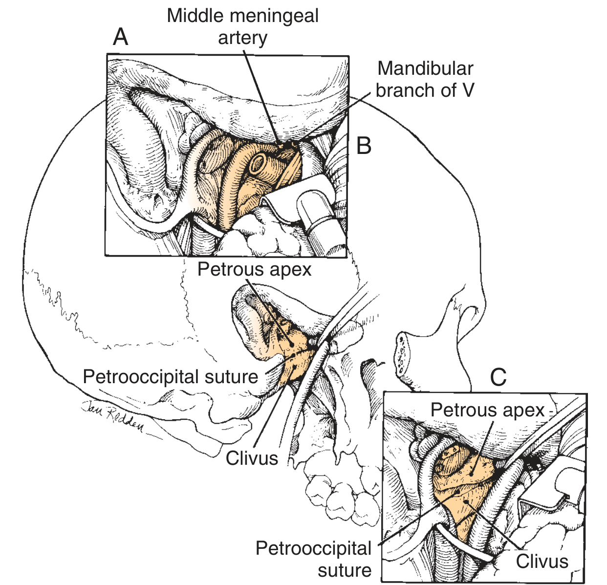
- Temporalis muscle is reflected (still attached to coronoid process and zygoma) allowing a retractor to expose the superior ITF
- The middle meningeal artery and mandibular branch of V (V3) are transected - this opens up the superior 4 cm of the ITF
- The ICA can be uncovered from its vertical segment to its anterior limit at the foramen lacerum, after separation from soft tissues around the eustachian tube
- The petrous apex is accessed with careful anterolateral retraction of the ICA
- Can be combined with temporal craniotomy for additional middle fossa and clival exposure
Fig. 178.27 - Fisch Type B: Elevation of the ICA permits access to petrous apex and clivus. Note middle meningeal artery and mandibular branch of V. - Cummings Otolaryngology
FISCH TYPE C APPROACH
Indications:
- Salvage surgery for squamous cell carcinoma
- Adenoid cystic carcinoma involving the eustachian tube region
- Advanced juvenile nasopharyngeal angiofibroma (JNA)
- Rostral clivus tumors, cavernous sinus lesions, sphenoid sinus tumors
- Parasellar tumors
Key additions beyond Type B:
- The type C approach is an anterior extension of the type B approach
- Access is gained to: rostral clivus, cavernous sinus, sphenoid sinus, peritubal space, pterygopalatine fossa, nasopharynx, and the full anterosuperior ITF
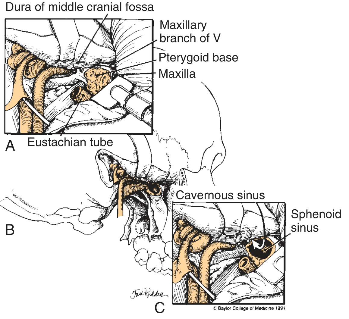
- Pterygoid base removal: The base of the pterygoid process is drilled/removed; this uncovers:
- V2 (maxillary nerve) at the foramen rotundum
- The inferior orbital fissure
- Entry into the sphenoid sinus and cavernous sinus
- Exposure extends anterior to the foramen lacerum up to the posterior wall of the maxillary sinus and nasopharynx
Fig. 178.29 - Fisch Type C: (A) Pterygoid base dissection with V2 exposed, Eustachian tube visible. (B/C) Sphenoid sinus entered, cavernous sinus accessible. - Cummings Otolaryngology
6. COMPARISON SUMMARY TABLE
| Feature | Type A | Type B | Type C |
|---|
| Access | Posterior ITF, jugular bulb, vertical ICA | Superior ITF, petrous apex, clivus | Anterior ITF, cavernous sinus, sphenoid sinus, nasopharynx |
| FN transposition | Yes (mandatory) | Usually not needed | Not needed |
| V3 transection | No | Yes | Yes |
| Middle meningeal artery | Preserved/sacrificed per need | Transected | Transected |
| Pterygoid base removal | No | No | Yes |
| Eustachian tube | Obliterated | Mobilized (ICA access) | Removed for ICA access |
| Key tumors | Paraganglioma, temporal bone SCC | Chordoma, chondrosarcoma, petrous apex | Advanced JNA, nasopharyngeal carcinoma, adenoid cystic |
7. COMPLICATIONS OF FISCH APPROACHES
- Facial nerve palsy - the most significant risk, especially in Type A where anterior transposition is performed. Significant permanent facial weakness can result
- Hearing loss - sacrifice of ossicular chain and middle ear; in type B/transcochlear the otic capsule may be removed (sensorineural loss)
- ICA injury - catastrophic; requires preoperative balloon occlusion testing
- Lower cranial nerve deficits (IX, X, XI, XII) - from cervical dissection and jugular foramen dissection
- CSF leak - from dural entry
- Sigmoid sinus/jugular vein thrombosis
- Eustachian tube dysfunction - managed by obliteration during surgery
8. ENDOSCOPIC ALTERNATIVES
Cummings notes that endoscopic endonasal approaches should be considered as an alternative, especially for paramedian lesions involving the clivus, parasellar area, pterygomaxillary fossa, nasopharynx, and occasionally the ITF - particularly when the primary lesion is medial to the ICA and accessible without lateral exposure. The Fisch lateral approaches and endoscopic endonasal approaches are often complementary.
Sources:
- Cummings Otolaryngology Head and Neck Surgery, 7th Ed., Chapters 175 and 178 (Anatomy of the Infratemporal Fossa; Infratemporal Fossa Approaches), pp. 3349-3351, 3416-3422
- K.J. Lee's Essential Otolaryngology, 11th Ed., Chapter 37 (Infratemporal Fossa; Surgery of the Infratemporal Fossa), pp. 703, 895-896