
Mullerian classification new updated
Mullerian duct anomalies classification updated ESHRE ESGE 2013 2023
Mullerian anomalies classification ESHRE ESGE uterine
https://pmc.ncbi.nlm.nih.gov/articles/PMC11832971
Mullerian duct anomalies classification diagram ESHRE ESGE uterine
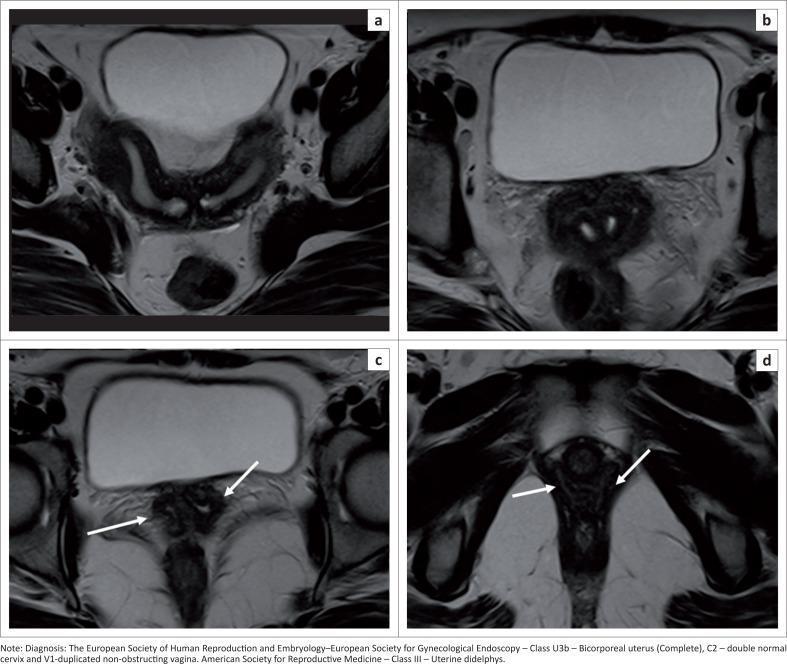
This set of axial T2-weighted MRI scans displays a case of Uterine Didelphys (ESHRE/ESGE Class U3b, C2, V1), a Müllerian duct anomaly. Images (a) and (b) show the uterine fundus and lower uterine segment, respectively. These views demonstrate a deep fundal external indentation that exceeds 50% of the uterine wall thickness, resulting in the complete division of the uterine corpus into two separate horns. Images (c) and (d) focus on the lower reproductive tract. Image (c) shows two distinct cervical openings, and image (d) highlights the presence of two separate vaginal cavities, indicated by white arrows. The diagnostic significance of these findings lies in the complete duplication of the uterus, cervix, and vagina, characteristic of a fusion defect during embryological development. These features are essential for accurate classification and management of female reproductive tract anomalies.
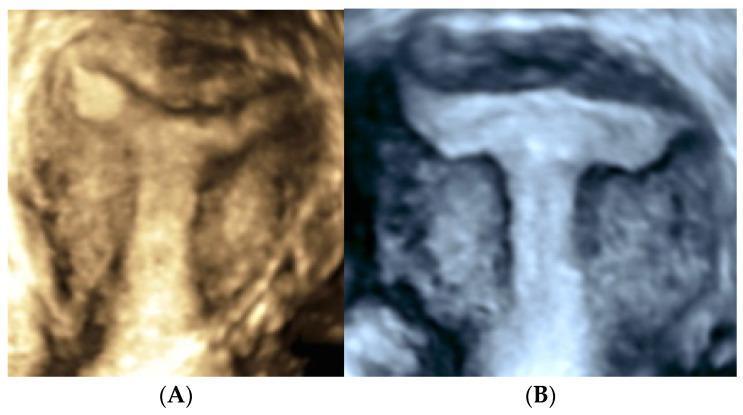
This diagnostic comparison chart features two side-by-side transvaginal three-dimensional (3D) ultrasound images of the coronal plane of the uterus, illustrating different Müllerian duct anomalies. Image (A) displays a Y-shaped uterine cavity, characterized by a smooth, wide bifurcation at the superior fundal region where the arms of the cavity extend diagonally. Image (B) demonstrates a T-shaped uterus (Class U1a according to ESHRE/ESGE classification), showing a narrow, tubular uterine body with a characteristic horizontal fundal configuration and lateral protrusions. The echogenic endometrial lining in image (B) is notably distinct against the darker myometrium, highlighting the constriction of the mid-segment. These images serve as educational references for gynecological radiology, focusing on the diagnosis of dysmorphic uteri and uterine malformations that may impact reproductive outcomes. The 3D reconstruction is essential for assessing the external fundal contour and internal cavity shape to differentiate between T-shaped, Y-shaped, and septate uterine variations.
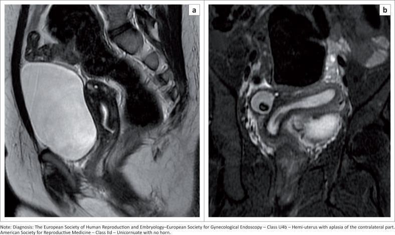
This diagnostic image consists of two MRI T2-weighted sequences (a and b) illustrating a Müllerian duct anomaly. Image (a) is a sagittal T2-weighted sequence showing the uterus where the hyperintense endometrial cavity does not extend to the fundal region, suggesting a structural malformation. Image (b) is a coronal oblique T2-weighted sequence demonstrating a unicornuate uterus (Class U4b hemi-uterus under ESHRE-ESGE classification). The functional uterine component shows a characteristic curved, 'banana-shaped' endometrial cavity displaced toward the left side of the pelvis. In the right adnexal region, a well-defined lesion consistent with a benign teratoma is visible. The imaging highlights the importance of true coronal and sagittal planes in evaluating uterine fundal contour and endometrial cavity morphology for the classification of congenital uterine anomalies. This content is relevant for radiology and obstetrics/gynecology trainees focusing on pelvic anatomy and congenital reproductive tract variations.
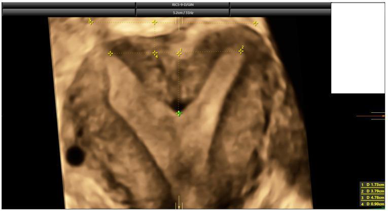
This diagnostic image is a 3D coronal ultrasound reconstruction of a human uterus, specifically highlighting a congenital uterine anomaly. The visual demonstrates a septate uterus, characterized by a prominent internal fundal indentation that divides the superior portion of the uterine cavity into two sections. Visible electronic calipers and numbered annotations (1–4) mark critical anatomical measurements used for classification according to ESHRE-ESGE and ASRM criteria. Measurement 1 (1.73 cm) indicates the depth of the fundal indentation, while Measurement 4 (0.90 cm) assesses the uterine wall thickness, resulting in a high indentation-to-wall-thickness (I:WT) ratio of 192%. The external fundal contour remains relatively flat or convex, a key feature distinguishing the septate uterus from a bicornuate uterus. This educational visual is used in reproductive medicine and radiology to teach the standardized assessment of Müllerian duct anomalies, focusing on the differentiation between septate, subseptate, and arcuate uterine morphologies through quantitative morphometric analysis.
Mullerian Duct Anomalies - Classification Systems (Updated)
Why Multiple Systems Exist
| System | Year | Status |
|---|---|---|
| AFS (American Fertility Society) | 1988 | Historical reference; widely taught |
| ESHRE/ESGE (CONUTA) | 2013 + 2016 update | Standard in Europe & radiology |
| ASRM 2021 | 2021 | Latest update; most widely used clinically |
| CUME (Ludwin et al.) | 2018 | Specialist use; expert-based |
1. AFS / Original Classification (1988) - Still Widely Taught
| Class | Anomaly | Subtype Notes |
|---|---|---|
| I | Segmental Mullerian Agenesis / Hypoplasia | A. Vaginal, B. Cervical, C. Fundal, D. Tubal, E. Combined |
| II | Unicornuate Uterus | A. With rudimentary horn (communicating / non-communicating / no cavity), B. No rudimentary horn |
| III | Uterus Didelphys | - |
| IV | Bicornuate Uterus | A. Complete (to internal os), B. Partial, C. Incomplete |
| V | Septate Uterus | A. Complete septum, B. Incomplete septum |
| VI | Arcuate Uterus | Internal luminal changes |
| VII | DES-related anomalies | T-shaped uterus, hypoplastic cavity |
2. ESHRE/ESGE Classification - CONUTA (2013, Updated 2016 - "Thessaloniki consensus")
Uterine Classes (U):
| Class | Name | Key Features |
|---|---|---|
| U0 | Normal uterus | Added to allow coding of C/V anomalies when uterus is normal |
| U1 | Dysmorphic uterus | No normal outer contour change; subclasses: U1a (T-shaped - thick lateral walls, 2/3 corpus: 1/3 cervix ratio), U1b (Infantilis - inverse ratio 1/3 corpus: 2/3 cervix), U1c (Others) |
| U2 | Septate uterus | Internal indentation >50% of wall thickness; U2a partial (above internal os), U2b complete (reaching internal os) |
| U3 | Bicorporeal uterus | External fundal indentation >50% of wall thickness; U3a partial, U3b complete (uterus didelphys), U3c bicorporeal septate |
| U4 | Hemi-uterus (Unicornuate) | One developed side; U4a with rudimentary (functional) cavity, U4b without rudimentary cavity |
| U5 | Aplastic uterus | U5a with rudimentary cavity, U5b without any uterine cavity |
| U6 | Unclassified | Does not fit above |
Cervical Classes (C):
| Class | Description |
|---|---|
| C0 | Normal cervix |
| C1 | Septate cervix |
| C2 | Double normal cervix (as in didelphys) |
| C3 | Unilateral cervical aplasia |
| C4 | Cervical aplasia (bilateral) |
Vaginal Classes (V):
| Class | Description |
|---|---|
| V0 | Normal vagina |
| V1 | Longitudinal non-obstructing vaginal septum |
| V2 | Longitudinal obstructing vaginal septum |
| V3 | Transverse vaginal septum / imperforate hymen |
| V4 | Vaginal aplasia |
3. ASRM 2021 Classification - Most Recent Update
- Remove class-based numbering
- Use 9 descriptive categories for clarity
- Include separate categories for cervical and vaginal anomalies
- Provide measurable morphometric criteria (updated from 2016 criteria)
The 9 Descriptive Categories:
- Mullerian agenesis (MRKH syndrome - bilateral aplasia)
- Cervical agenesis
- Unicornuate uterus
- Uterus didelphys
- Bicornuate uterus
- Septate uterus
- Longitudinal vaginal septum (LVS)
- Transverse vaginal septum
- Complex anomalies (combinations not fitting above)
Key Morphometric Criteria (ASRM 2021 vs ESHRE/ESGE):
| Feature | ESHRE/ESGE 2013 | ASRM 2021 |
|---|---|---|
| Septate vs Arcuate | Indentation >50% of uterine wall thickness = septate | Indentation depth >1.5 cm from interostial line = septate |
| Arcuate uterus | Classified as U0 (normal variant) or U2 | Recognized as a distinct normal variant |
| Bicornuate vs Septate | External indentation >50% = bicorporeal | External fundal cleft >1 cm differentiates |
| Measurement tool | Relative ratios (3D USS preferred) | Absolute measurements (MRI preferred) |
Key Clinical Points
- ESHRE/ESGE tends to over-diagnose septate uterus compared to ASRM 2021, because its threshold (>50% wall thickness) is less stringent than ASRM's absolute 1.5 cm criterion.
- Arcuate uterus: Now considered a normal variant in ASRM 2021 (no longer a separate class needing treatment).
- MRI is the gold standard imaging for classification (preferred over 3D USS for surgical planning).
- Renal anomalies co-exist in 30-50% of cases - always image kidneys.
- Vertebral anomalies occur in up to 29% of MDA patients.
- The septate uterus is the most common MDA (35%), and is the most surgically correctable (hysteroscopic metroplasty).
MRI Images for Reference
Mullerian classification new updated 2021
ASRM 2021 Mullerian anomalies classification 9 categories morphometric criteria full details
uterine anomalies classification ASRM 2021
ASRM 2021 Mullerian duct anomalies classification categories diagram
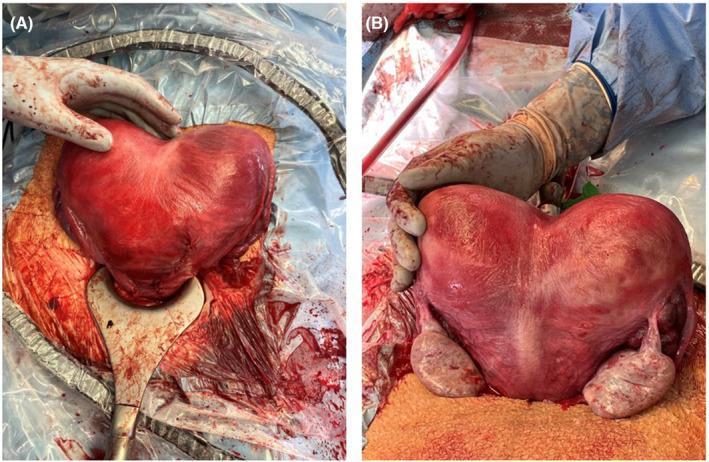
Clinical photograph of a human uterus post-cesarean delivery and hysterotomy repair, demonstrating a bicornuate bicollis anomaly (ASRM classification). The composite image shows (A) an anterior view and (B) a posterior view of the heart-shaped uterus. A deep midline serosal indentation exceeding 1 cm is clearly visible at the fundus, indicative of failed fusion of the Müllerian ducts. The uterine serosa appears erythematous and congested, a physiological finding typical of immediate postpartum and surgical manipulation. In the posterior view (B), both ovaries and fallopian tubes are visible flanking the two distinct uterine horns. The image demonstrates the surgical appearance of a dicavitary uterine anomaly following a twin pregnancy. It serves as an educational reference for Mullerian duct anomalies and their intraoperative presentation during obstetric surgery.
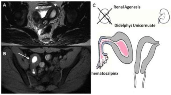
This composite educational material demonstrates Mullerian duct anomalies (MDA). Figure A is an axial T2-weighted MRI scan showing an asymmetrical uterine morphology with heterogeneous signal intensity, indicating a deviated endometrial cavity. Figure B is an axial fat-saturated T1-weighted MRI scan revealing a localized area of high signal intensity (hyperintense) within a rudimentary uterine horn, consistent with methemoglobin from blood degradation products (hematometra). Figure C is a clinical illustration summarizing the findings: a didelphys/unicornuate uterus with an obstructed, non-communicating right horn leading to hematosalpinx. The diagram also correlates this genital anomaly with ipsilateral renal agenesis, a common associated finding in Herlyn-Werner-Wunderlich syndrome or related MDA classifications. This content is designed for medical education regarding diagnostic imaging of congenital female reproductive tract malformations and the importance of screening for associated renal variations.
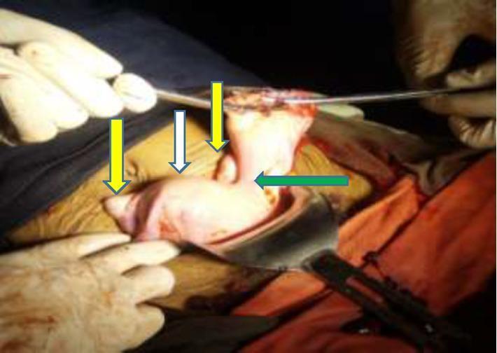
This clinical photograph shows an intraoperative view of a female reproductive system following the resection of a ruptured ectopic pregnancy. The image illustrates a congenital Mullerian duct anomaly, specifically a unicornuate uterus with a non-communicating rudimentary horn. A white arrow identifies the main unicornuate uterine body, while a green arrow points to the left-sided rudimentary horn. The connection between the two structures is visible as a bridge of dense fibro-muscular tissue. Yellow arrows indicate the bilateral ovaries, which appear in their relative anatomical positions. The tissues are fleshy and vascularized, consistent with a surgical environment. This case demonstrates the anatomical relationships associated with Class II Mullerian anomalies according to the American Society for Reproductive Medicine (ASRM) classification, highlighting the risk of obstetric complications such as rudimentary horn pregnancy and rupture.
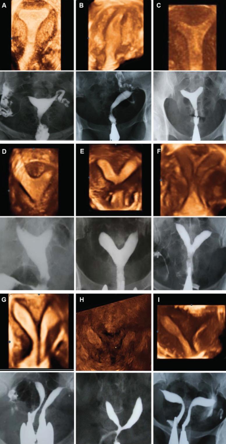
This composite educational graphic provides a side-by-side comparison of 3D ultrasound (coronal view) and Hysterosalpingography (HSG) imaging for the classification of Mullerian Duct Anomalies (MDAs) according to American Fertility Society (AFS) criteria. The panel features nine clinical cases (A-I), each illustrating a specific uterine morphology. (A) Normal uterus with a convex fundal contour and triangular cavity. (B) Unicornuate uterus demonstrating a single uterine horn and fallopian tube. (C) Arcuate uterus characterized by a mild fundal indentation. (D-G) Subtypes of septate uterus, ranging from partial to complete septation, where the 3D ultrasound clarifies the external fundal contour (straight or convex) versus the internal division. (H) Bicornuate uterus showing a prominent external fundal cleft and two divergent horns. (I) Uterus didelphys displaying two completely separate uterine bodies. The dual-modality approach highlights how 3D ultrasound assesses both the external serosal contour and internal cavity, while HSG provides high-contrast visualization of the internal lumen and tubal patency.
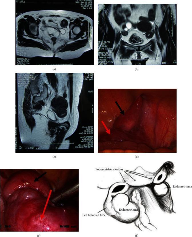
This composite figure illustrates the diagnostic and surgical findings of a patient with Müllerian duct anomalies, specifically Mayer-Rokitansky-Küster-Hauser (MRKH) syndrome type II with functional rudimentary uteri. Panels (a)-(c) present multi-planar pelvic MRI scans (axial, coronal, and sagittal) demonstrating bilateral rudimentary uteri with visible endometrial signal and associated low-signal adnexal cystic masses. Panels (d) and (e) are intraoperative laparoscopic photographs showing the reddish, smooth-surfaced rudimentary uterine horns (black arrows) and large ovarian chocolate cysts (red arrows), consistent with pelvic endometriosis. Panel (f) provides a labeled anatomical diagram summarizing the surgical findings: bilateral residual uteri of disparate sizes (left 6-7 cm, right 2-3 cm), bilateral ovarian endometriomas, and the left fallopian tube. This collection serves as an educational tool for identifying ectopic endometrial tissue and functional uterine remnants in the context of congenital vaginal agenesis and secondary periodic pelvic pain.
https://integration.reproductivefacts.org/globalassets/_asrm…
ASRM Mullerian Anomaly Classification 2021 (MAC2021)
Why MAC2021 Was Needed
- Focusing only on the uterus (excluded cervix and vagina)
- No clear measurable diagnostic criteria
- Inability to classify complex/combined anomalies
- Class numbers were confusing and not intuitive
The 9 Descriptive Categories (No More Class Numbers)
| # | Category | Key Features / Subtypes |
|---|---|---|
| 1 | Mullerian Agenesis | Absent uterus ± vaginal aplasia (MRKH syndrome); may have atrophic uterine remnant with or without functional endometrium |
| 2 | Cervical Agenesis | Absent cervix; uterus may be present; also includes distal cervical agenesis |
| 3 | Unicornuate Uterus | One developed horn; subtypes: unicornuate alone / + distal atrophic remnant / + remnant with functional endometrium / + communicating horn at cervix level |
| 4 | Uterus Didelphys | Two completely separate uteri with two cervices; ± longitudinal vaginal septum; ± obstructed hemivagina (OHVIRA syndrome) |
| 5 | Bicornuate Uterus | Two horns with serosal (external) indentation >1 cm; subtypes: bicornuate unicollis / bicornuate bicollis / bicornuate with communicating tract / combined bicornuate-septate |
| 6 | Septate Uterus | Internal endometrial septum >1 cm from bicornual line, septum angle <90°; subtypes: partial / complete / complete with duplicated cervices / Robert's uterus |
| 7 | Longitudinal Vaginal Septum (LVS) | Variable length; may be obstructing (hemivagina) or non-obstructing; often co-exists with didelphys or septate uterus |
| 8 | Transverse Vaginal Septum | Can be mid-vaginal or distal; includes distal vaginal agenesis |
| 9 | Complex Anomalies | Combinations not fitting single category; cross-referenced across categories |
Critical Morphometric Criteria (The Most Important Change in 2021)
Septate vs. Arcuate/Normal Uterus:
| Parameter | Septate Uterus | Arcuate / Normal |
|---|---|---|
| Septum length (from bicornual line) | >1 cm | ≤1 cm |
| Angle at apex of septum | <90° | >90° (blunt/flat) |
| External fundal contour | Flat or convex (no deep indentation) | Flat or convex |
Key point: Arcuate uterus is now grouped with normal - it is NOT a separate pathological category in MAC2021. This avoids unnecessary intervention.
Bicornuate vs. Septate Uterus:
| Feature | Bicornuate | Septate |
|---|---|---|
| External (serosal) indentation | >1 cm (deep fundal cleft) | None or <1 cm |
| Internal indentation | Present but due to 2 horns | Septum >1 cm, angle <90° |
| Intercornual angle (HSG) | >105° | <105° |
| Morphology | Two well-formed cornua with convex fundal contour in each horn | Single uterine body with internal septum |
Subcategories in Detail
1. Mullerian Agenesis
- Mullerian agenesis (classic MRKH - Type I)
- Mullerian agenesis + R/L atrophic uterine remnant
- Mullerian agenesis + R/L remnant with functional endometrium (causes cryptomenorrhea/hematometra - requires surgery)
2. Cervical Agenesis
- Complete cervical agenesis
- Distal cervical agenesis
3. Unicornuate Uterus
- R/L unicornuate uterus (alone)
-
- distal atrophic uterine remnant
-
- distal uterine remnant with functional endometrium (obstructed - can cause hematometra, ectopic risk)
-
- associated atrophic remnant
-
- uterine horn communicating at cervix level
4. Uterus Didelphys
- Uterus didelphys ± longitudinal vaginal septum (variable length)
- Uterus didelphys + obstructed R/L hemivagina (OHVIRA / Herlyn-Werner-Wunderlich syndrome)
5. Bicornuate Uterus
- Bicornuate unicollis (one cervix)
- Bicornuate bicollis (two cervices)
- Bicornuate + R/L communicating tract
- Combined bicornuate-septate uterus
6. Septate Uterus
- Partial septate uterus
- Complete septate uterus (septum to internal os)
- Complete septate + duplicated cervices + obstructed R/L hemivagina (Robert's uterus)
- Complete septate + duplicated cervices + longitudinal vaginal septum
- Complete septate with septate cervix + longitudinal vaginal septum
7. Longitudinal Vaginal Septum
- LVS of variable length (non-obstructing)
- LVS + uterus didelphys
- Obstructed R/L hemivagina + uterus didelphys (OHVIRA)
8. Transverse Vaginal Septum
- Mid-vaginal septum
- Distal vaginal agenesis (includes imperforate hymen spectrum)
9. Complex Anomalies
- Any anomaly that does not clearly fit one category
- Combined anomalies cross-referenced between categories
- Allows flexibility for the continuum of Mullerian development
What Changed From AFS 1988 vs. MAC2021
| Feature | AFS 1988 | ASRM MAC2021 |
|---|---|---|
| Format | 7 numbered classes | 9 descriptive categories (no numbers) |
| Cervix | Not classified | Included (cervical agenesis = separate category) |
| Vagina | Not classified | Included (LVS + transverse septum = categories) |
| Arcuate uterus | Class VI (pathological) | Grouped with normal - no treatment needed |
| Septate criteria | No clear measurement | Septum >1 cm, angle <90° |
| Bicornuate criteria | No clear measurement | Serosal indentation >1 cm |
| DES-related (T-shaped) | Class VII | Falls under complex anomalies |
| Complex/combined | Cannot classify | Dedicated category with cross-referencing |
| Imaging modality | Not specified | Any modality acceptable (2D/3D USS, MRI, SHG, hysteroscopy) |
Clinical Relevance of Key Changes
- Arcuate = Normal: Stops overdiagnosis and unnecessary hysteroscopic surgery. Most arcuate uteri carry pregnancy without complication.
- Septate stricter threshold (>1 cm): Reduces overdiagnosis compared to ESHRE/ESGE (which used relative ratios, leading to ~3x more septate diagnoses).
- Robert's uterus recognized: A complete septate uterus with obstructed hemicavity - previously under-recognized, now explicitly included.
- OHVIRA syndrome recognized: Obstructed hemivagina + ipsilateral renal agenesis + uterus didelphys - now explicitly in the classification.
- Functional endometrium in remnants: Emphasizes that even small remnant horns with functional endometrium require surgical attention (hematometra, endometriosis, ectopic pregnancy risk).
Imaging Reference