
Rule of 12 hr in thanatology
Reading File
The "Rule of 12" in thanatology refers to the approximate timing of rigor mortis - it develops over 12 hours, is fully established by 12 hours, and resolves over the next 12 hours (total cycle ~36 hours). Let me search the textbooks for the precise details.
Searching sources...
Finding Sources
Finding Sources
Found the key forensic medicine textbook references. Let me read both of them for full content.
Reading File
Reading File
Now let me get more detail on the rule of 12 in thanatology - searching for rigor mortis content from the forensic medicine book specifically.
Finding Sources
Searching the Web
rule of 12 thanatology forensic medicine rigor mortis march of rigor
I now have comprehensive information to give a thorough answer. Let me compile the full response.
Rule of 12 in Thanatology (March of Rigor)
What Is the "Rule of 12"?
The Rule of 12 (also called the "March of Rigor") is a classical forensic guideline describing the time course of rigor mortis - the postmortem stiffening of muscles caused by depletion of ATP, which prevents the actin-myosin cross-bridges from being released.
The Classic Rule (Temperate Climate)
The rule is built on three successive 12-hour windows:
| Phase | Timing | Description |
|---|---|---|
| Onset / Development | 0 - 12 hrs after death | Rigor begins in small facial muscles (~3 hrs), progresses to arms (~6 hrs), and is fully established throughout the body, including the legs, by ~12 hrs |
| Full Establishment | 12 - 24 hrs after death | Rigor is maximally developed and maintained |
| Resolution / Passing off | 24 - 36 hrs after death | Rigor disappears in the same sequence it appeared (face first, then down the body), due to autolysis and putrefaction |
Total cycle: ~36 hours (12 + 12 + 12 hrs)
Mason JK described it succinctly: rigor presents in the face at 3 hours, arms at 6 hours, complete in legs at 12 hours - remains well marked for 12 hours - then disappears over the next 12 hours in the order in which it appeared.
Nysten's Law (Direction of March)
Rigor mortis follows Nysten's Law - it appears and disappears in a craniocaudal direction:
- Appears: Face/jaw → neck → upper limbs → trunk → lower limbs
- Disappears: In the same downward sequence
This is the basis for using rigor to estimate the time since death (postmortem interval, PMI).
Mechanism (Why Rigor Occurs)
- After death, aerobic respiration stops, ATP production ceases
- Without ATP, actin-myosin cross-bridges cannot be broken
- Muscles become locked in a contracted state → stiffness
- As autolysis progresses (protein breakdown), the cross-bridges eventually dissolve → rigor resolves
Why the Rule of 12 is NOT Applicable in Tropical Countries
This is a key exam point. The textbook The Essentials of Forensic Medicine and Toxicology (36th ed., 2026) explicitly states: "The so-called rule of 12 (march of rigor) is not applicable in tropical countries."
In India and other tropical regions:
- Rigor begins 1-2 hours after death (much earlier)
- Takes only 1-2 more hours to fully develop
- Well-established throughout the body in 6 hours in summer
- Begins to disappear in ~12 hours in summer
- Lasts only 18-36 hours in summer vs. 24-48 hours in winter
The high temperature accelerates both the onset and resolution dramatically.
Factors That Modify the Rule
Accelerate onset and shorten duration:
- High environmental temperature
- Physical exertion / struggle before death
- Convulsions, electrocution, hyperpyrexia
- Diseases causing muscle wasting or exhaustion (cholera, typhoid, TB, cancer)
- Organophosphate poisoning
- HCN and strychnine poisoning (also prolongs duration)
- Children and elderly (rigor is feeble and rapid)
Delay onset and prolong duration:
- Cold environment (rigor lasts 2-3 days in temperate regions)
- Deaths from asphyxia, severe hemorrhage, apoplexy, nervous diseases
- CO poisoning (delays disappearance)
- CO poisoning - well-developed musculature
Special cases:
- Cadaveric spasm (instantaneous rigor): occurs at the moment of death in cases of extreme emotion, nerve stimulation, or certain toxins - important for establishing position of body at time of death
- Rigor may disappear very rapidly with gas gangrene (bacterial putrefaction)
- Rigor mortis timing table (Mallach data from Essentials of Forensic Medicine):
| Stage | Lower limit (hrs) | Upper limit (hrs) |
|---|---|---|
| Beginning | 0.5 | 7 |
| Maximum | 2.0 | 20 |
| Re-establishment after breaking | 2.0 | 8 |
Medico-legal Significance
- Estimation of PMI: The most widely used postmortem sign for rough time-of-death estimation in the field
- Body position at death: If the body is found in an unusual posture and rigor has fixed it, it indicates the original position of death (important if the body was moved)
- Re-establishment of rigor: If rigor is broken (e.g., body moved within the first 6-8 hours), it re-establishes in the same position - this indicates the body was moved while rigor was still developing
- Murder vs. natural death: Absence of rigor in a fresh body or unusually early disappearance may point to systemic infection or tampering
Limitations of the Rule
The NCBI StatPearls article on postmortem changes and case data confirm that the Rule of 12 is a rough guide only - rigor was complete in 98% of cases by 12 hours in one series, but was already complete in 14% at just 3 hours. The rule is "open to considerable variation and the sequence is by no means constant" (Mason JK). Temperature, humidity, and individual factors make it unreliable as a precise PMI tool.
Sources:
- The Essentials of Forensic Medicine and Toxicology, 36th ed. (2026) - Muscular Changes After Death
- Mason JK, as cited in Juniper Publishers forensic review
- IJCMR study on rigor mortis and time since death
Pugilistic attitude Boxer attitude Defence attitude In thanatology
The pugilistic/boxer/defence attitude is a postmortem body posture seen in burn victims, caused by heat-induced muscle contracture. Let me pull the full textbook content on this.
Searching sources...
Finding Sources
Searching the Web
pugilistic attitude boxer attitude defence attitude thanatology forensic burns
Excellent results. Now let me read the detailed content from the key textbooks.
Reading File
Reading File
Reading File
Let me also fetch the Parikh's textbook image of the pugilistic attitude and get a bit more content including the differences from rigor mortis.
Reading File
Loading Image
I now have all the information needed for a thorough, well-sourced answer.
Pugilistic / Boxer's / Defence Attitude in Thanatology
Definition and Synonyms
All three terms refer to the same postmortem body posture seen in bodies exposed to intense heat or fire. The names come from the position the body adopts:
| Term | Source |
|---|---|
| Pugilistic attitude | From Latin pugil (boxer) - most common forensic term |
| Boxer's attitude | Describes the raised-fists appearance |
| Defence / Fencing attitude | Describes the overall posture resembling a defensive stance |
Cause / Mechanism
The posture results from heat-induced coagulation and contraction of muscle proteins - specifically:
- High temperatures cause denaturation and coagulation of muscle proteins (myosin and actin)
- Flexor muscles are larger and bulkier than extensor muscles in both the upper and lower limbs
- Therefore, heat causes disproportionate contraction of flexors over extensors
- The net effect is flexion at all major joints
This is distinctly different from rigor mortis, which involves ATP depletion and actin-myosin cross-bridge locking.
Posture Described
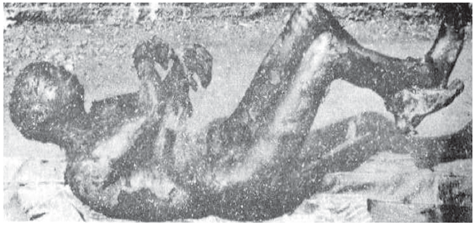
Typical pugilistic attitude - the body is slightly bent, limbs are flexed, and the skin is tense with frequent splits (Parikh's Textbook of Medical Jurisprudence)
The body adopts:
- Trunk: slightly flexed / bent
- Lower limbs: hips and knees flexed
- Upper limbs: elbows flexed, arms held out in front of the body
- Fingers: hooked like claws, resembling a fist
- Skin: tense, leathery, hard, with frequent heat rupture splits
The Most Important Medico-legal Point
The pugilistic attitude is present whether a LIVING or DEAD body is burnt.
This is the single most examined fact about this condition. Because it occurs in both antemortem and postmortem burning, it has NO medico-legal significance in determining whether the person was alive or dead at the time the fire started. It tells you only that the body was exposed to sufficient heat - nothing more.
- Parikh's Textbook: "This attitude is present whether a living or dead body is burnt and has, therefore, no medicolegal significance."
- PC Dikshit: "It is not an antemortem phenomenon"
- DiMaio's Forensic Pathology: "Assumption of the pugilistic attitude is unrelated to whether the individual was alive or dead prior to the fire."
Difference from Rigor Mortis
This distinction is frequently asked in exams:
| Feature | Pugilistic Attitude | Rigor Mortis |
|---|---|---|
| Cause | Heat coagulation of muscle proteins | ATP depletion; actin-myosin cross-bridge locking |
| Onset | During or after exposure to heat | 1-6 hrs after death |
| Duration | Permanent - does not pass off | Temporary - resolves in 24-48 hrs |
| Reversibility | Irreversible | Reversible (passes off with putrefaction) |
| Proteins involved | Different proteins from those in rigor | Actin-myosin complex |
| Occurs in live persons? | Yes (if burnt alive) | No - only after death |
| Medico-legal value | None (no sign of ante/postmortem burning) | Helps estimate PMI |
As Parikh's states: "The condition is due to coagulation of proteins other than those affected by rigor mortis. It differs from rigor mortis in that it is permanent and does not pass off."
Associated Findings in Burned Bodies
Along with the pugilistic attitude, the forensic examiner should note:
-
Heat rupture splits in the skin - due to tension from the contracted, tense skin; these can be mistaken for incised or lacerated wounds. Distinguished by:
- Intact nerves, blood vessels, and connective tissue bridging the split
- No clotted blood in fissures (heat coagulates blood in vessels)
- No bruising or vital reaction at the margins
-
Heat fractures - produced by sudden violent muscle contraction from heat
-
Postmortem epidural thermal hematoma - chocolate-brown, crumbly/honeycomb appearance; must not be confused with antemortem epidural hematoma from trauma
-
Soot in airways / cherry-red lividity (CO) - indicate the person was alive when the fire started
-
Buttocks and armpits typically spared from burns (protected areas)
Summary for Exams
- What is it? A heat-stiffening posture with flexion of all limbs, arms raised in front, fingers curled
- Caused by? Coagulation of muscle proteins by heat; flexors > extensors
- Also called? Boxer's attitude / Defence attitude / Fencing posture
- Medico-legal significance? NONE - occurs in both antemortem and postmortem burns
- Permanent? YES - unlike rigor mortis, it is irreversible
- Diagnostic of burning? Yes - but does NOT help determine if the person was alive or dead when burnt
Sources: Parikh's Textbook of Medical Jurisprudence, p. 374 | PC Dikshit Textbook of Forensic Medicine, p. 244 | DiMaio's Forensic Pathology 3rd ed., p. 377 | The Essentials of Forensic Medicine and Toxicology 36th ed.
Postmortem skin bloster in thanatology explain
Postmortem skin blisters are fluid-filled bullae that form on the skin after death, mainly as a result of putrefaction or heat. They differ importantly from antemortem burn blisters. Let me pull full textbook content on this.
Searching sources...
Finding Sources
Finding Sources
Multiple excellent sources found. Let me read them all in parallel.
Reading File
Reading File
Reading File
Reading File
Now let me read the full comparison table from Parikh's and fetch an image of postmortem blisters.
Reading File
Reading File
Loading Image
I now have comprehensive data from four authoritative forensic textbooks. Here is the complete answer:
Postmortem Skin Blisters in Thanatology
Overview
Postmortem skin blisters (also called bullae or blebs) are fluid- or gas-filled elevations of the skin that develop after death. They are a critical medico-legal topic because they can be easily confused with antemortem (burn) blisters, leading to false conclusions about whether a person was alive or dead when exposed to heat or fire.
There are two distinct types of postmortem blisters:
- Heat-induced postmortem blisters (from burns applied to a dead body)
- Putrefactive blisters (from decomposition / putrefaction)
Type 1: Heat-Induced Postmortem Blisters
Mechanism
When heat is applied to the skin of a dead body, the epidermis separates from the dermis (corium) - this is a purely physical/chemical effect of heat, with no vital (inflammatory) reaction involved.
Characteristics
| Feature | Description |
|---|---|
| Appearance | Pale yellow, raised dome |
| Contents | Contains air, or if fluid is present, it is thin and clear |
| Fluid chemistry | Practically non-albuminous (low/no protein) |
| Chlorides | Absent |
| Blood corpuscles | Absent |
| Base | Dry, hard and yellow (not red/injected) |
| Line of hyperaemia | Absent around the blister |
| Vital reaction | None - no cellular exudation, no enzyme increase |
| Size | Limited in size |
As Parikh's Textbook states: "Heat applied to the skin of a dead body may loosen the epidermis from dermis and produce a postmortem blister... A postmortem blister is limited in size, contains air, or if it contains fluid, it is practically non-albuminous and without chlorides and blood corpuscles, there is no line of hyperaemia round the blister, and its base is not injected."
The "Red Rim" Trap
An important exam point from Essentials of Forensic Medicine (36th ed., 2026): sometimes a red rim can be seen around postmortem blisters. This happens because contraction of dermal capillaries forces liquid blood to the periphery of the burn or blister. This must NOT be misinterpreted as an antemortem vital reaction (inflammation).
Type 2: Putrefactive Blisters
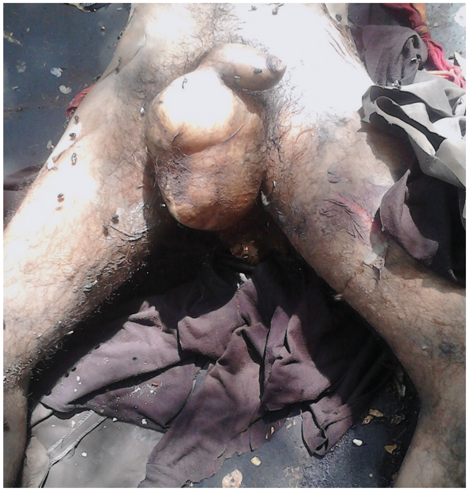
Mechanism
During putrefaction, bacteria produce gases (H₂S, NH₃, methane, CO₂) that accumulate under the skin, lifting the entire epidermis off the dermis.
Timing
- Appear approximately 36-48 hours after death in average conditions (sooner in hot/humid environments)
Characteristics
- The entire epidermis is elevated from the corium (unlike antemortem blisters where only part of the epidermis may be lifted)
- Contents: mainly putrefactive gas + a small amount of reddish-coloured fluid
- When punctured: gas escapes (distinguishes from antemortem blisters)
- No vital reaction whatsoever
- Fluid contains no albumin, no chlorides
Important Pitfall
When a putrefactive blister ruptures/breaks, it leaves behind patches of raw skin that can closely simulate fresh scalds - this is a well-recognised forensic trap that can mislead investigators into thinking the person was scalded.
Comparison: Antemortem vs. Postmortem Blisters
The most important table in this topic - from multiple textbooks:
| Feature | Antemortem Blister | Postmortem Blister |
|---|---|---|
| Line of redness | Present (inflammatory hyperaemia) | Absent |
| Contents | Serous fluid with protein (albumin) and chlorides | Air or thin, clear, non-albuminous fluid |
| Base | Red and inflamed | Dry, hard and yellow |
| Vital reaction | Marked cellular exudation; reactive tissue changes | Absent |
| Enzymes | Peripheral zone shows increased SH groups, ATP, esterases, acid/alkaline phosphatases | No enzyme increase |
| Blood corpuscles in fluid | Present | Absent |
| Chlorides in fluid | Present | Absent |
| Infection/healing | Pus, sloughing, granulation tissue | None |
| Soot in airways | Present (if fire) | Absent |
| Carboxyhaemoglobin | Present (if CO exposure while alive) | Absent |
Medico-legal Significance
1. Determining Antemortem vs. Postmortem Burns
The nature of the blister is a key tool to answer: "Was the person alive or dead when burnt?"
- Antemortem blisters with albuminous fluid + vital reaction = person was alive
- Postmortem blisters with only air/non-albuminous fluid = person was dead when burnt
2. Distinguishing Putrefactive Blisters from Scalds
When a putrefactive blister ruptures, the raw skin surface can mimic scalds - this could wrongly implicate someone for assault. Key differentiator: no vital reaction histologically.
3. Histological Confirmation
When doubt exists, Parikh's Textbook recommends: "In case of doubt, it is best to excise the vesicle and its related tissues and examine histologically for evidence of a tissue reaction." The presence or absence of cellular inflammatory reaction is the definitive answer.
4. Burns Shortly Before or After Death
Essentials of Forensic Medicine (36th ed.) makes a critical point: "Burns produced shortly before or after death cannot be distinguished either by naked eye or by microscopic examination." This is the boundary zone where forensic certainty fails.
Summary for Exams
| Point | Fact |
|---|---|
| What causes postmortem heat blister? | Heat loosening epidermis from dermis in dead body |
| Contents? | Air, or non-albuminous fluid without chlorides/blood cells |
| Base character? | Dry, hard, yellow |
| Red rim in postmortem blister? | Can occur - due to capillary contraction, NOT vital reaction |
| Putrefactive blister timing? | ~36-48 hours after death |
| Putrefactive blister - when broken? | Mimics fresh scalds (forensic trap) |
| Definitive test to differentiate? | Histology - look for vital reaction |
| Burns just before or after death? | Cannot be distinguished (antemortem from postmortem) |
Sources: Parikh's Textbook of Medical Jurisprudence, pp. 374, 377 | The Essentials of Forensic Medicine and Toxicology 36th ed., pp. 320-321 | PC Dikshit Textbook of Forensic Medicine, p. 164
Pox virisae explain
Poxviridae are large, double-stranded DNA viruses that replicate in the cytoplasm and cause characteristic skin lesions. Key members include Variola (smallpox), Vaccinia, Monkeypox, and Molluscum contagiosum. Let me pull full textbook content.
Searching sources...
Finding Sources
Finding Sources
Excellent. Multiple authoritative sources found. Let me read the key sections in parallel.
Reading File
Reading File
Reading File
Rich data from multiple textbooks. Let me now get the classification table from Jawetz and the Fitzpatrick content + a key image.
Reading File
Reading File
Reading File
I now have rich content from multiple authoritative textbooks. Let me also get images for the EM of vaccinia and the Jawetz chapter summary section.
Reading File
Loading Image
I have comprehensive data from Jawetz, Goldman-Cecil, and Dermatology textbooks. Here is the complete, well-structured answer:
Poxviridae - Complete Overview
Electron Micrograph
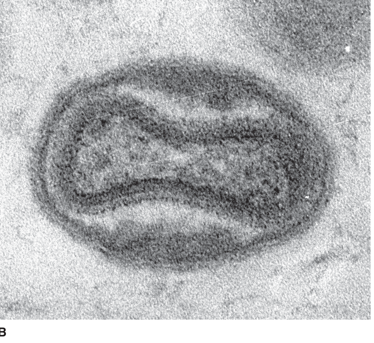
Electron micrograph of Vaccinia virion - the brick/ovoid shape with a central biconcave core and two lateral bodies is characteristic of the entire family. (Jawetz Medical Microbiology 28e)
Basic Properties (Virology)
| Property | Detail |
|---|---|
| Family | Poxviridae |
| Subfamily | Chordopoxvirinae (vertebrate poxviruses) |
| Shape | Brick-shaped or ovoid |
| Size | Largest animal viruses (~300 × 200 nm) - visible under light microscope |
| Genome | Double-stranded DNA (linear, ~130-375 kb) |
| Envelope | Enveloped |
| Replication site | Cytoplasm (unique among DNA viruses - all others replicate in nucleus) |
| GC content | Orthopoxvirus ~33%; Yatapoxvirus ~32%; Molluscum ~60%; Parapoxvirus ~63% |
| Structure | Central biconcave core + 2 lateral bodies + outer lipoprotein membrane |
Key Unique Feature: Poxviruses are the only DNA viruses that replicate entirely in the cytoplasm. They carry their own RNA polymerase within the virion to transcribe early mRNA without entering the nucleus.
Classification - Genera and Human Pathogens
| Genus | Virus | Primary Host | Human Disease |
|---|---|---|---|
| Orthopoxvirus | Variola (major & minor) | Humans only | Smallpox (eradicated 1980) |
| Vaccinia | Humans | Localized lesion; used for vaccination | |
| Monkeypox (Mpox) | Rodents, monkeys | Generalized vesicular disease | |
| Cowpox | Cows | Localized ulcerating lesion | |
| Buffalopox | Water buffalo | Rare human localized lesion | |
| Parapoxvirus | Orf | Sheep/goats | Localized nodule (ecthyma contagiosum) |
| Pseudocowpox (Milker's nodule) | Cows | Nodular lesion in milkers | |
| Bovine papular stomatitis | Cows | Rare | |
| Molluscipoxvirus | Molluscum contagiosum | Humans only | Multiple benign skin nodules |
| Yatapoxvirus | Tanapox | Monkeys | Rare localized lesion |
| Yabapox | Monkeys | Very rare accidental skin tumors |
Only 2 viruses are exclusively human pathogens: Variola (smallpox) and Molluscum contagiosum. All others are zoonoses.
Replication Cycle (Cytoplasmic)
Step 1 - Attachment & Penetration
- Virus fuses with cell membrane
- Viral cores released into cytoplasm
Step 2 - Early Transcription (inside viral core)
- A virus-encoded RNA polymerase (carried inside the virion) transcribes ~50% of the genome into early mRNA
- Early transcription occurs inside the viral core - not affected by inhibitors of protein synthesis
- Early proteins include: DNA polymerase, thymidine kinase, immune-evasion proteins, and the "uncoating protein"
Step 3 - Uncoating (unique 2-step process)
- The uncoating protein (an early protein) acts on cores to release viral DNA
- Requires both RNA and protein synthesis - distinguishes poxvirus uncoating from all other viruses
Step 4 - DNA Replication & Late Transcription
- Occurs 2-6 hours post-infection in cytoplasmic "factories" (viral inclusion bodies)
- Late mRNAs translated into structural proteins (major components)
- High rate of homologous recombination occurs within infected cells
Step 5 - Maturation & Release
- Complex assembly in cytoplasmic factories
- Most particles remain intracellular (unlike most viruses)
- Some particles released by budding from cell membrane
Non-Genetic Reactivation (Important Concept)
Heat-inactivated poxviruses can be reactivated by co-infection with another viable poxvirus - the heat-killed virus provides the DNA template while the live virus provides the enzymes for transcription. Any vertebrate poxvirus can reactivate any other vertebrate poxvirus.
Individual Members - Clinical Details
1. Variola (Smallpox) - Orthopoxvirus
- Eradicated in 1980 - last natural case in Somalia in 1977
- Transmission: inhalation of respiratory droplets (large airborne); also contact with lesions/fomites
- Secondary attack rate: ~60% in unvaccinated contacts; ~4% in vaccinated contacts
- Variola major: CFR ~25-30%; Variola minor (alastrim): CFR ~1%
- Rash: synchronous (all lesions at same stage) - centrifugal distribution (face and extremities > trunk)
- Prodrome: fever, headache, backache 1-4 days before rash
- Now a bioterrorism threat - stocks exist only at CDC (Atlanta) and VECTOR (Russia)
- Variolation (deliberate infection with mild disease) preceded Jenner's vaccination
2. Vaccinia - Orthopoxvirus
- The virus used in smallpox vaccine (distinct species from cowpox - exact origin unknown)
- Broader host range than variola
- Contraindicated in: immunodeficiency, immunosuppression, malignancies, pregnancy, eczema
- Used as a vector for recombinant vaccines
3. Mpox (Monkeypox) - Orthopoxvirus
- Zoonosis from rodents (primary reservoir) and monkeys in Africa
- Two clades: Congo Basin clade (more severe, CFR up to 10%) and West African clade (milder)
- 2022 international outbreak - treated with Tecovirimat and JYNNEOS vaccine (Modified Vaccinia Ankara)
- Congo Basin clade is a select agent in the USA
4. Molluscum Contagiosum - Molluscipoxvirus
- Most common poxvirus infection in humans after smallpox eradication
- Only replicates in human epidermis
- Two subtypes (MCV I and MCV II) - indistinguishable clinically
- Lesions: firm, umbilicated, pearly papules with waxy surface - characteristic "central dell"
- Distribution: skin folds, lateral trunk, thighs, genital region, face
- Occurs in: children (skin-to-skin contact), adults (STI), immunocompromised (widespread/large lesions in HIV)
- Self-limited; giant/deforming lesions suggest AIDS
5. Orf (Ecthyma Contagiosum) - Parapoxvirus
- From sheep/goats - affects farmers, shepherds, veterinarians
- Distinctive EM: crisscross (criss-cross) surface pattern of virion (unique among poxviruses)
- Localized nodular lesion on hands; self-limiting
- GC content ~63% (highest among poxviruses)
6. Milker's Nodule (Pseudocowpox) - Parapoxvirus
- From dairy cattle; affects milkers
- Localized firm nodules on hands/forearms
Immune Evasion - Key Feature
Poxviruses encode numerous immune evasion proteins:
- Virokines (viral cytokine mimics)
- Inhibitors of complement
- Inhibitors of apoptosis
- Inhibitors of interferon signaling
- TNF receptor homologs
Cross-reactive immunity exists within a genus (e.g., vaccinia protects against variola, monkeypox) but NOT across genera - vaccination with vaccinia affords no protection against molluscum contagiosum or orf.
Diagnosis
- Electron microscopy: Demonstrates brick-shaped virions (can identify as poxvirus but not species)
- PCR and DNA sequencing: Most precise method for genus/species/strain identification
- Cell culture: Cytopathic effect with cytoplasmic inclusion bodies (Guarnieri bodies / A-type inclusions)
- Guarnieri bodies: Eosinophilic cytoplasmic inclusion bodies seen in smallpox/vaccinia (composed of viral factories)
- Paschen bodies: Elementary bodies of poxviruses seen on Giemsa/Gutstein stain
Treatment
| Drug | Indication |
|---|---|
| Tecovirimat | FDA-approved for smallpox; expanded to mpox and other orthopoxvirus infections |
| Brincidofovir | FDA-approved for smallpox (all ages including neonates ≥13 kg) |
| Cidofovir | Off-label; used in severe vaccinia complications |
| JYNNEOS (MVA) | Smallpox + mpox vaccine (non-replicating Modified Vaccinia Ankara) |
| ACAM2000 | Live replicating vaccinia vaccine for smallpox |
Key Summary Points for Exams
- Largest animal viruses; only DNA virus replicating in cytoplasm
- Carry their own RNA polymerase - transcribe early genes without host enzymes
- Smallpox eradicated 1980; last natural case Somalia 1977
- Guarnieri bodies = cytoplasmic inclusion bodies (viral factories)
- Only variola and molluscum contagiosum are purely human pathogens
- Molluscum contagiosum = most common poxvirus in humans today
- Orf virus EM shows distinctive crisscross surface pattern
- Cross-immunity within genus only - not cross-genus
- Tecovirimat + JYNNEOS vaccine used in 2022 mpox outbreak
- Bioterrorism risk: human population has no smallpox immunity since vaccination stopped
Sources: Jawetz Melnick & Adelberg's Medical Microbiology 28e, Ch. 34 | Goldman-Cecil Medicine, Ch. 343 | Dermatology 2-Volume Set 5e, Ch. 81
This is a shared conversation. Sign in to Orris to start your own chat.