
Patient ke kamar me dard rahta h , aur pair me bhe dard rahta h aur pair me jalan rahta h , age 60 year male
low back pain leg pain elderly diagnosis treatment
lumbar spinal stenosis MRI diagram back pain leg pain
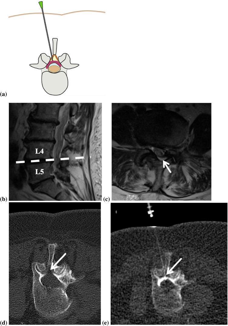
This composite educational graphic illustrates a CT-guided lumbar epidural steroid injection procedure. Panel (a) provides an anatomical diagram in the axial plane showing the spinal needle entering the posterior epidural space with tip placement in the dorsal epidural fat triangle. Panel (b) is a sagittal T2-weighted MRI of the lumbar spine of a 70-year-old female, highlighting multisegmental degenerative changes at L4 and L5. Panel (c) shows a corresponding axial T2-weighted MRI at the L4/L5 level, with an arrow indicating significant spinal canal stenosis. Panels (d) and (e) are axial helical planning and intra-procedural CT scans, respectively. Panel (d) identifies the target zone in the epidural fat (arrow). Panel (e) demonstrates the final needle tip position within the dorsal epidural space during the infiltration. The series illustrates the clinical pathway from diagnostic imaging of lumbar stenosis to the interventional management of chronic back pain using CT fluoroscopy navigation for precise needle localization.
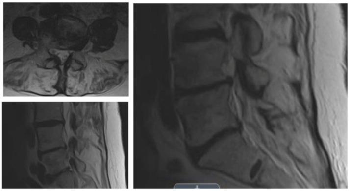
Diagnostic imaging series of the lumbar spine, featuring T1- or T2-weighted MRI scans in both axial (top left) and sagittal (bottom left and right) planes. The sagittal views demonstrate the vertebral bodies, intervertebral discs, and the spinal canal. Notable findings include degenerative changes characterized by reduced disc height and signal intensity variations. There is evidence of moderate-to-severe lateral recess and foraminal stenosis at a single lumbar level, as indicated by the narrowing of the neural exit pathways. The axial view provides a cross-sectional perspective of the vertebral body, facet joints, and the spinal canal, showing narrowing that suggests compression of the neural elements within the lateral recess. These findings are clinically relevant for the diagnosis of neurogenic claudication in patients presenting with progressive leg and back pain. The images serve as an educational example of degenerative spinal pathology and the radiological assessment of foraminal narrowing.
🩺 Clinical Assessment: 60 Saal, Male - Kamar Dard + Pair Dard + Jalan
📋 Sambhavit Diagnoses (Differential Diagnosis)
1. 🥇 Lumbar Spinal Stenosis (Neurogenic Claudication) - SABSE COMMON IS UMAR MEIN
- Kamar mein dard jo pair mein failta hai (dono ya ek taraf)
- Chalna ya khada rehne se dard badh jaata hai
- Aage jhukne ya baith jaane se aram milta hai
- Pair mein jalan, kamzori, sunn ho jaana
- Raat ko bhi dard rehta hai
"The diagnosis of lumbar spinal stenosis is most often suspected when a history of neurogenic claudication is elicited. The diagnosis is best confirmed by MRI." - Firestein & Kelley's Textbook of Rheumatology
2. 🥈 Sciatica / Lumbar Radiculopathy (L4-L5, L5-S1)
- Kamar se shuru hokar ek pair mein electric shock jaisa dard
- Pair mein burning/jalan - dermatome follow karta hai
- Seedha lait ke pair upar uthane se dard (Straight Leg Raise positive)
- Ek taraf zyada affected
"Sciatica: Constant or intermittent, burning or aching, shooting or electric shock-like pain; may follow dermatome; leg pain > back pain" - Tintinalli's Emergency Medicine
3. 🥉 Diabetic Peripheral Neuropathy
- Dono pairon mein symmetric burning jalan (stocking pattern)
- Raat ko dard zyada hota hai
- Sunn ho jaana, jhunjhunahat
- Kamar dard bhi ho sakta hai (diabetic amyotrophy mein)
"Painful diabetic neuropathy is usually intermittent and worse at night. It can be experienced variably as pins and needles, throbbing, burning, aching, or cramping." - Tintinalli's Emergency Medicine
"Diabetic amyotrophy is a lumbosacral plexopathy - presents with back pain followed by progressive leg weakness." - Tintinalli's EM
4. Peripheral Vascular Disease (PVD) - Vascular Claudication
- Chalne se pair mein dard/jalan, ruk ke aram milta hai
- Dono paon thande rehte hain
- Pulse kamzor ya absent
- Risk factors: smoking, hypertension, diabetes
🔍 Zaruri Sawaal (History Lena Zaroori Hai)
| Sawaal | Kyu Important |
|---|---|
| Kya diabetes hai? | Neuropathy diagnose karne ke liye |
| Dard kab zyada hota hai? Chalne se ya araam se? | Stenosis vs vascular |
| Aage jhukne se aram milta hai? | Spinal stenosis |
| Dono pair mein hai ya ek mein? | Radiculopathy vs neuropathy |
| Sunn ho jaana ya kamzori? | Nerve compression |
| Bowel/bladder problem? | Cauda Equina - Emergency! |
| Raat ko dard zyada? | Neuropathic ya serious cause |
| Weight loss, bukhar? | Malignancy ya infection rule out |
🚨 RED FLAGS - TURANT DOCTOR KE PAAS JAAYEIN
- Bowel ya bladder mein control khatam (potty-peshab par control na rahe) → Cauda Equina Syndrome - SURGICAL EMERGENCY
- Dono pairon mein kamzori
- Bukhar ke saath dard
- Raat ko bahut zyada dard
- Sudden weight loss
🧪 Kaun Se Tests Karne Chahiye
| Test | Kyu |
|---|---|
| X-Ray Lumbar Spine (AP + Lateral) | Spondylosis, disc space, alignment dekhne ke liye |
| MRI Lumbar Spine | Gold standard - stenosis, disc herniation, nerve compression |
| Blood Sugar (Fasting + HbA1c) | Diabetes check |
| CBC, ESR, CRP | Infection/inflammation rule out |
| Nerve Conduction Study (NCS/EMG) | Neuropathy confirm karne ke liye |
| Doppler Ultrasound legs | PVD rule out karne ke liye |
💊 Ilaaj (Treatment)
Dawa (Medication)
- Neuropathic pain ke liye: Pregabalin (Lyrica) ya Gabapentin - strong evidence hai - Tintinalli's EM
- Duloxetine, Amitriptyline bhi use hoti hai
- NSAIDs: Diabetes mein avoid karein (kidney aur heart ke liye haanikaarak)
Physiotherapy
- Hot fomentation (garaan sikhai)
- Lumbar extension exercises
- Traction (doctor ki salah se)
- TENS (electrical stimulation)
Lifestyle
- Wajan kam karein
- Chalne mein stick ka use karein
- Sone ka sahi tarika (knees ke neechay cushion rakhein)
Surgery (Agar conservative treatment fail ho)
- Spinal canal narrow ho to Laminectomy/Decompression surgery
- Disc herniation mein Microdiscectomy
⚕️ Kya Karein - ABHI
- Pehle doctor se milein - proper physical examination zaroori hai (Straight Leg Raise test, neurological exam)
- Blood sugar test karaaein - diabetes rule out karein
- X-Ray lumbar spine - starting point
- MRI lumbar spine - agar X-ray se confirm na ho
⚠️ Disclaimer: Yeh information sirf educational purpose ke liye hai. Patient ka proper clinical examination aur doctor ki salah zaroor lein. Koi bhi dawa shuru karne se pehle qualified doctor se milein.
Inko kya kya blood test karwana chaiye..?
🩸 Blood Tests - 60 Saal Male (Kamar + Pair Dard + Jalan)
✅ GROUP 1: SABSE PEHLE YE TESTS (Mandatory - Pehli Visit)
🔴 1. Blood Sugar Tests - MOST IMPORTANT
| Test | Kyu Zaruri |
|---|---|
| Fasting Blood Sugar (FBS) | Diabetes check - pair ki jalan ka sabse bada karan |
| HbA1c (Glycated Hemoglobin) | Pichhle 3 mahine ka blood sugar ka average - diabetes confirm karta hai |
| Post Prandial Blood Sugar (PPBS) | Khana khane ke 2 ghante baad - hidden diabetes pakadta hai |
Pair mein jalan + 60 saal + male = diabetes ruled out karna PEHLI priority hai. Diabetic neuropathy mein dono pairon mein raat ko burning hoti hai - Tintinalli's Emergency Medicine
🟠 2. Complete Blood Count (CBC)
- Haemoglobin, WBC, Platelets check hote hain
- Anemia (khoon ki kami) se bhi pair mein dard/kamzori hoti hai
- WBC badha hua ho to infection/inflammation ki taraf ishara
🟡 3. Kidney Function Test (KFT / RFT)
- Serum Creatinine, Urea, eGFR
- Kidney kharab hone se bhi neuropathy hoti hai
- Dawa dene se pehle kidney status pata hona zaroori hai (NSAIDs kidney kharab kar sakti hain)
🟢 4. Uric Acid
- Gout (joint mein uric acid ka jamna) se pair mein dard + jalan ho sakti hai
- 60 saal ke mard mein bahut common
🔵 5. ESR (Erythrocyte Sedimentation Rate) + CRP (C-Reactive Protein)
- Inflammation/infection hai ya nahi
- Ankylosing Spondylitis, Rheumatoid Arthritis ya koi infection rule out karta hai
- Agar kaafi badha hua ho to cancer (myeloma) bhi sochna padta hai
✅ GROUP 2: YE BHI KARWAAYEIN (Important 2nd Line)
6. Liver Function Test (LFT)
- Dawa metabolism ke liye baseline banana zaroori
- Kuch medicines liver se hoke jaati hain
7. Thyroid Function Test (TSH, T3, T4)
- Hypothyroidism (thyroid slow hona) se pair mein dard, kamzori aur jalan hoti hai
- 60 saal ke mard mein common - aksar miss ho jaata hai!
8. Serum Vitamin B12
- Vitamin B12 ki kami se pair mein jalan, sunn hona, kamzori - Neuropathy hoti hai
- Vegetarian hain to zyada risk
- Metformin leta ho (diabetes ki dawa) to bhi B12 kam hoti hai
9. Serum Calcium + Phosphorus
- Calcium kam hone se muscles mein dard/cramping hoti hai
- Bone problems (osteoporosis, Paget's disease) bhi check hoti hai
10. Lipid Profile (Cholesterol, Triglycerides, LDL, HDL)
- Peripheral Vascular Disease (khoon ki nali band hona) se pair mein dard + jalan
- 60 saal mein heart aur peripheral artery disease ka risk
✅ GROUP 3: AGAR DOCTOR ZARURI SAMJHE (Special Conditions)
11. PSA (Prostate Specific Antigen)
- 60 saal ke mard mein prostate cancer ho sakta hai jo kamar mein failta hai
- Agar kamar dard bahut zyada ho aur raat ko bhi hota ho - zaroor karwana chahiye
12. Serum Protein Electrophoresis (SPEP)
- Multiple Myeloma (blood cancer) rule out karne ke liye
- Agar kamar bahut dard kare, anemia ho, kidney problem ho
13. ANA, RA Factor, Anti-CCP
- Autoimmune disease (Rheumatoid Arthritis, Lupus) sochna ho tab
- Agar subah akkahat (morning stiffness) zyada ho
14. Vitamin D (25-OH Vitamin D)
- Kami hone se haddiyon mein dard, muscles mein kamzori
- India mein bahut common deficiency hai
📋 Summary Table - Priority Order
| Priority | Test | Kuan Sa Condition |
|---|---|---|
| ⭐⭐⭐ | FBS + HbA1c | Diabetic Neuropathy |
| ⭐⭐⭐ | CBC | Anemia, Infection |
| ⭐⭐⭐ | KFT | Kidney Disease Neuropathy |
| ⭐⭐⭐ | ESR + CRP | Inflammation/Arthritis |
| ⭐⭐⭐ | Uric Acid | Gout |
| ⭐⭐ | TSH | Hypothyroid Neuropathy |
| ⭐⭐ | Vitamin B12 | B12 Deficiency Neuropathy |
| ⭐⭐ | Lipid Profile | Vascular Disease |
| ⭐⭐ | LFT | Dawa ke liye baseline |
| ⭐⭐ | Calcium | Bone/Muscle issues |
| ⭐ | PSA | Prostate Cancer rule out |
| ⭐ | Vitamin D | Deficiency |
| ⭐ | SPEP | Myeloma rule out |
💡 Practical Tip
- FBS + HbA1c
- CBC
- KFT
- Uric Acid
- TSH + Vitamin B12
⚠️ Note: Blood tests ke saath X-Ray Lumbar Spine bhi zaroor karwaayein. Aur agar dono pairon mein control khatam ho (potty/peshab) to turant emergency mein jaayein - ye Cauda Equina Syndrome ki emergency ho sakti hai!