I now have all the content and images needed for a comprehensive answer. Here is the full organized response:
Macro and Microscopic Features of Adult Cortical Bone
Macroscopic Features
General Definition and Distribution
Cortical bone (also called compact or lamellar bone) represents approximately 80% of the total skeletal mass. It forms the outer shell (cortex) of all bones and constitutes the bulk of the diaphysis (shaft) of long bones. Its proportion varies by site: the radial diaphysis cortex is ~95% cortical bone; the femoral head cortex ~50%; and vertebral bodies are only ~33% cortical. - Imaging Anatomy, Vol. 3
Gross Anatomy of a Long Bone
The long bone is organized into three regions:
| Region | Description |
|---|
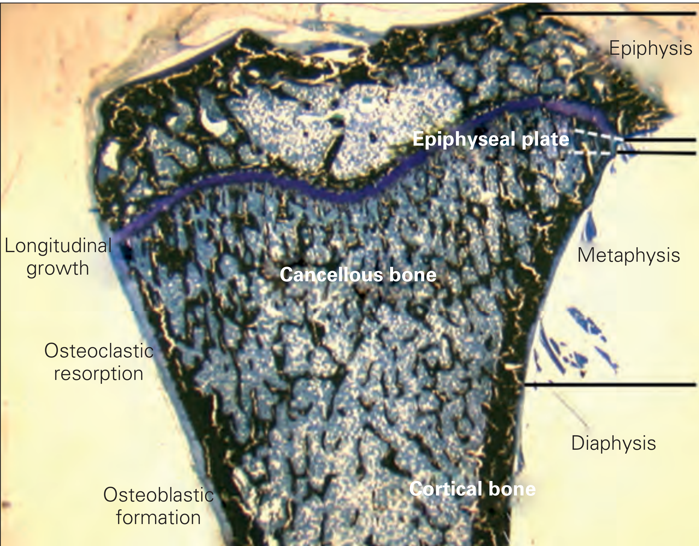
| Diaphysis | Tubular shaft; thick cortex surrounding a central medullary (marrow) cavity |
| Metaphysis | Flared transition zone; thin cortical shell surrounding cancellous bone |
| Epiphysis | Rounded end; thin cortical shell with cancellous interior; capped by articular cartilage |
In adults, the epiphyseal plate is fused (seen as the epiphyseal line). The medullary cavity contains yellow (fatty) marrow in adult diaphyses; red marrow persists in the epiphyses and metaphyses of flat bones (sternum, iliac crest).
Cross-section showing the epiphysis, metaphysis, and diaphysis with cortical and cancellous bone regions (McNeal tetrachrome stain). - Rheumatology, 2-Volume Set
Surfaces and Covering Layers
- Periosteum - dense fibrous connective tissue covering the outer surface of all cortical bone except articular surfaces. Contains osteoblasts, osteoclasts, blood vessels, and nerve fibers. Attached tightly to cortex via thick collagenous Sharpey's fibers. Does not extend over joint surfaces.
- Endosteum - thin single-cell layer lining the inner (medullary) surface of cortical bone, all trabecular surfaces, Haversian canals, and Volkmann's canals. Contains osteoprogenitor cells and bone-lining cells. Has higher bone formation activity than the periosteal surface. - Imaging Anatomy, Vol. 3
Microscopic Features
The Osteon (Haversian System) - Fundamental Unit
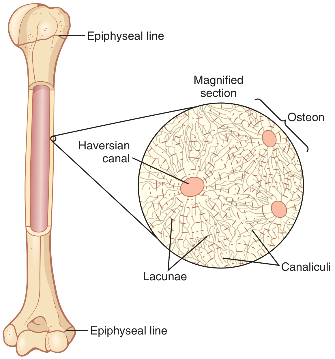
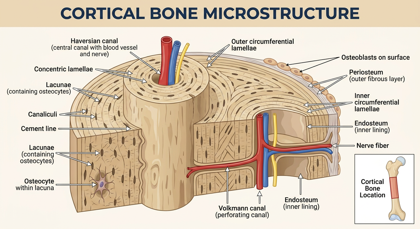
The fundamental structural unit of cortical bone is the osteon (Haversian system): a cylindrical column approximately 4 mm long and running along the long axis of the bone, with its components including:
Structure of cortical bone. - Guyton and Hall Textbook of Medical Physiology
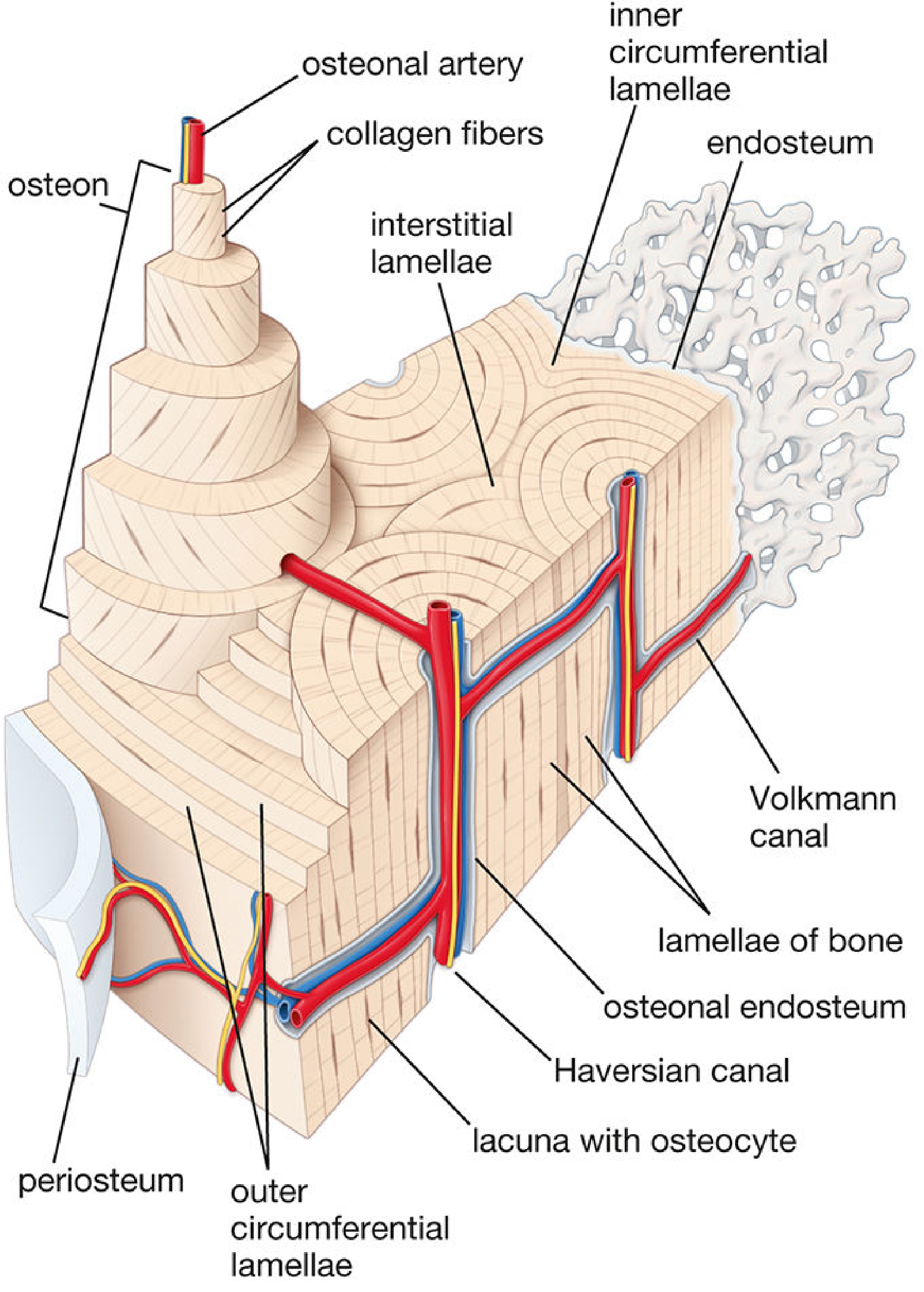
Diagram of compact bone structure showing all lamellar types and vascular channels. - Histology: A Text and Atlas (Ross)
Components of the Osteon
| Structure | Description |
|---|
| Haversian canal | Central canal, 50-90 µm in diameter. Contains a pair of fenestrated capillaries (no smooth muscle), lymphatics absent; accompanied by 2-7 unmyelinated/poorly myelinated sensory and sympathetic nerve fibers ~60% of the time |
| Concentric lamellae | 4-20 rings of mineralized collagen matrix surrounding the canal; collagen fibrils in adjacent lamellae are oriented in alternating directions (like plywood), maximizing mechanical strength |
| Lacunae | Small lens-shaped spaces between lamellae containing osteocytes |
| Canaliculi | Tiny radiating channels connecting lacunae to each other and to the Haversian canal; transmit cellular processes of osteocytes; facilitate Ca²⁺ transfer (osteocytic osteolysis) |
| Cement line | Peripheral boundary of the osteon; composed largely of sulfated glycosaminoglycans; arrests crack propagation and prevents micro-fractures from growing to critical size |
- Rheumatology (Firestein & Kelley); Histology: A Text and Atlas; Medical Physiology (Boron & Boulpaep)
The Vascular Supply - Canals
- Haversian canals - run longitudinally along the long axis of the bone, supplying each osteon
- Volkmann's (perforating) canals - run transversely/obliquely, connecting Haversian canals to each other and to the periosteal and endosteal surfaces; carry vessels from the periosteum into the cortex. Unlike Haversian canals, Volkmann canals are NOT surrounded by concentric lamellae.
Types of Lamellae in Cortical Bone
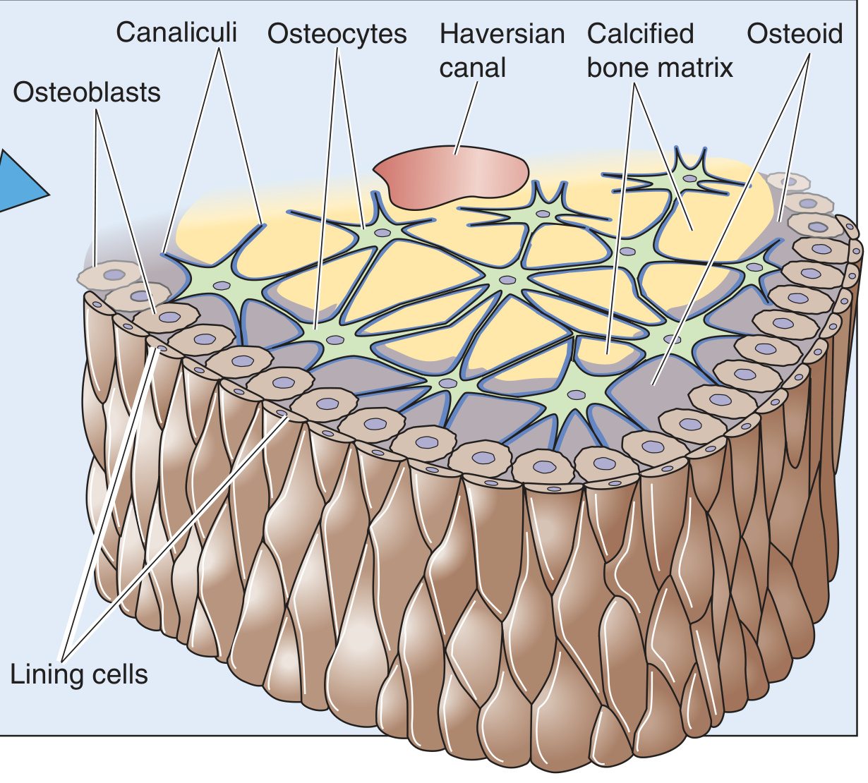
Osteon cross-section detailing cell types and matrix layers. - Medical Physiology (Boron & Boulpaep)
Three distinct lamellar systems are present in adult cortical bone:
- Osteonal (Haversian) lamellae - concentric rings within each secondary osteon; product of bone remodeling (secondary bone)
- Circumferential lamellae
- Outer circumferential lamellae - below the periosteum, encircling the entire bone shaft; laid down by periosteal osteoblasts
- Inner circumferential lamellae - lining the endosteal surface; laid down by endosteal osteoblasts
- Interstitial lamellae - irregular angular fragments filling spaces between osteons; these are remnants of previously remodeled (partially resorbed) older osteons
Bone Cells
| Cell | Location | Function |
|---|
| Osteoblasts | Bone surfaces, Haversian canals | Secrete osteoid (unmineralized matrix); stimulated by PTH, mechanical load |
| Osteocytes | Lacunae within matrix | Derived from osteoblasts; mechanosensing via canalicular processes; regulate remodeling via sclerostin, Wnt ligands, osteocalcin |
| Lining cells | Haversian canal lining | Quiescent osteoblasts covering surfaces |
| Osteoclasts | Resorption surfaces | Multinucleated; ruffled border; resorb bone via acid + proteases; controlled by RANKL/OPG |
Bone Matrix Composition
Bone matrix is composed of:
- Mineral - ~65-67% by weight; hydroxyapatite [Ca₁₀(PO₄)₆(OH)₂] nanocrystals deposited in the hole zones between collagen fibrils (D-periodic 67 nm spacing)
- Organic matrix - ~22-25%; type I collagen (~90% of organic fraction) composed of two α1 chains + one α2 chain; also minor collagens (types III, V) and noncollagenous proteins (osteocalcin, osteopontin, bone sialoprotein)
- Water - ~10%
The N-telopeptide and C-telopeptide at each end of the collagen molecule are cleaved during resorption and serve as biochemical markers of bone turnover. - Rheumatology (Firestein & Kelley)
Primary vs. Secondary Bone
| Feature | Primary Osteon | Secondary Osteon (Haversian System) |
|---|
| Origin | Vascular channels that "filled in" with lamellae | Product of remodeling - existing bone resorbed then replaced |
| Cement line | Absent (no definable cement line) | Present - clearly demarcated |
| Size | Smaller, fewer concentric lamellae | Larger; Haversian canal 50-90 µm |
| Clinical relevance | Cannot be reconstructed after loss (contributes to irreversible trabecular perforation in osteoporosis) | Normal turnover in adult cortex |
Woven vs. Lamellar Bone
| Feature | Lamellar Bone (normal adult cortex) | Woven Bone (abnormal in adults) |
|---|
| Collagen arrangement | Alternating parallel orientations | Random/disorganized |
| Strength | High (plywood-like) | Low |
| Seen in | All normal adult cortex | Fracture callus, Paget's disease, hyperparathyroidism, primary bone formation |
Summary
Adult cortical bone is a dense, organized tissue (~80% of skeletal mass) providing mechanical strength for weight-bearing. Macroscopically it forms the outer shell of all bones, covered externally by periosteum (with Sharpey's fibers) and internally by endosteum. Its microscopic hallmark is the secondary osteon (Haversian system): concentric lamellae of alternating-direction collagen around a central vascular canal, bounded by a crack-arresting cement line, and interconnected via Volkmann's canals. Three lamellar systems - osteonal, circumferential, and interstitial - fill the cortex and reflect cycles of lifelong remodeling. - Imaging Anatomy Vol. 3; Junqueira's Basic Histology; Histology: A Text and Atlas (Ross); Guyton & Hall Medical Physiology; Rheumatology (Firestein & Kelley)