
Popliteal fossa
Reading File
Now let me search the library for popliteal fossa content in parallel:
Finding Sources
Finding Sources
Excellent results. Now let me read the main sections from Gray's and Thieme Atlas in parallel:
Reading File
Reading File
Now let me read more Gray's content and fetch the key images in parallel:
Reading File
Reading File
Loading Image
Loading Image
Now let me fetch one more image (the visualizing contents diagram) and the Baker's cyst / clinical info:
Loading Image
Reading File
I now have comprehensive information to provide a detailed answer. Let me compile everything.
Popliteal Fossa
The popliteal fossa is a diamond-shaped space behind the knee joint, serving as the major route by which neurovascular structures pass between the thigh and the leg.
Overview Diagram
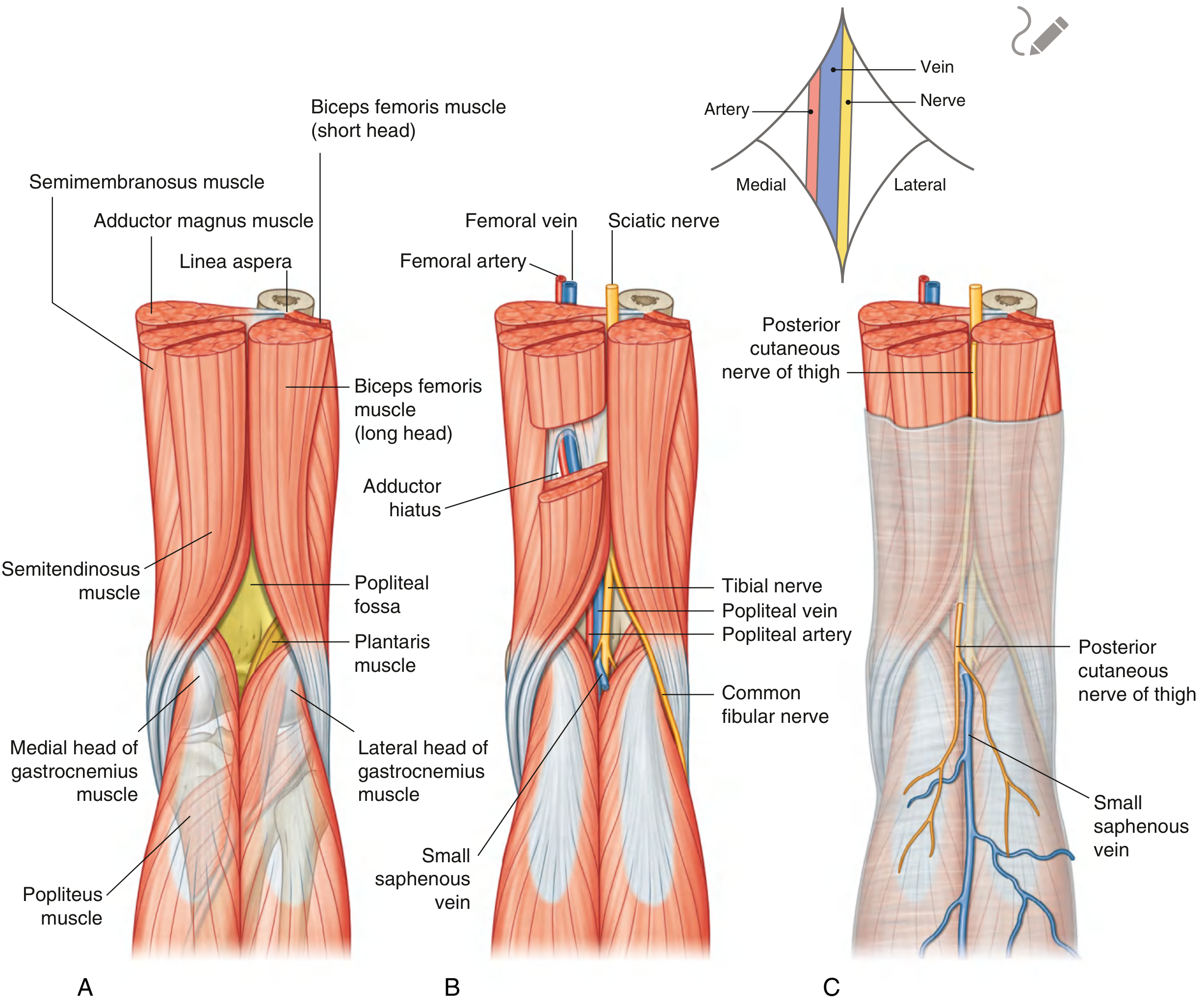
Fig. 6.85 Popliteal Fossa: (A) Boundaries, (B) Nerves and vessels, (C) Superficial structures - Gray's Anatomy for Students
Shape and Boundaries
The fossa is diamond-shaped with an upper and lower triangle:
| Part | Medial margin | Lateral margin |
|---|---|---|
| Upper (larger) triangle | Semitendinosus + semimembranosus | Biceps femoris |
| Lower (smaller) triangle | Medial head of gastrocnemius | Plantaris + lateral head of gastrocnemius |
- Floor - formed by the posterior capsule of the knee joint, adjacent surfaces of the femur and tibia, and inferiorly the popliteus muscle
- Roof - deep fascia continuous with fascia lata above and leg fascia below, covered by superficial fascia and skin
- Gray's Anatomy for Students, p. 720
Visualizing the Contents
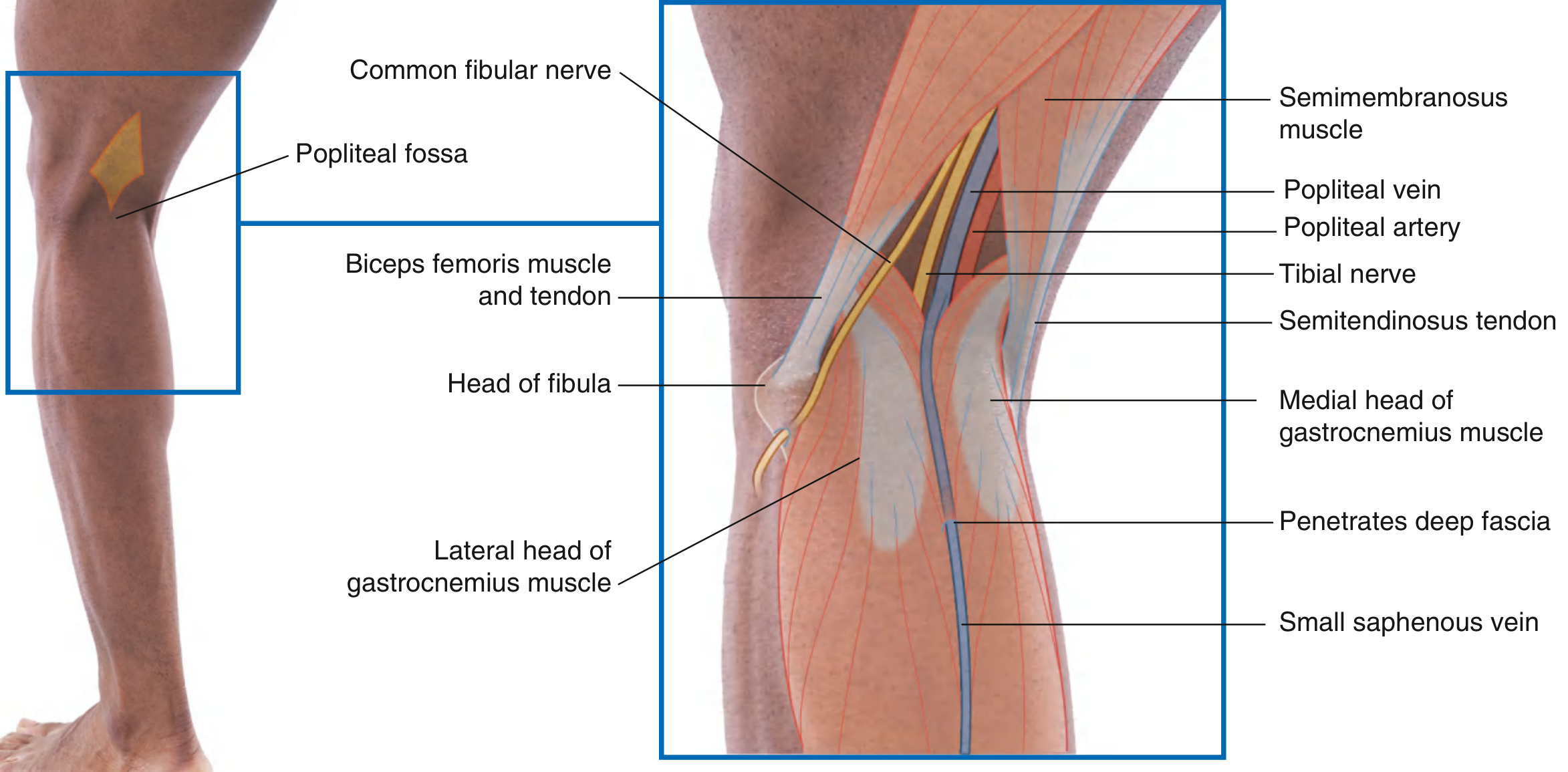
Fig. 6.134 Visualizing the Contents of the Popliteal Fossa - Gray's Anatomy for Students
Contents
The four major contents, listed superficial to deep:
1. Nerves (most superficial)
Both nerves enter the popliteal fossa from above, under the margin of biceps femoris:
- Tibial nerve - descends vertically through the fossa, exits deep to the margin of the plantaris muscle, continues into the posterior compartment of the leg
- Common fibular (peroneal) nerve - exits by following the biceps femoris tendon over the lower lateral margin of the fossa, wraps around the neck of the fibula, and enters the lateral compartment of the leg
The posterior cutaneous nerve of the thigh also passes through the roof of the fossa and continues with the small saphenous vein to innervate skin on the upper half of the back of the leg.
2. Popliteal vein
Superficial to the popliteal artery; travels with it and exits superiorly through the adductor hiatus to become the femoral vein.
3. Popliteal artery (deepest)
The popliteal artery:
- Enters the fossa through the adductor hiatus from the adductor canal
- Appears on the upper medial side under the margin of semimembranosus
- Descends obliquely with the tibial nerve
- Is the deepest structure in the fossa - difficult to palpate, but a pulse can be detected by deep palpation near the midline
- Terminates at the lower border of the popliteus muscle by dividing into the anterior and posterior tibial arteries
- Gray's Anatomy for Students, p. 721
Branches of the Popliteal Artery in the Fossa
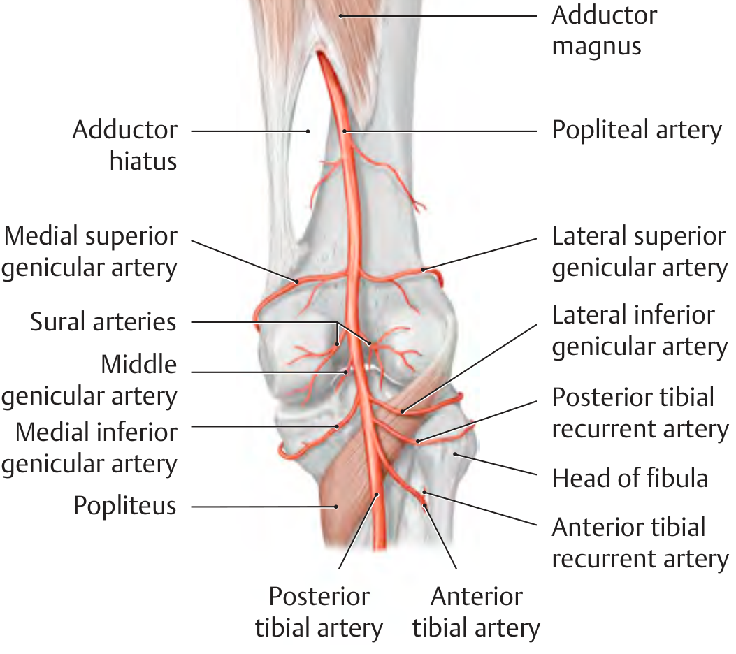
Branches of the popliteal artery - THIEME Atlas of Anatomy
Five genicular branches arise to supply the knee joint and form the periarticular arterial rete:
- Medial superior genicular artery
- Lateral superior genicular artery
- Middle genicular artery - pierces the joint capsule at the oblique popliteal ligament to supply the cruciate ligaments
- Medial inferior genicular artery
- Lateral inferior genicular artery
Paired sural arteries supply the two heads of gastrocnemius. The anterior and posterior tibial recurrent arteries also contribute to the articular rete.
- THIEME Atlas of Anatomy, General Anatomy & Musculoskeletal System
Roof - Superficial Structures
- Small (short) saphenous vein - ascends from the lateral side of the dorsal venous arch of the foot, runs vertically in superficial fascia up the back of the leg, penetrates the deep fascia forming the roof of the fossa, and joins the popliteal vein
- Posterior cutaneous nerve of the thigh - passes through the roof and descends alongside the small saphenous vein
Neurovascular Order (Lateral to Medial at Mid-fossa)
From lateral to medial: Common fibular nerve → Tibial nerve → Popliteal vein → Popliteal artery
A useful mnemonic: "VAN" from deep to superficial - artery deepest, then vein, then nerves most superficial.
Clinical Relevance
Baker's Cyst (Popliteal Synovial Cyst)
- An enlarged synovial sac formed by the fusion of the semimembranosus bursa with the subtendinous gastrocnemius bursa
- Located in the medial popliteal fossa at the level of the medial posterior femoral condyle
- Develops secondary to chronic inflammation (e.g., rheumatoid arthritis), which drives excess synovial fluid through a channel into the fused bursa
- Causes painful swelling in the posterior knee
- Best diagnosed with MRI (shows the cyst communicating with the joint as a bright T2 signal) or ultrasound with duplex Doppler
- THIEME Atlas of Anatomy, p. 463
Popliteal Artery Aneurysm
- Defined as diameter > 7 mm
- Often associated with femoral artery or aortic aneurysms - the entire arterial tree must be investigated when one is found
- Complications are mainly thromboembolic (distal embolization, ischemia, possible amputation) rather than rupture
- Managed with surgical repair
- Gray's Anatomy for Students, p. 722
Common Fibular Nerve Injury
- The common fibular nerve is vulnerable where it curves around the neck of the fibula, which can be used as a palpable landmark to locate it
- Injury here causes foot drop (loss of dorsiflexion and eversion)
Popliteal Artery Pulse
- Because the artery is the deepest structure, the popliteal pulse requires firm, deep palpation with the knee slightly flexed, pressing near the midline of the fossa
Sources:
- Gray's Anatomy for Students, pp. 720-722
- THIEME Atlas of Anatomy: General Anatomy and Musculoskeletal System
This is a shared conversation. Sign in to Orris to start your own chat.