
from Guyton and Hall textbook of medical physiology, 4th South Asia edition, write a 10-mark long answer on Clotting-define; Mechanism of Blood Coagulation-flowchart; Anticoagulants-name
Reading File
Finding Sources
Finding Sources
Reading File
Reading File
Reading File
Reading File
Loading Image
Loading Image
Reading File
Reading File
Finding Sources
Loading Image
I now have all the textbook content and diagrams needed. Let me compile the comprehensive 10-mark answer.
Blood Coagulation (Clotting)
Source: Guyton and Hall Textbook of Medical Physiology, 4th South Asia Edition - Chapter 37
I. DEFINITION OF CLOTTING (Blood Coagulation)
Blood coagulation (clotting) is the process by which blood changes from a liquid to a gelatinous, solid state - forming a blood clot. It is the third step of hemostasis and represents the body's response to vascular injury.
When a blood vessel is severed or ruptured, the following sequential hemostatic events occur:
- Vascular constriction
- Formation of a platelet plug
- Blood coagulation (clot formation)
- Growth of fibrous tissue into the clot to permanently seal the vessel
A clot is a meshwork of fibrin fibers running in all directions and entrapping blood cells, platelets, and plasma. The fibrin fibers also adhere to damaged surfaces of the blood vessels.
II. MECHANISM OF BLOOD COAGULATION
General Mechanism
More than 50 important substances that cause or affect blood coagulation have been identified. These are broadly classified as:
- Procoagulants - promote coagulation
- Anticoagulants - inhibit coagulation
In the normal bloodstream, anticoagulants predominate. When a vessel ruptures, activated procoagulants override the anticoagulants and the clot develops.
Three Essential Steps of Clotting
Clotting takes place in three essential steps:
| Step | Event |
|---|---|
| 1 | A cascade of chemical reactions involving 12+ clotting factors produces Prothrombin Activator |
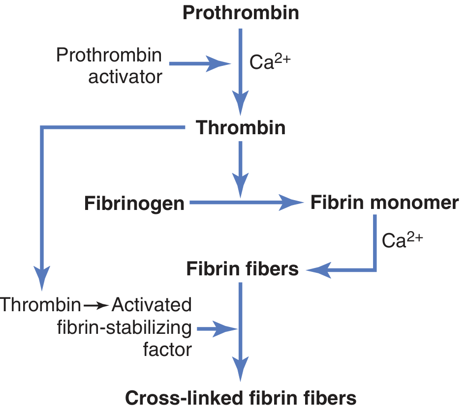
| 2 | Prothrombin activator catalyzes conversion of Prothrombin → Thrombin |
| 3 | Thrombin converts Fibrinogen → Fibrin fibers forming the clot |
FLOWCHART: Mechanism of Blood Coagulation
STEP 1 - FINAL COMMON PATHWAY (Prothrombin → Thrombin → Fibrin)
STEP 2A - EXTRINSIC PATHWAY (Rapid: 15 seconds)
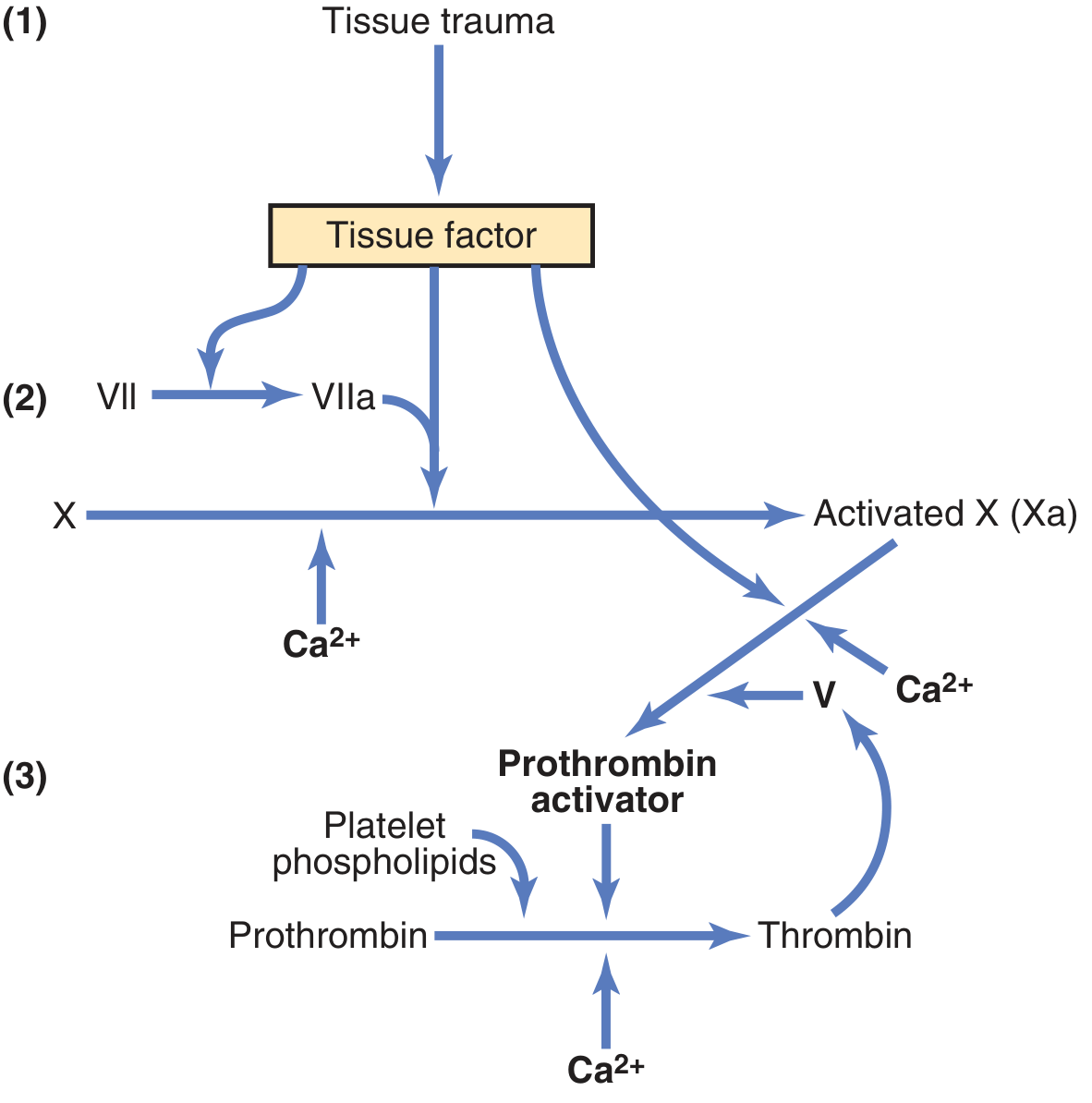
Steps of the Extrinsic Pathway:
- Tissue trauma → Release of Tissue Factor (TF / Tissue Thromboplastin) - composed of phospholipids from tissue membranes + a lipoprotein complex
- TF + Factor VII + Ca²⁺ → Activation of Factor X (Xa)
- Xa + Factor V + Ca²⁺ + Platelet phospholipids → Prothrombin Activator
- Prothrombin Activator + Ca²⁺ → Prothrombin → Thrombin
- Note: Thrombin activates Factor V (positive feedback), accelerating the process further
STEP 2B - INTRINSIC PATHWAY (Slower: 1-6 minutes)
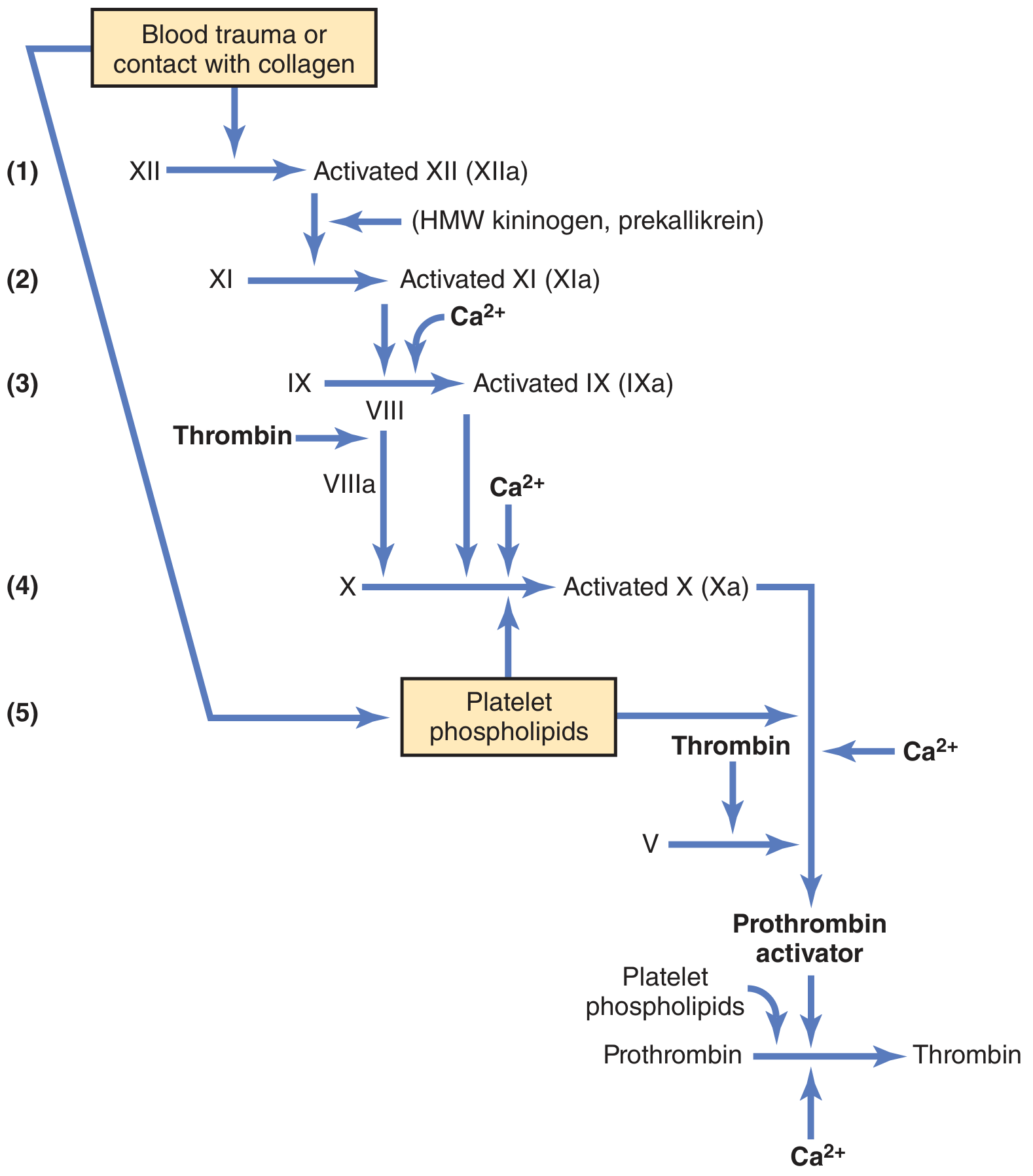
Steps of the Intrinsic Pathway:
- Blood trauma or contact with collagen → Factor XII → XIIa (assisted by HMW kininogen + prekallikrein)
- XIIa → Factor XI → XIa
- XIa + Ca²⁺ → Factor IX → IXa
- IXa + Factor VIIIa + Ca²⁺ + Platelet phospholipids → Factor X → Xa
- Xa + Factor Va + Ca²⁺ + Platelet phospholipids → Prothrombin Activator → Prothrombin → Thrombin
- Note: Thrombin activates Factor VIII (positive feedback), amplifying the cascade
STEP 3 - FIBRIN CLOT FORMATION (Common Final Step)
Thrombin (proteolytic enzyme)
        ↓
Fibrinogen (MW ~340,000) → Fibrin monomers
        ↓  (polymerization, seconds)
Fibrin fibers (weak, H-bonds)
        ↓  (Thrombin activates fibrin-stabilizing factor + Ca²⁺)
Activated Fibrin-Stabilizing Factor (Factor XIIIa)
        ↓
Cross-linked fibrin fibers → BLOOD CLOT (stable meshwork)
The clot entraps red blood cells, platelets, and plasma proteins. Within 30-60 minutes, clot retraction occurs driven by thrombosthenin (contractile protein in platelets), pulling vessel edges together.
POSITIVE FEEDBACK of Clot Formation
Once thrombin is formed, it acts on:
- Prothrombin (direct proteolytic effect - more thrombin)
- Factors VIII, IX, X, XI, XII (activation)
- Platelet aggregation
This creates a positive feedback loop that grows the clot until bleeding ceases.
III. ANTICOAGULANTS
A. Intravascular (Natural) Anticoagulants
1. Endothelial Surface Factors
- Smoothness of endothelial surface - prevents contact activation of the intrinsic clotting system
- Glycocalyx (mucopolysaccharide layer) on endothelium - repels clotting factors and platelets
- Thrombomodulin (membrane-bound protein) - binds thrombin, removing it; the thrombomodulin-thrombin complex activates Protein C, which inactivates activated Factors V and VIII
- Prostacyclin (PGI₂) and Nitric Oxide (NO) - released by intact endothelial cells; both inhibit platelet aggregation and clot initiation
2. Fibrin and Antithrombin III
- During clot formation, 85-90% of thrombin formed is adsorbed onto fibrin fibers, preventing spread of thrombin into surrounding blood
- Antithrombin III (an α-globulin = antithrombin-heparin cofactor) - removes remaining free thrombin within 12-20 minutes
3. Heparin
- Highly negatively charged conjugated polysaccharide
- By itself has little anticoagulant activity
- When combined with antithrombin III, potentiates its effect 100-1000 fold
- Also inactivates activated Factors IX, X, XI, XII
- Produced naturally by basophilic mast cells in pericapillary connective tissue (especially lungs and liver); also by blood basophils
B. Clinical / Pharmacological Anticoagulants
| Anticoagulant | Mechanism |
|---|---|
| Heparin (therapeutic) | Potentiates antithrombin III; rapid onset (IV/SC) |
| Warfarin / Vitamin K antagonists | Inhibit hepatic synthesis of Factors II, VII, IX, X (vitamin K-dependent factors) |
| Citrate / Oxalate (in vitro) | Chelate Ca²⁺, preventing coagulation in stored blood |
| EDTA | Chelates Ca²⁺ |
| Direct thrombin inhibitors (e.g., hirudin) | Directly block thrombin activity |
| Protein C | Inactivates activated Factors V and VIII |
C. Fibrinolytic System (Clot Dissolution)
Plasminogen (euglobulin, present in clot) is activated by tissue plasminogen activator (t-PA) released by injured tissues and vascular endothelium. This converts plasminogen → Plasmin, which digests fibrin fibers and other coagulation factors (fibrinogen, Factor V, Factor VIII, prothrombin, Factor XII), thereby lysing the clot after healing is complete.
Summary Table of Clotting Factors
| Factor | Name |
|---|---|
| I | Fibrinogen |
| II | Prothrombin |
| III | Tissue Factor (Tissue Thromboplastin) |
| IV | Calcium ions (Ca²⁺) |
| V | Labile factor (Proaccelerin) |
| VII | Stable factor (Proconvertin) |
| VIII | Antihemophilic factor A |
| IX | Christmas factor (Antihemophilic factor B) |
| X | Stuart-Prower factor |
| XI | Plasma thromboplastin antecedent |
| XII | Hageman factor |
| XIII | Fibrin-stabilizing factor |
Reference: Guyton and Hall Textbook of Medical Physiology, 4th South Asia Edition, Chapter 37 - Hemostasis and Blood Coagulation, pp. 481-490
This is a shared conversation. Sign in to Orris to start your own chat.