
Visual pathway
Reading File
The visual pathway runs from the retina through the optic nerves, optic chiasm, optic tracts, lateral geniculate nucleus (LGN), optic radiations, and finally to the primary visual cortex (V1) in the occipital lobe.
Searching sources...
Finding Sources
Finding Sources
Good results. Now let me read the most comprehensive sources in parallel - Gray's Anatomy for the central visual pathway, Kandel's Principles of Neural Science, and Guyton for physiology.
Reading File
Reading File
Reading File
Excellent content. Now let me fetch the key images and get more on the LGN, optic radiations, visual cortex, and lesion effects.
Loading Image
Loading Image
Reading File
Reading File
Now let me get the correct image URL from Gray's and fetch the key diagram of visual field deficits, plus read the Kandel section on LGN and optic radiations.
Loading Image
Loading Image
Reading File
Excellent - I have the key diagrams and comprehensive content. Now let me also get the LGN layer structure and M/P pathway details from Kandel.
Reading File
I now have comprehensive content from multiple authoritative textbooks. Let me compile the full answer.
Visual Pathway
The visual pathway is the neural route by which light signals captured by the retina are transmitted to the visual cortex for conscious perception. It is a sequential, point-by-point (retinotopic) relay with partial decussation at the optic chiasm, ensuring the contralateral hemisphere processes each visual hemifield.
1. Retina - The Starting Point
Light strikes photoreceptors (rods and cones), which convert it into neural signals via phototransduction. The circuitry within the retina involves:
- Photoreceptors (rods/cones) → Bipolar cells → Ganglion cells (the output neurons)
- Horizontal cells: lateral inhibition in the outer plexiform layer
- Amacrine cells: lateral signal modulation in the inner plexiform layer
- Interplexiform cells: retrograde signals from inner to outer plexiform layer (inhibitory, for contrast control)
Transmission within the retina is mostly electrotonic (graded potentials), not action potentials. The exception is retinal ganglion cells (RGCs), which fire true action potentials all the way to the brain via the optic nerve. Glutamate is the main neurotransmitter at photoreceptor-bipolar synapses; amacrine cells use GABA, glycine, dopamine, and acetylcholine.
Two parallel streams leave the retina:
- M (Magnocellular) pathway: large RGCs, fast conduction (2-5x faster), motion/depth/low-spatial-frequency contrast
- P (Parvocellular) pathway: smaller RGCs, slower conduction, color and fine detail
(Guyton and Hall Textbook of Medical Physiology; Kandel, Principles of Neural Science, 6th ed.)
2. Optic Nerve (CN II)
Axons from all RGCs converge at the optic disc (the blind spot - no photoreceptors here). They exit the eye, acquire a myelin sheath from oligodendrocytes (not Schwann cells), and are covered by cranial meninges - making the optic nerve a true extension of the CNS, not a peripheral nerve.
(Gray's Anatomy for Students)
3. Optic Chiasm
Located anterior to the infundibular stalk. Here, partial decussation occurs:
| Fiber origin | Destination |
|---|---|
| Nasal (medial) retina - sees temporal visual field | Crosses → contralateral optic tract |
| Temporal (lateral) retina - sees nasal visual field | Stays ipsilateral → ipsilateral optic tract |
Result: Each optic tract carries information from the contralateral visual hemifield of both eyes.
4. Optic Tract
Courses around the midbrain. Most fibers proceed to the lateral geniculate nucleus (LGN) of the thalamus. A small contingent branches off to:
- Pretectal area and superior colliculus → mediates the pupillary light reflex and orienting reflexes (NOT conscious vision)
5. Lateral Geniculate Nucleus (LGN) of Thalamus
The main relay station. Organized into 6 layers:
- Layers 1 & 2 = Magnocellular layers (M pathway: motion, contrast, depth)
- Layers 3-6 = Parvocellular layers (P pathway: color, fine detail)
- Layers 1, 4, 6 receive input from the contralateral eye
- Layers 2, 3, 5 receive input from the ipsilateral eye
The LGN is not merely a relay - it also receives feedback from the visual cortex and signals from arousal systems.
6. Optic Radiations (Geniculocalcarine Tract)
Axons from LGN neurons fan out as the optic radiations toward the primary visual cortex. They travel in two divisions:
| Division | Path | Visual field represented |
|---|---|---|
| Upper fibers (parietal route) | Directly through parietal lobe | Lower contralateral visual field |
| Lower fibers - Meyer's loop | Arc anteriorly into the temporal lobe around the inferior horn of the lateral ventricle, then back | Upper contralateral visual field |
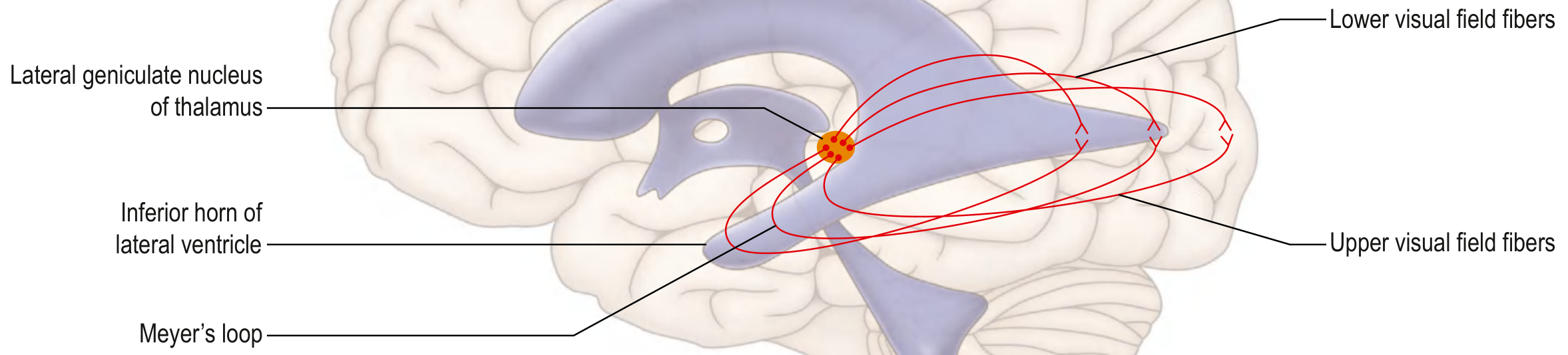
(eFig. 9.66 - Gray's Anatomy for Students: Thalamocortical fibers from LGN showing Meyer's loop)
7. Primary Visual Cortex (V1 / Striate Cortex / Brodmann Area 17)
Located in the occipital lobe, in the walls and floor of the calcarine sulcus. It is retinotopically organized:
- Upper bank of calcarine sulcus → lower visual field
- Lower bank → upper visual field
- Posterior pole (occipital pole) → foveal (central) vision - disproportionately large representation (cortical magnification)
V1 contains orientation columns, ocular dominance columns, and "blob" regions for color processing.
8. Beyond V1 - Higher Visual Processing
From V1, two major streams diverge:
| Pathway | Route | Function |
|---|---|---|
| Dorsal ("where/how") stream | V1 → V2 → V5/MT → Parietal lobe | Spatial location, motion, visuomotor guidance |
| Ventral ("what") stream | V1 → V2 → V4 → Inferior temporal cortex | Object recognition, color, faces, form |
Damage to the dorsal pathway → unilateral neglect, optic ataxia. Damage to the ventral pathway → prosopagnosia (inability to recognize faces), object agnosia.
(Kandel, Principles of Neural Science, 6th ed.)
Lesion Localization - Visual Field Defects
This is the most clinically tested aspect of the visual pathway:
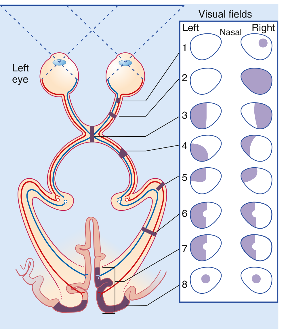
(eFig. 9.68 - Gray's Anatomy for Students: Visual field deficits at each level)
| Site of Lesion | Visual Field Defect |
|---|---|
| 1. Optic nerve (retina) | Ipsilateral monocular scotoma |
| 2. Optic nerve (complete) | Ipsilateral monocular blindness |
| 3. Optic chiasm (crossing fibers) | Bitemporal hemianopia (classic pituitary adenoma sign) |
| 4. Optic tract | Contralateral homonymous hemianopia |
| 5. Meyer's loop (temporal lobe) | Contralateral homonymous superior quadrantanopia ("pie in the sky") |
| 6-7. Optic radiation / V1 | Contralateral homonymous hemianopia with macular sparing (dual blood supply to macular cortex) |
| 8. Bilateral occipital poles | Bilateral central scotoma |
Summary Flowchart
Photoreceptors (rods/cones)
       ↓
Bipolar cells
       ↓
Retinal Ganglion Cells
       ↓
Optic Nerve (CN II)  ← blind spot at optic disc
       ↓
Optic Chiasm  ← nasal fibers cross / temporal fibers stay
       ↓
Optic Tract  ← side branch to pretectum (pupillary reflex)
       ↓
Lateral Geniculate Nucleus (Thalamus)
       ↓
Optic Radiations (Meyer's loop = lower fibers through temporal lobe)
       ↓
Primary Visual Cortex V1 (Calcarine sulcus, occipital lobe)
       ↓
    /       \
Dorsal     Ventral
(where)    (what)
Sources:
- Gray's Anatomy for Students - Central Visual Pathway, p. 1346-1348
- Kandel, Principles of Neural Science, 6th ed. - Visual Processing and the Geniculostriate Pathway, p. 544-550
- Guyton and Hall Textbook of Medical Physiology - Neural Function of the Retina, p. 640
This is a shared conversation. Sign in to Orris to start your own chat.