I now have comprehensive content from Cummings Otolaryngology, KJ Lee's Essential Otolaryngology, and Mulholland & Greenfield's Surgery. Let me compile the full detailed answer.
Nasal Framework Deformities, Saddle Nose, and Rhinoplasty - A Complete Guide
(Based on Cummings Otolaryngology Head & Neck Surgery, KJ Lee's Essential Otolaryngology, and Mulholland & Greenfield's Surgery)
PART 1: DEFORMITIES OF THE NASAL FRAMEWORK
The nasal framework is divided into three zones - upper third (bony vault), middle third (cartilaginous vault/upper lateral cartilages), and lower third (tip/lower lateral cartilages). Deformities in each zone look and behave differently.
Upper Third Deformities (Bony Vault)
- Nasal hump (dorsal hump): Excess bone +/- cartilage at the nasal bridge. Can be purely bony, purely cartilaginous, or combined. Very common in Middle Eastern and Southern European patients.
- Crooked/deviated nose: The bony pyramid is off midline. Usually due to trauma or septal deviation. Corrected with osteotomies.
- Open roof deformity: When a large dorsal hump is removed without closing the roof back, a flat, wide-looking dorsum results. Requires spreader grafts + medial and lateral osteotomies to close.
Middle Third Deformities (Cartilaginous Vault - Upper Lateral Cartilages)
- Saddle nose: Collapse/depression of the dorsum due to loss of support (see dedicated section below).
- Inverted-V deformity: The upper lateral cartilages (ULCs) drop inferiorly after rhinoplasty, visible as a dark V-shadow on the dorsum. Usually from failure to reattach ULCs after hump reduction.
- Middle vault narrowing/pinching: Due to ULC collapse inward, narrowing the internal nasal valve (INV). Causes nasal obstruction.
Lower Third Deformities (Tip/Lower Lateral Cartilages)
- Pollybeak deformity: Fullness/bump in the supratip area, giving a parrot-beak appearance. Caused by:
- Underreduction of dorsal cartilage/anterior septal angle
- Paradoxical pollybeak from excessive dorsal resection causing a soft tissue void that scars
- Tip deprojection (loss of tip support) leaving relative supratip excess
- Alar retraction: The alar rim rides too high, showing excessive nostril on frontal view.
- Supra-alar pinching: Over-resection of lateral crura leaves a pinched-in look.
- Tip asymmetry / bossae: Cartilage knuckling visible under thin skin.
- Bulbous tip: Dome too wide and rounded, no definition.
- Ptotic tip (drooping tip): Tip rotates downward - often from septal hematoma, aging, or previous surgery.
PART 2: SADDLE NOSE DEFORMITY
What is it?
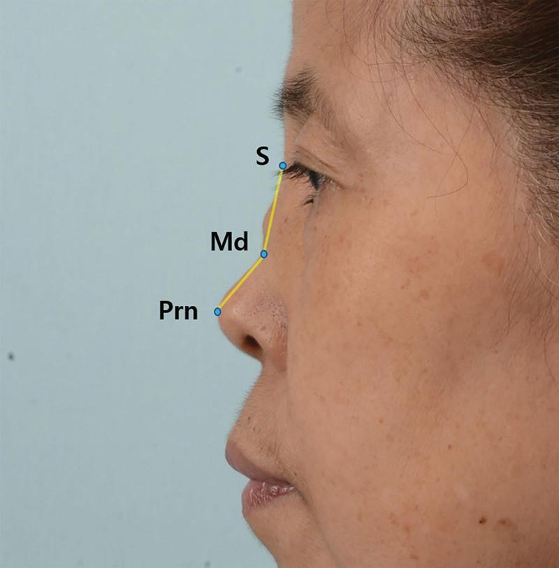
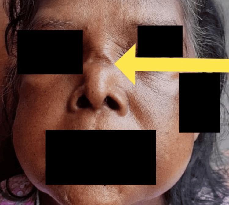
A saddle nose is a depression/collapse of the nasal dorsum - the bridge looks scooped in like a horse's saddle. It involves loss of support of the nasal framework, leading to dorsal collapse.
Causes (Etiology)
Think: "Trauma, Infection, Autoimmune, Iatrogenic, Congenital"
| Category | Examples |
|---|
| Iatrogenic | Over-resection of dorsal cartilage, destabilization of dorsal/caudal L-strut during septoplasty, previous rhinoplasty |
| Traumatic | Nasal fracture, septal hematoma (untreated hematoma leads to avascular necrosis of cartilage) |
| Infectious | Syphilis (classic cause - saddle nose + septal perforation), tuberculosis, leprosy |
| Autoimmune | Granulomatosis with polyangiitis (GPA/Wegener's), relapsing polychondritis |
| Drugs | Cocaine use (septal perforation → saddle) |
| Malignancy | NK/T-cell lymphoma, other midline destructive lesions |
| Congenital | Congenital syphilis, Down syndrome |
Classification (What collapses?)
- Isolated cartilaginous dorsal collapse - only the cartilaginous vault sinks
- Combined bony + cartilaginous deficiency - the entire dorsum is low
- Over-resection of underlying cartilaginous framework - iatrogenic
- Loss of dorsal septal support - due to necrosis, infection, trauma, autoimmune, cocaine, or septal hematoma
Clinical Features
- Concave nasal profile (the hallmark)
- Columellar retraction (the columella withdraws inward)
- Short nose (vertical height loss)
- Associated septal perforation (especially in syphilis, cocaine, GPA)
- Nasal obstruction
- External and internal deformities
Determining Appropriate Dorsal Height
This is nuanced:
- In men: dorsum should be straight or slightly convex at the rhinion
- In women: dorsum should be straight or slightly concave
- A low radix can make the dorsum look convex even when it is not - augmenting the radix may be the right answer rather than reducing the dorsum
- Tip projection directly affects how high the dorsum should be - if the tip is under-projected, the dorsum looks too prominent even when normal
PART 3: MANAGEMENT OF SADDLE NOSE DEFORMITY
Step 1 - Analysis
Before operating, analyze WHICH components are involved - bony vault, cartilaginous vault, columella, tip - so a complete reconstructive plan can be made preoperatively.
Step 2 - Primary Repair Principle
Re-establish septal support and strengthen the septal L-strut. The L-strut is the "backbone" of the nose - it consists of the dorsal strut (top of the L) and the caudal strut (front of the L). This is the most important step.
Step 3 - Graft Materials Used
Autogenous (Gold Standard - Preferred)
| Source | Use |
|---|
| Septal cartilage | First choice; limited supply |
| Auricular (ear) cartilage | Small defects; unpredictable warping |
| Costal (rib) cartilage | Best for major structural support; unlimited supply; risk of warping (minimize by carving central core) |
| Iliac crest bone | Bony pyramid augmentation |
| Calvarial bone / Rib bone | Bony vault augmentation |
| Temporalis fascia / Dermis | Filling, contouring, softening; camouflaging irregularities in thin skin |
| Diced cartilage + fascia (DCF) | Wrapped diced cartilage in fascia; excellent for dorsal contour when structural support is not needed |
Alloplastic Materials (If autogenous unavailable/refused)
| Material | Notes |
|---|
| Gore-Tex (ePTFE) | Preferred alloplast for dorsal augmentation; minimal fibrous ingrowth = removable; taper edges to prevent visibility |
| Medpor (porous polyethylene) | Good tissue ingrowth |
| Silicone | Higher extrusion rate, artificial feel in lower third; still used in Asian rhinoplasty for dorsal upper 2/3 |
| Mersilene mesh | Partially incorporated into surrounding tissue |
Key principle: Autogenous > alloplastic. Alloplastic materials have risk of rejection, infection, inflammation, extrusion. Silicone is most common but most problematic at tip region.
Homologous Materials (Now largely abandoned)
Irradiated/lyophilized cartilage, bone, acellular dermis - unpredictable long-term resorption and buckling.
Step 4 - Surgical Techniques for Saddle Nose
For Small Defects
- Auricular cartilage grafts are sufficient
- Rolled fascia + diced cartilage can augment dorsum when structural support is not needed
For Major Defects
- Costal cartilage is most effective for establishing structural support of the dorsal septum
- Extended spreader grafts (costal cartilage) attached to a caudal septal replacement graft re-create the L-strut
- Grafts should be canoe-shaped:
- Cephalic margin: snug in a subperiosteal pocket extending to the desired nasal starting point
- Caudal margin: terminates in the supratip
- Lateral margins: beveled to minimize visibility
Skin contracture (common in revision cases)
- When skin contracts over a saddle deformity, it resists expansion
- Dorsal augmentation grafts are needed for both support AND to expand contracted skin
- A piece of temporalis fascia draped over the entire nasal framework can camouflage edge irregularities - placed through intercartilaginous incision or open approach
Staging
- When extensive septal reconstruction + osteotomies are required, stage the procedure: do septal work first, place implants at a second operation
- This reduces the size of implant needed (straighter nose = less augmentation needed)
PART 4: RHINOPLASTY - TYPES AND APPROACHES
What is Rhinoplasty?
Surgery to reshape or reconstruct the nose. It is truly a "game of millimeters" - meticulous planning and execution are non-negotiable. It addresses both aesthetics and function. As KJ Lee states: "Preserve and conserve. Do not resect and regret."
TYPES OF RHINOPLASTY
| Type | Definition |
|---|
| Primary rhinoplasty | First-time rhinoplasty on an unoperated nose |
| Revision/Secondary rhinoplasty | Re-operation after a prior rhinoplasty to correct unsatisfactory results |
| Augmentation rhinoplasty | Increasing the size/projection of the nose (adding tissue) |
| Reduction rhinoplasty | Reducing the size of the nose (removing tissue) |
| Functional rhinoplasty | Improving nasal airflow - addresses septum, nasal valves, turbinates |
| Reconstructive rhinoplasty | Rebuilding the nose after trauma, tumor resection, or major deformity |
| Ethnic/Non-Caucasian rhinoplasty | Tailored approach for non-Caucasian patients - usually augmentation dominant |
RHINOPLASTY APPROACHES
1. Closed (Endonasal) Approach
All incisions are inside the nose. Two subtypes:
A. Non-delivery technique
- Only intercartilaginous incisions (between upper and lower lateral cartilage)
- Lower lateral cartilage (LLC) is NOT delivered out
- Advantages: No disruption of major tip support, no external scar, intact caudal rim
- Disadvantages: Minimal exposure of LLC, limited tip modification, potential valve scar
B. Delivery technique
- Combines marginal + intercartilaginous (or transcartilaginous) incisions
- LLC is delivered out as a bilaterally pedicled chondrocutaneous flap
- Advantages: Better visualization of entire LLC, good tip/dome access, no external incision
- Disadvantages: Disrupts one major tip support (LLC-ULC attachment), limited tip modification possible, higher revision rates than open approach
2. Open (External) Approach
- Combines a transcolumellar incision (inverted-V or stair-step) + bilateral marginal incisions
- The entire skin-soft tissue envelope (SSTE) is elevated off the nose
- Advantages:
- Complete exposure of all nasal structures
- Precise graft and suture placement
- Best for complex tip work, revision cases, cleft rhinoplasty
- Lower revision rates than endonasal
- Ideal for teaching
- Disadvantages:
- External scar (transcolumellar - usually fades well)
- Disruption of SSTE (minor tip support mechanism)
- More postoperative edema and numbness
- Longer recovery of tip definition (up to 1 year)
Rhinoplasty Incisions Summary
| Incision | Description |
|---|
| Marginal | Along caudal edge of lower lateral cartilage |
| Intercartilaginous | Between lower and upper lateral cartilage |
| Trans/intracartilaginous | Through the lower lateral cartilage |
| Transcolumellar | Across the columella (inverted-V or stair-step) - for open approach |
| Transfixion | Between caudal septum and medial crura, through-and-through |
| Hemitransfixion | Same, but one side only |
PART 5: AUGMENTATION RHINOPLASTY
What is it?
Adding volume to the nose - to raise the dorsum, project the tip, or lengthen a short nose. Dominant in Asian, African, and Middle Eastern patients. In Asia, it is the most requested rhinoplasty type.
Indications
- Low dorsum (flat nose) - most common in Asian, African patients
- Saddle nose deformity
- Post-traumatic nasal collapse
- Underprojected tip
- Short nose needing lengthening
- After major reduction where too much tissue was removed
Surgical Approach Choice
- Only dorsal work needed? → Endonasal (intercartilaginous or extended septoplasty incision)
- Tip work also needed? → External/open approach - offers best control for combined dorsal + tip augmentation + lengthening
Augmentation Materials (already detailed above - same principles apply)
The critical decision: autogenous vs. alloplastic
Alloplastic Implants for Augmentation
- L-shaped silicone strut - traditional Asian rhinoplasty; augments dorsum + lengthens nose. But tension on the tip → risk of skin breakdown and extrusion, especially at the tip
- Modern approach in Asia: Alloplastic for dorsal upper 2/3 + autologous tissue for tip (tip grafts, cap grafts, columellar struts, caudal extension grafts)
- Straight silicone implant for dorsum only
- Gore-Tex (ePTFE): preferred over silicone for dorsum if autologous not available; softer, less palpable, minimal fibrous ingrowth = removable
- Medpor: more tissue integration, harder to remove
- Key technical point: Bevel/taper all implant edges; fix with sutures to avoid slippage; irrigate copiously with antibiotic solution; avoid undue tension
Autologous Tissue for Augmentation
- Stacked cartilage grafts (septal, ear, rib) - layers sutured together
- Diced cartilage wrapped in fascia (DCF) - excellent dorsal contour, no edge visibility
- Articulated costal cartilage - for major augmentation needs
- Iliac crest / calvarial / rib bone - for bony vault augmentation
Special Considerations: Non-Caucasian (Asian/African/Middle Eastern)
- Asian nose: Wide dorsum, flat, low radix, bulbous tip, thick skin. Despite appearing wide, lateral osteotomies are rarely needed - midline augmentation creates illusion of narrowing
- Tip augmentation: Thick skin in Asian patients camouflages minor irregularities but demands high structural support from the cartilaginous framework
- Warping of rib graft: Avoid by carving the central core of the rib or cutting obliquely into symmetric cortical portions
- Middle Eastern patients requesting hump reduction: Strong dorsum has cultural significance - careful preoperative counseling needed; scooping the dorsum may be culturally unacceptable
Complications of Augmentation Rhinoplasty
- Implant extrusion (especially silicone at tip)
- Infection
- Migration/slippage
- Skin breakdown over the tip
- Visible edges of implant (thin-skinned patients)
- Long-term: warping of rib grafts, resorption of cartilage
PART 6: FUNCTIONAL RHINOPLASTY
What is it?
Rhinoplasty aimed at improving nasal airflow - addressing structural causes of nasal obstruction. It targets the septum, nasal valves, and lower lateral cartilages.
Preoperative Assessment
- Complete nasal history: obstruction, allergies, trauma, prior surgery
- Identify which structures cause obstruction:
- Deviated septum
- Inferior turbinate hypertrophy
- Internal nasal valve (INV) collapse
- External nasal valve (ENV) collapse
- Paradoxical inferior turbinate
- Cottle maneuver: lateral cheek/lateral nasal wall displacement to open the valve - improvement = positive test (but unreliable alone; gentle lateral displacement of ULC is a better predictor)
Structure 1: The Nasal Valve
Internal Nasal Valve (INV)
- The narrowest part of the nasal airway
- Formed by the angle between the dorsal septum and the caudal edge of the upper lateral cartilage (ULC)
- Normal angle: 10-15 degrees
- Surrounded by: septum medially, ULC superolaterally, inferior turbinate/nasal floor inferiorly
- This is the "keystone" - stenosis here dramatically increases airflow resistance
External Nasal Valve (ENV)
- The nasal vestibule/nostril opening
- Supported by: lower lateral cartilages, soft tissue of alar lobule, columella
- Collapse here causes visible nostril collapse on inspiration
Structure 2: Septoplasty for Functional Rhinoplasty
The deviated septum is the most common correctable cause of obstruction. Septoplasty:
- Releases ULCs from dorsal septum (improves valve)
- Corrects septal deviations deforming the INV
- In a severely deviated nose - bilateral ULC release + spreader graft + osteotomies as needed
Structure 3: Spreader Grafts (Middle Vault / INV Repair)
The primary workhorse for INV collapse
What they do:
- Maintain or improve nasal valve patency
- Align dorsal aesthetic lines and establish middle vault width
- Straighten a deviated dorsal septum
- Reconstruct an open roof deformity
- Reestablish contour between middle vault and bony vault
Shape: Rectangular in cross section
Placement:
- Sit at the apex of the INV, between the ULC and dorsal septum
- Can extend cephalically under the nasal bones, or caudally to splint a caudal septal extension graft
Technique (open or endonasal):
- ULCs are detached from the dorsal septum
- Spreader grafts placed into desired position between ULC and dorsal septum
- Secured with resorbable mattress sutures
- A "clocking suture" can subtly correct a tilted septum; major deviations need septal reconstruction
For deviated dorsal septum:
- Thicker graft on the concave side, thinner on the convex side - the asymmetry straightens the dorsum
Submucosal placement (alternative):
- Spreader grafts placed into submucosal pockets at the apex of the INV WITHOUT detaching ULCs
- Viable when no significant dorsal septal deviation or ULC displacement
- Acts as a cantilever to expand the INV
Structure 4: Batten Grafts (External Nasal Valve)
- Alar batten grafts bolster the lateral nasal wall and external valve
- Used when LLC over-resection or aging has removed lateral wall support
- Cartilage grafts (septal preferred) sewn to the undersurface of the lateral crura
Causes of INV Collapse Specifically
- Malpositioned or migrated spreader grafts (post-rhinoplasty)
- Scarring at INV from endonasal incisions
- Inferomedial displacement of the ULC (after rhinoplasty without ULC reattachment)
- Dorsal septal deviations
- High septal bowing
- Inferior turbinate hypertrophy
- Paradoxical inferior conchal curve
Causes of ENV Collapse
- Previous rhinoplasty with over-resection or morselization of LLCs
- Natural aging - the scroll between ULC and LLC unfurls, losing support
- Cartilage/soft tissue losing inherent stiffness
Tip Modification for Functional AND Cosmetic Purposes
Goal
Improve tip definition and shape while maintaining or improving tip support - cartilage excision should be minimized.
For Bulbous Tip
- Cephalic trim: Remove a strip from the cephalic edge of the LLC; always leave 8-10 mm of alar cartilage width for support
- Suture tip contouring: Transdomal + interdomal sutures improve definition without excision
- Lateral crural strut grafts: Sewn to undersurface of lateral crura - flatten contour AND strengthen the lateral crura to prevent external valve collapse
- Vertical lobule (dome) division: Excise a portion of intermediate crura and reapproximate
To Increase Tip Projection
- Transdomal/interdomal sutures
- Lateral crural steal
- Shield graft, cap graft
- Columellar strut or caudal septal extension graft
- Septocolumellar suture
Tip Models (understanding rotation and projection)
- Tripod Model: Conjoined medial crura + lateral crura on each side = three legs of a tripod; changing the length of any leg alters tip rotation and projection
- M-Arch Model: Incorporates the intermediate crural curvature for more accurate tip mechanics
Osteotomies in Rhinoplasty
| Osteotomy | Purpose |
|---|
| Medial osteotomy | Done first; separates nasal bones from the perpendicular plate of ethmoid; frees nasal bones from septum |
| Lateral osteotomy | Low-to-high cut along nasomaxillary groove; closes an open roof, narrows a wide upper third, or straightens a crooked nose. Initiated ABOVE the level of the inferior turbinate to prevent nasal obstruction |
| Intermediate osteotomy | For excessively wide or convex nasal bones, or asymmetric sidewall |
Indications for osteotomies:
- Close an open roof defect (after dorsal hump reduction)
- Straighten a crooked nose
- Narrow a broad upper third
Crooked Nose Management
| Zone | Treatment |
|---|
| Upper third (bony) | Osteotomies |
| Middle third (cartilaginous) | Septoplasty + septal scoring + asymmetric spreader grafts + onlay grafts |
| Lower third (tip area) | Septoplasty + caudal septal repositioning + modification of lower lateral cartilages + camouflage grafts |
PART 7: POSTOPERATIVE CARE
- External nasal splint for 10-14 days to maintain nasal bone and cartilage position
- Head elevation + ice packs for 2-3 weeks
- Swelling, pain, bruising expected for 2-3 weeks
- Periorbital ecchymosis if osteotomies performed
- Full 1 year needed for complete resolution of nasal tip swelling (especially after open rhinoplasty)
- Approximately 10% of primary rhinoplasty patients will require operative revision
- Revision should NOT occur for at least 1 year to allow all swelling and tissue remodeling to stabilize
QUICK MEMORY AID
NASAL FRAMEWORK DEFORMITIES
├── Upper 1/3 (Bony): Hump, Crooked, Open roof
├── Middle 1/3 (Cartilaginous): SADDLE, Inverted-V, INV collapse
└── Lower 1/3 (Tip): Pollybeak, Alar retraction, Bulbous, Ptotic

SADDLE NOSE CAUSES: TIACCD
T - Trauma/septal hematoma
I - Iatrogenic (over-resection)
A - Autoimmune (GPA, relapsing polychondritis)
C - Cocaine/Congenital
C - Cocaine → septal perforation
D - Disease (syphilis, TB, leprosy)

RHINOPLASTY TYPES:
Cosmetic = Primary / Reduction / Augmentation
Functional = septum + valves + turbinates
Ethnic = usually Augmentation

APPROACHES:
CLOSED = No scar, limited exposure
   └── Non-delivery (intercartilaginous only)
   └── Delivery (LLC brought out)
OPEN = Transcolumellar scar, full exposure, best for revision/tip work

SPREADER GRAFT = Fix for INV collapse, placed BETWEEN ULC and septum
BATTEN GRAFT = Fix for ENV collapse, placed UNDER lateral crura
Sources: Cummings Otolaryngology Head & Neck Surgery (6th ed.), Ch. 29, 32, 33, 34 | KJ Lee's Essential Otolaryngology (11th ed.) | Mulholland & Greenfield's Surgery (7th ed.)