
Anatomy of eye lide
I'll look up the anatomy of the eyelid from the medical library for you.
Searching sources...
Reading File
Finding Sources
Searching Images
eyelid anatomy layers cross section diagram
Reading File
Finding Sources
Reading File
Loading Image
Reading File
Loading Image
Now I have comprehensive information. Let me also grab the posterior lamella and tarsal plate sections:
Reading File
I now have everything needed for a thorough answer. Here is the complete anatomy of the eyelid:
Anatomy of the Eyelid (Palpebra)
The eyelids are the protective coverings of the eye. Each eyelid is organized into two main divisions called lamellae, separated by the eyelid retractors.
The Two Lamellae
| Lamella | Contents |
|---|---|
| Anterior lamella | Skin + Orbicularis oculi muscle |
| Posterior lamella | Tarsus (tarsal plate) + Conjunctiva |
The two lamellae have separate blood supplies - an important consideration in eyelid reconstruction.
Layer-by-Layer Structure (Anterior to Posterior)
1. Skin
- The eyelid skin is the thinnest skin in the body, with almost no reticular dermis
- It is highly vascular, which allows rapid healing with minimal scarring
- It thickens rapidly as it approaches the orbital rim
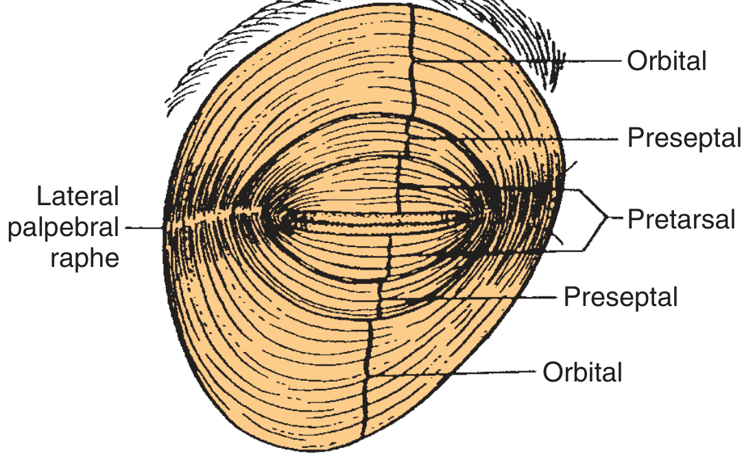
2. Orbicularis Oculi Muscle
The orbicularis acts as the sphincter of the eyelid, innervated by the temporal and zygomatic branches of the facial nerve (CN VII).
It is divided into three concentric segments:
| Segment | Location | Function |
|---|---|---|
| Pretarsal | Overlies the tarsal plate | Involuntary blinking; drives lacrimal pump |
| Preseptal | Overlies the orbital septum | Both involuntary and voluntary closure |
| Orbital | Over the orbital rim | Voluntary forced eye closure (winking) |
All three segments fuse laterally to form the crura of the lateral canthal tendon, which inserts at the Whitnall tubercle to maintain globe apposition.
The orbicularis is also essential for the lacrimal pump - propelling tears into the puncta, canaliculi, and lacrimal sac. Facial nerve paralysis disrupts this and causes epiphora (watering eye).
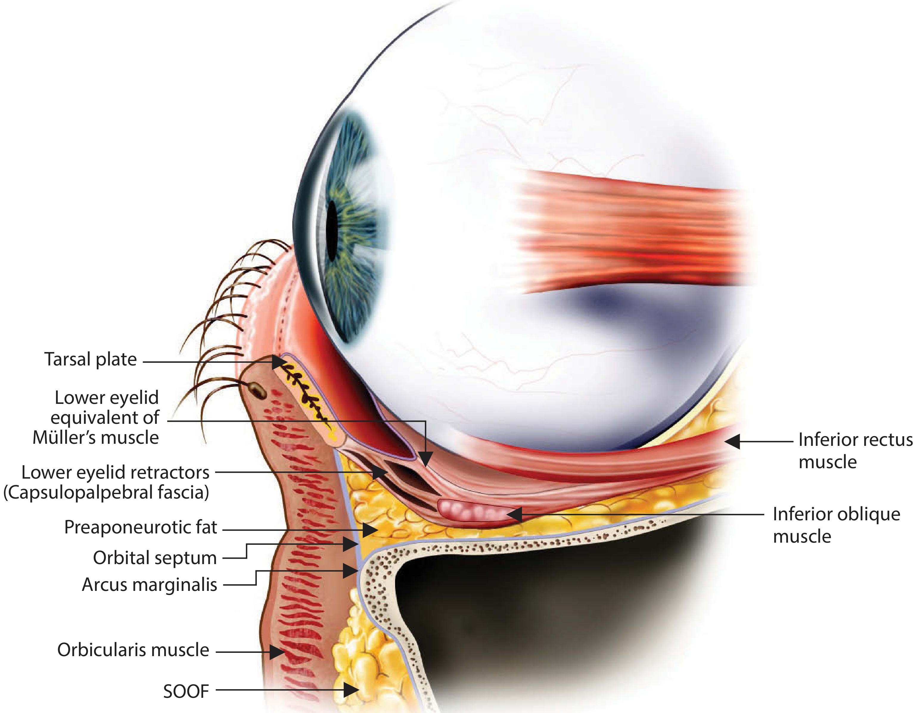
3. Orbital Septum
- A fibrous membrane deep to the orbicularis
- Forms the anterior border of the orbit and confines the orbital fat
- Arises from the orbital periosteum at the arcus marginalis at the orbital rim
- In upper eyelids (in whites): fuses with the levator aponeurosis ~3 mm above the superior tarsal border
- In Asian upper eyelids: this fusion occurs more inferiorly (below the tarsal border), allowing fat to prolapse anteriorly - this prevents a prominent upper eyelid crease
4. Eyelid Retractors
Upper eyelid retractors:
- Levator palpebrae superioris - the main elevator; a striated muscle innervated by CN III (oculomotor nerve). Its aponeurosis inserts into the anterior surface of the upper tarsal plate and into the skin to create the upper eyelid crease
- Müller's (superior tarsal) muscle - arises from the undersurface of the levator aponeurosis and inserts on the superior tarsal plate. Sympathetically innervated; contributes 2-3 mm of lid elevation. Loss (e.g., Horner's syndrome) causes mild ptosis
Lower eyelid retractors:
- Capsulopalpebral fascia - the equivalent of the levator aponeurosis in the lower lid; originates from the inferior rectus muscle fascia and inserts on the inferior tarsal border
- Inferior tarsal muscle - the sympathetically innervated equivalent of Müller's muscle in the lower lid
5. Orbital Fat Pads
- Pre-aponeurotic (preaponeurotic) fat lies between the orbital septum and the levator aponeurosis (upper lid) or capsulopalpebral fascia (lower lid)
- The upper eyelid has 2 fat compartments: nasal and central
- The lower eyelid has 3 fat compartments: nasal, central, and temporal
6. Tarsal Plates (Tarsus)
- Dense fibrous connective tissue forming the structural skeleton of the eyelids
- Horizontal dimension: 25-30 mm (both lids)
- Vertical height: 10-12 mm (upper lid) vs. 3-5 mm (lower lid)
- Contains the Meibomian (tarsal) glands - modified sebaceous glands that secrete the lipid layer of the tear film
7. Conjunctiva
- The innermost layer in contact with the globe
- Composed of nonkeratinized stratified squamous epithelium with goblet cells that secrete the mucous layer of the tear film
- Palpebral conjunctiva lines the eyelid; bulbar conjunctiva lines the globe; they meet at the conjunctival fornices
Surface Anatomy
| Measurement | Normal Value |
|---|---|
| Horizontal palpebral fissure | 28-30 mm |
| Vertical palpebral fissure | 9-10 mm |
| Intercanthal distance | 25-30 mm |
| Upper lid crease (women) | 10-12 mm above lid margin |
| Upper lid crease (men) | 7-8 mm above lid margin |
- The lateral canthal angle is more acute and lies 2 mm higher than the medial canthal angle
- The upper lid margin normally rests midway between the superior limbus and the pupil
- The lower lid margin abuts the inferior corneal limbus
Canthal Tendons
- Medial canthal tendon: has anterior and posterior limbs anchoring the medial ends of both tarsal plates to the lacrimal crest
- Lateral canthal tendon: formed by the fusion of the three orbicularis segments; inserts into the Whitnall tubercle of the zygomatic bone, ensuring eyelid-globe apposition
Blood Supply
- Anterior lamella: supplied by branches of the facial artery (external carotid system)
- Posterior lamella: supplied by branches of the ophthalmic artery (internal carotid system) via medial and lateral palpebral arteries, forming the marginal and peripheral arcades
Sources: Cummings Otolaryngology Head and Neck Surgery, Chapter 26 (Eyelid Anatomy); Scott-Brown's Otorhinolaryngology Head & Neck Surgery
This is a shared conversation. Sign in to Orris to start your own chat.