
Flat feet anatomy with images
Reading File
Now let me search the medical library for flat feet anatomy content and images in parallel.
Finding Sources
Finding Sources
Searching the Web
flat feet pes planus anatomy bones ligaments arches explained
Good results. Now let me read the main textbook sections and fetch relevant images in parallel.
Reading File
Reading File
Reading File
Loading Image
Now let me get the key anatomy content from Bailey & Love and fetch more images in parallel.
Reading File
Reading File
Loading Image
Loading Image
Now let me fetch the textbook images and also get the PTTD anatomy content.
Loading Image
Finding Sources
Finding Sources
Let me fetch the key anatomy arch images from the textbooks.
Loading Image
Reading File
Reading File
Let me now fetch the arch support diagram from Gray's Anatomy for Students, and the foot images from Bailey & Love.
Loading Image
Loading Image
Loading Image
I now have comprehensive content and images. Let me compile the full answer.
Flat Feet (Pes Planus) - Anatomy with Images
Flat feet, or pes planus, is defined as a partial or complete loss of the medial longitudinal arch of the foot, causing the arch to contact or nearly contact the ground during weight-bearing. About 15% of adults are affected, and it is essentially universal in children under 3 years of age.
1. Normal Foot Architecture - What Is Lost in Flat Feet
The foot has three arches that together form a structural tripod:
| Arch | Bones Involved | Key Support |
|---|---|---|
| Medial longitudinal arch | Calcaneus - talus - navicular - cuneiforms - 1st-3rd metatarsals | Spring ligament, tibialis posterior tendon |
| Lateral longitudinal arch | Calcaneus - cuboid - 4th-5th metatarsals | Long & short plantar ligaments |
| Transverse arch | Bases of metatarsals, cuneiforms, cuboid | Fibularis longus tendon |
In flat feet, the medial longitudinal arch collapses - this is the defining anatomical event.
Normal foot vs. flat foot - bone anatomy comparison:
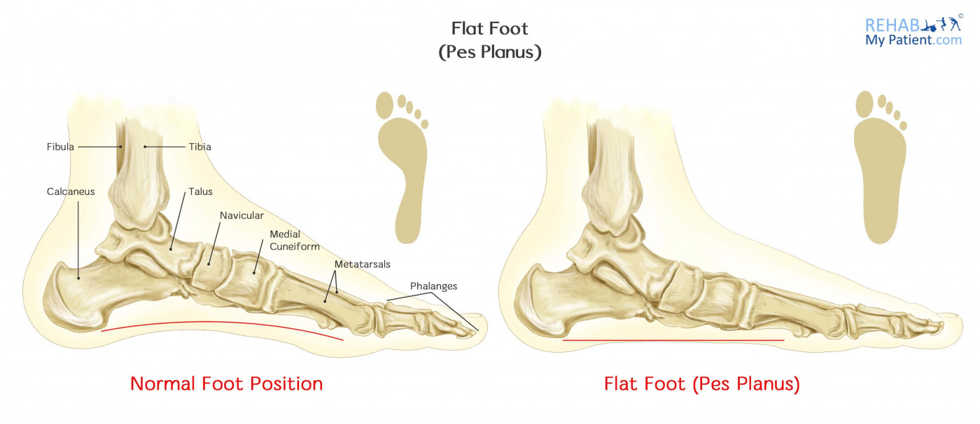
Normal foot (left) vs. pes planus (right). Note the collapsed medial arch in the flat foot. - RehabMyPatient
2. Bony Anatomy of Flat Feet
Key bones involved:
- Calcaneus (heel bone) - the largest tarsal bone; in flat feet it tilts into valgus (everts outward relative to the talus)
- Talus - articulates with the tibia above and calcaneus below at the subtalar joint; in flat feet the head of the talus drops medially and plantarward, causing talonavicular subluxation
- Navicular - key "keystone" bone; undergoes dorsal subluxation on the talus, producing midfoot sag
- Cuneiforms (medial, intermediate, lateral) - lose their elevated position as the arch flattens
- Metatarsals - 1st metatarsal angle increases as the arch drops
Radiographic measurement - the Feiss line:

The Feiss line connects the medial malleolus to the 1st metatarsophalangeal joint. In flat feet, the navicular tuberosity drops below this line. - Bloomsbury Osteopathy
On a standing lateral X-ray, the talus-first metatarsal angle (Meary's angle) is normally 0-10°. An increased angle indicates loss of the medial longitudinal arch. - Campbell's Operative Orthopaedics, 15th Ed.
X-ray showing flat foot (standing lateral view):
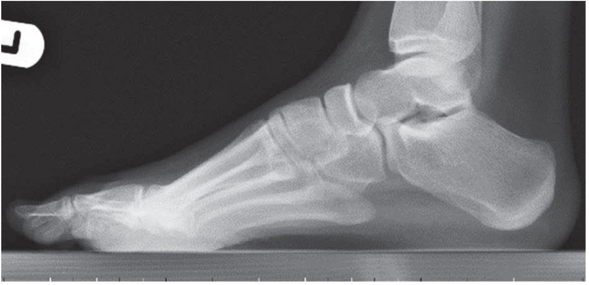
Normal medial longitudinal arch on lateral radiograph. - Bailey & Love's Short Practice of Surgery, 28th Ed.
3. Soft Tissue Anatomy - The Arch Support System
The arch is maintained by a layered system of static (passive) and dynamic (active) supports:
Static (Ligamentous) Supports
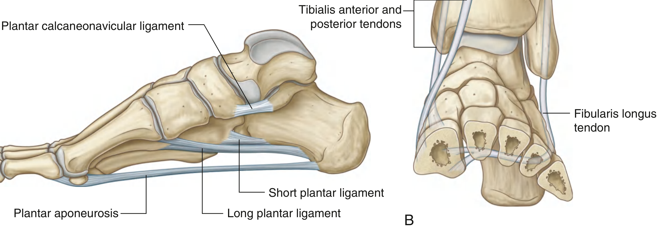
Ligaments supporting the foot arches, medial view. - Gray's Anatomy for Students
- Plantar calcaneonavicular (spring) ligament - the most important static stabilizer; runs from the sustentaculum tali of the calcaneus to the navicular and directly supports the head of the talus from below. Rupture of this ligament is a direct cause of acquired flat foot.
- Long plantar ligament - runs from the calcaneus to the cuboid and metatarsals; key for lateral arch support
- Short plantar ligament (plantar calcaneocuboid) - shorter, deeper than the long plantar ligament
- Plantar aponeurosis - a thick band anchored to the medial calcaneal tuberosity that fans out to the toes; supports the longitudinal arch via the "windlass mechanism"
Dynamic (Muscular) Supports
| Tendon/Muscle | Role in Arch Support |
|---|---|
| Tibialis posterior | Primary dynamic support; inverts foot and maintains medial arch |
| Tibialis anterior | Dynamic support for medial arch |
| Fibularis (Peroneus) longus | Supports transverse arch; stabilizes 1st metatarsal |
| Flexor hallucis longus | Supports medial longitudinal arch |
| Intrinsic foot muscles | Active load-sharing during stance phase |
4. The Cross-Sectional Anatomy of Flat Foot Deformity

Pes planus side cross-section - collapsed medial arch with key structures labeled.
5. Types of Flat Foot
Both types share a flattened medial arch and a valgus heel, but differ in behavior:
| Feature | Flexible Flat Foot | Rigid Flat Foot |
|---|---|---|
| Arch on tiptoe | Returns (subtalar joint corrects) | Fails to return; heel stays in valgus |
| Subtalar movement | Full, pain-free | Restricted, often painful |
| Cause | Normal variant, ligament laxity | Tarsal coalition, inflammatory arthritis |
| Treatment | Usually none needed; orthoses for symptoms | Surgical management often required |
- Bailey & Love's Short Practice of Surgery, 28th Ed.
Clinical photo - flat foot appearance:
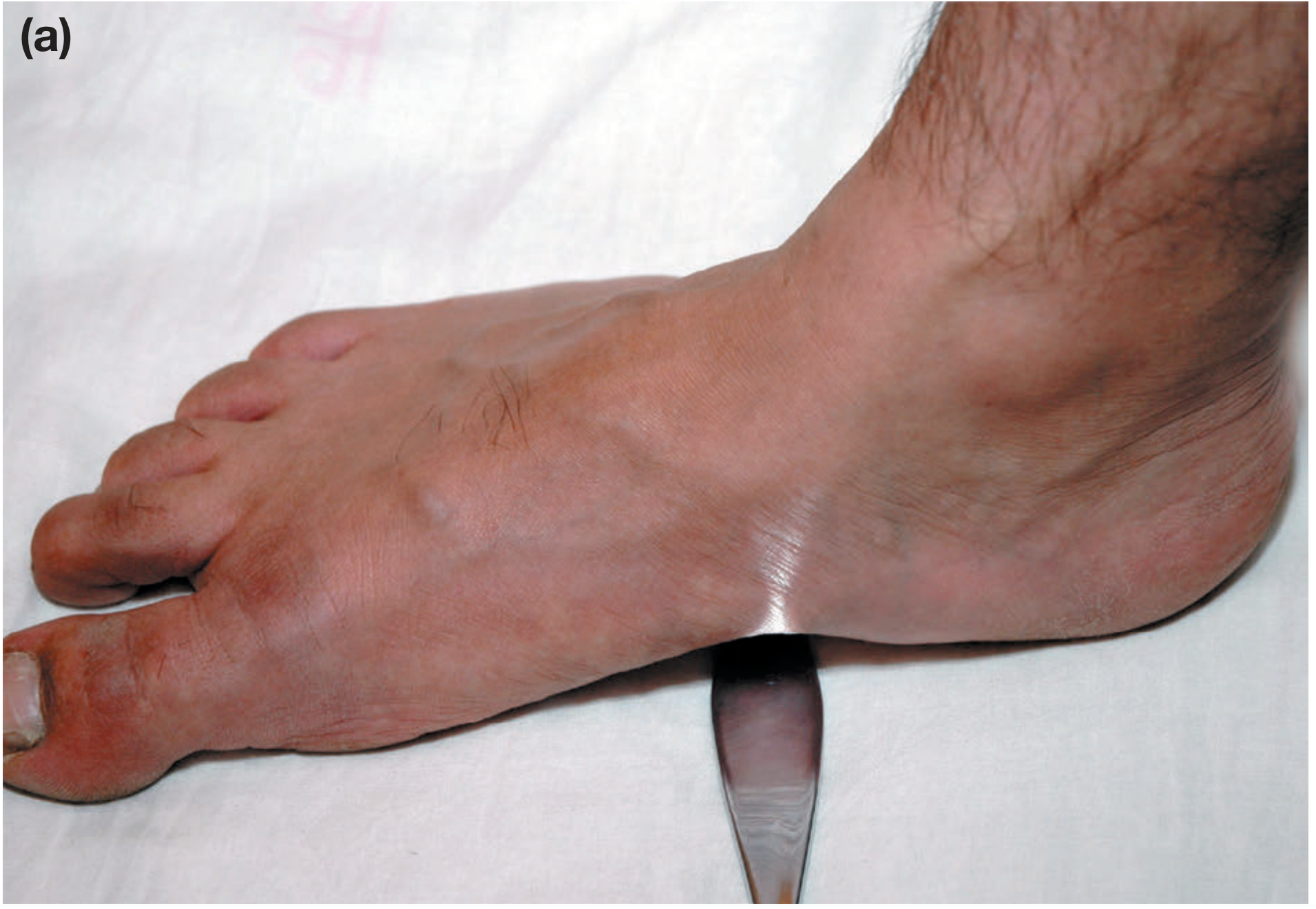
Clinical photo showing a flat foot with instrument passed under the absent arch. - Bailey & Love's Short Practice of Surgery, 28th Ed.
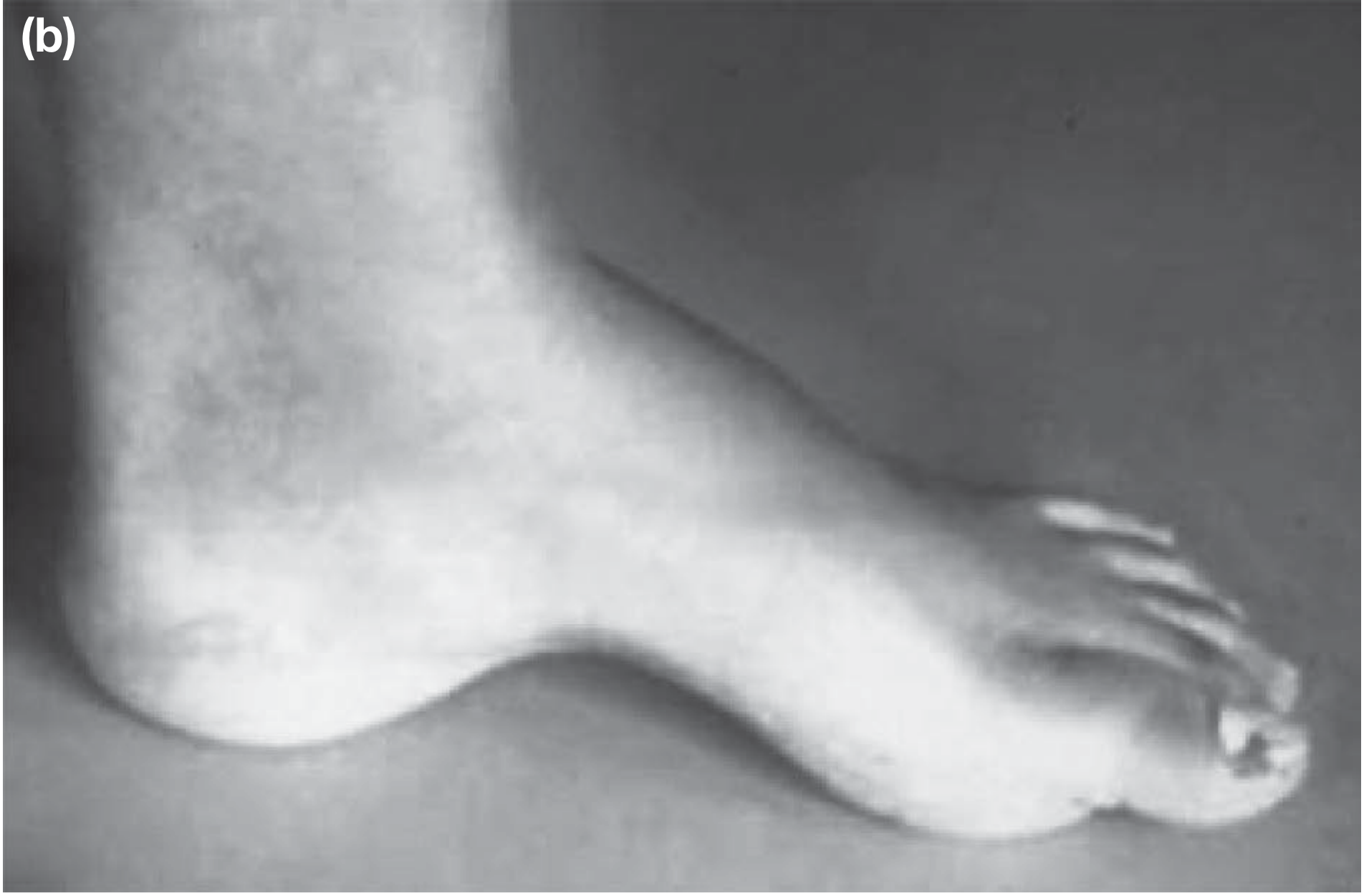
Pes planus - complete loss of medial arch visible clinically. - Bailey & Love's Short Practice of Surgery, 28th Ed.
6. Causes of Flat Foot (Pes Planus)
Congenital / developmental:
- Normal variant in infants (fat pad obscures arch before age 3)
- Familial ligamentous laxity / hypermobility syndromes (e.g. Marfan syndrome, Ehlers-Danlos)
- Tarsal coalition (failure of segmentation of mesenchymal tissue between tarsal bones) - causes a rigid, painful flat foot; affects ~1 in 100 people; 50% bilateral; presents in adolescence
Acquired Adult Flat Foot Deformity (AAFD):
- Posterior tibial tendon dysfunction (PTTD) - by far the most common cause in adults; the tibialis posterior is the primary dynamic arch support. Failure (common in overweight individuals and those with pre-existing flat feet) leads to progressive arch collapse. Key sign: inability to perform single-leg tiptoe.
- Spring ligament rupture - causes acute flat foot, especially in young athletes
- Tarsometatarsal arthritis / Lisfranc injury
- Charcot neuroarthropathy (e.g. diabetes)
- Inflammatory / degenerative arthritis of subtalar, talonavicular, or naviculocuneiform joints
- Bailey & Love's Short Practice of Surgery, 28th Ed.
- Miller's Review of Orthopaedics, 9th Ed.
7. Biomechanical Consequences
When the medial longitudinal arch collapses:
- The calcaneus moves into valgus (eversion) and external rotation relative to the talus
- Talonavicular subluxation occurs throughout the stance phase - a major biomechanical consequence
- The lateral column appears short relative to the medial column
- The navicular drops, causing midfoot sag
- The forefoot pronates
- There is increased stress on the spring ligament, deltoid ligament complex, and plantar fascia
- Downstream effects include altered knee, hip, and lumbar spine biomechanics
Radiographic evidence: On X-ray, an incomplete calcaneonavicular bar (a tarsal coalition) looks like an elongated anterior process of the calcaneus on lateral views. The "C-sign" or dorsal talar beaking suggests middle facet coalition.
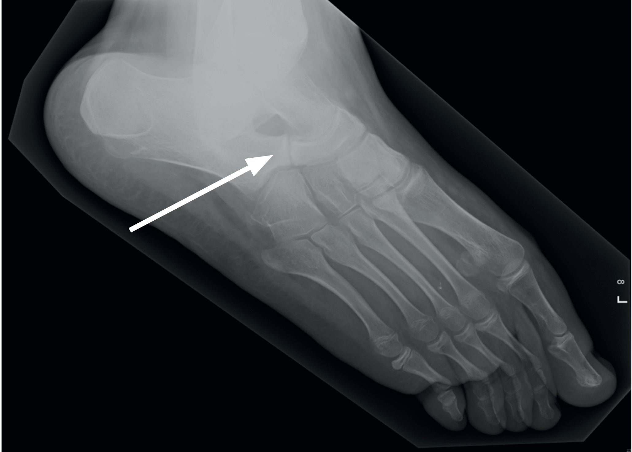
Oblique radiograph showing a calcaneonavicular bar (arrow) - the most common form of tarsal coalition causing rigid flat foot. - Bailey & Love's Short Practice of Surgery, 28th Ed.
Summary of Key Anatomical Structures in Flat Foot
| Structure | Normal Role | What Happens in Flat Foot |
|---|---|---|
| Medial longitudinal arch | Spring-like shock absorption | Collapses to ground |
| Talus | Sits atop calcaneus | Head drops medially/plantarward |
| Calcaneus | Heel foundation | Everts into valgus |
| Spring (calcaneonavicular) ligament | Cradles talus head | Overstretched or torn |
| Tibialis posterior tendon | Dynamic arch support | Dysfunctional or ruptured |
| Plantar aponeurosis | Static arch tie-bar | Overstrained |
| Navicular | Arch keystone | Subluxes dorsally on talus |
This is a shared conversation. Sign in to Orris to start your own chat.