
Why is there cymba concha tenderness in acute mastoiditis
cymba concha tenderness acute mastoiditis anatomy reason
cymba concha overlies mastoid antrum anatomy ENT otology
cymba concha mastoid anatomy auricle diagram
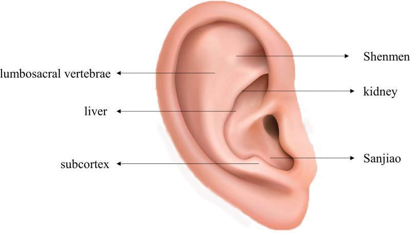
Anatomical diagram of a right human auricle (external ear) identifying specific acupoints used in auricular acupoint pressure therapy. The illustration shows a realistic depiction of ear landmarks including the helix, antihelix, concha, and lobe. Six specific acupoints are labeled with arrows pointing to their topographical locations: Shenmen (located in the triangular fossa), Kidney (superior concha), Sanjiao (inferior concha near the intertragic notch), Subcortex (inner aspect of the antitragus), Liver (cymba conchae), and Lumbosacral Vertebrae (antihelix crus). This visual serves as a clinical guide for the placement of pressure beans (Semen vaccariae) in Traditional Chinese Medicine protocols for systemic conditions. The image is categorized under clinical imaging and skull-related anatomy, intended for medical education on non-pharmacological interventions.
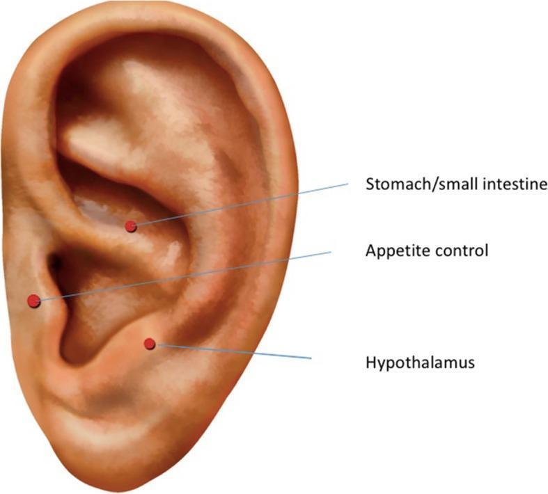
Anatomical diagram of a human right auricle illustrating specific auricular acupuncture points. The image highlights three targeted areas within the concha of the ear, marked with red circular dots and corresponding text labels. The 'Stomach/small intestine' point is located in the superior portion of the concha, near the crus of the helix. The 'Appetite control' point is positioned centrally within the auricular concha. The 'Hypothalamus' point is located in the inferior concha, adjacent to the tragus and the external auditory canal entrance. This diagram serves as a clinical reference for Auricular Electroacupuncture (AEA) protocols, demonstrating the topographic relationship between external ear anatomy (helix, antihelix, concha, and lobe) and traditional acupuncture sites used for physiological regulation, such as managing appetite or hormonal symptoms.
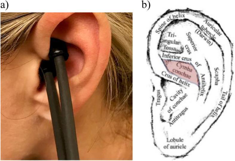
Educational visual regarding transcutaneous auricular vagus nerve stimulation (ta-VNS). Panel (a) is a clinical photograph showing a hand-held bipolar sensor probe applied to a human left ear. The probe's tips are positioned within the cymba conchae, the superior portion of the concha located above the crus of helix. Conductive gel is visible at the point of contact to facilitate electrical transmission. Panel (b) is a detailed anatomical diagram of the left auricle (pinna) labeling major landmarks: helix (spine, tail, and tubercle), antihelix (superior and inferior crus), scapha, triangular fossa, tragus, antitragus, cavity of conchae, and lobule. The cymba conchae is highlighted in red, illustrating its boundaries between the inferior crus of the antihelix and the crus of helix. This area is the primary anatomical target for ta-VNS due to the dense distribution of the auricular branch of the vagus nerve (ABVN). The content is designed for medical training in neuromodulation techniques and clinical neuroanatomy.
https://www.theentresident.com/ent-notes-lectures/external-e…
Why Cymba Concha is Tender in Acute Mastoiditis
The Key Anatomical Relationship
How This Causes Tenderness in Mastoiditis
- Infection spreads from the middle ear into the mastoid air cells via the aditus ad antrum.
- Inflammatory edema and pus build up in the mastoid air cell system, including and especially the mastoid antrum.
- The infection spreads to the periosteum overlying the mastoid cortex via emissary veins (periostitis stage) or by direct bony erosion (osteitis/coalescent stage).
- Since the mastoid antrum is directly deep to the cymba concha, the inflamed and edematous periosteum is immediately beneath the skin and cartilage at that point.
- Pressure applied to the cymba concha during examination is transmitted directly to this inflamed periosteum, producing tenderness.
Contrast with Postauricular Tenderness
| Site | What it overlies | Clinical significance |
|---|---|---|
| Cymba concha | Mastoid antrum (suprameatal triangle) | Tender in early mastoiditis (periostitis stage); accessible without displacing the pinna |
| Postauricular region | Mastoid cortex (outer surface) | Tender/swollen/erythematous when subperiosteal abscess forms; pinna is displaced forward and downward |
Summary
- The cymba concha overlies the suprameatal (MacEwen's) triangle, which itself directly overlies the mastoid antrum
- Infected, inflamed periosteum in mastoiditis is thus immediately deep to this auricular recess
- Pressing on the cymba concha compresses this inflamed periosteum, eliciting pain
- This is the same anatomical rationale behind using the suprameatal triangle as the surgical entry point in cortical mastoidectomy
Make a clinical reference PDF on mastoiditis diagnosis: signs, imaging findings, and antibiotic management