
What is dilatation and curettage? What are instruments required for this surgery? How do you position the patient on OT table?
dilatation and curettage D&C instruments uterus
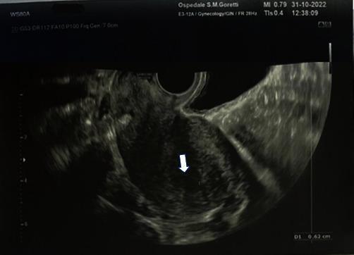
This diagnostic image is a transvaginal ultrasound (TVUS) of the uterus in a 36-year-old patient, performed as a post-procedural evaluation following dilatation and curettage (D&C) for a partial molar pregnancy. The uterus is visualized in a sagittal or longitudinal plane, appearing as a roughly pear-shaped or fan-shaped structure with mixed mottled echogenicity of the myometrium. A white arrow points to the endometrial cavity, which reveals a thin, regular, and linear hyperechoic (bright) endometrial stripe. This finding is clinically significant as it confirms the successful evacuation of gestational tissue and the absence of retained products of conception (RPOC). The image serves as an educational example of normal post-operative uterine morphology and the baseline appearance of the endometrium during the follow-up of gestational trophoblastic disease (GTD).
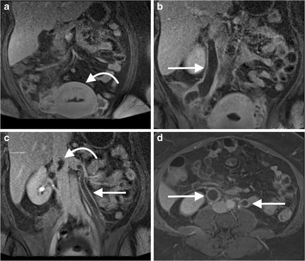
This diagnostic imaging panel consists of coronal (a-c) and axial (d) images from a post-contrast venous phase fat-saturated T1-weighted MR venogram of the abdomen. The case illustrates bilateral ovarian (gonadal) vein thrombosis in a 35-year-old post-partum patient following dilatation and curettage. Image (a) identifies the post-partum uterus (curved arrow). Images (b) and (c) demonstrate marked dilatation of the right and left gonadal veins (straight arrows) with prominent central filling defects, indicating intraluminal thrombus. In image (c), a curved arrow highlights the superior extension of the thrombus reaching the juxtarenal inferior vena cava (IVC). The axial image (d) provides a cross-sectional view of the expanded gonadal veins bilaterally, clearly showing the low-signal intensity central thrombi surrounded by peripheral contrast enhancement (straight arrows). This study demonstrates a rare but serious post-partum complication involving extensive pelvic venous thrombosis and its pathway toward the central venous system.
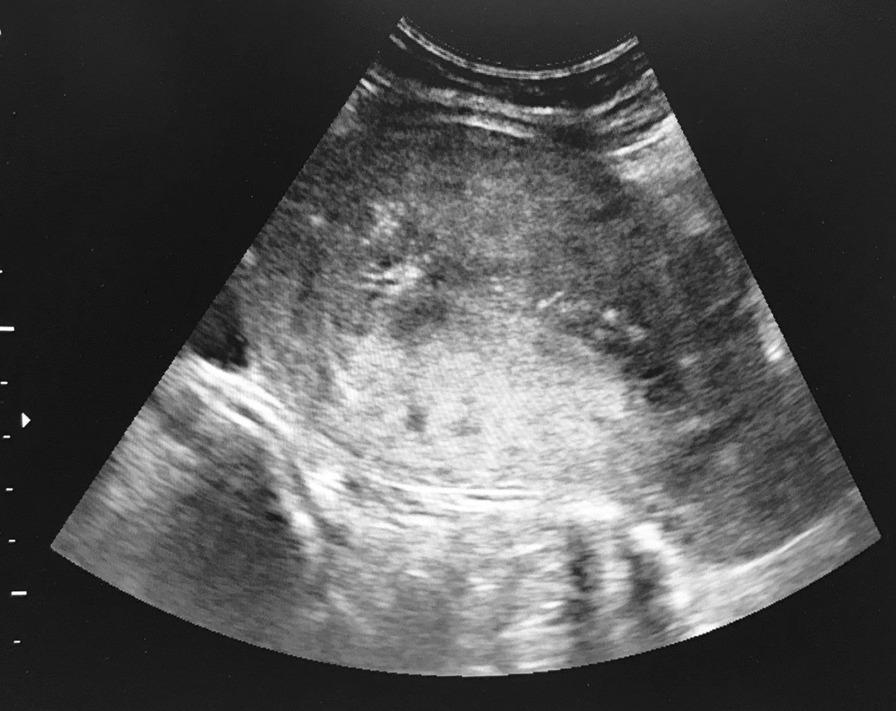
This diagnostic image is a transabdominal ultrasound of the pelvis, focusing on the uterus. The scan reveals a significantly enlarged uterine cavity filled with complex, heterogeneous tissue. The echotexture is characterized by a 'snowstorm' appearance, consisting of numerous small, hypoechoic (cystic) spaces interspersed with hyperechoic (echogenic) areas, suggesting solid components or blood clots. The margins of the intrauterine mass are irregular and poorly defined, with no evidence of a healthy gestational sac or normal endometrial strip. This visual presentation is highly characteristic of gestational trophoblastic disease (GTD), such as a hydatidiform mole. The image demonstrates the clinical utility of real-time ultrasound guidance during a dilatation and curettage (D&C) procedure to monitor the evacuation of abnormal uterine contents and ensure the safety of the uterine wall. The primary educational focus is the sonographic identification of abnormal molar pregnancy tissue and the management of high-beta-hCG pelvic masses.
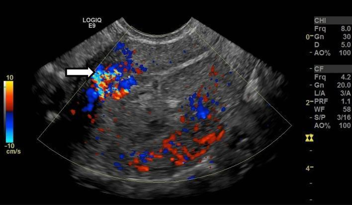
This color Doppler ultrasound image in the longitudinal plane displays the uterus of a 28-year-old female patient with a history of recent dilation and curettage (D&C). The image demonstrates enhanced myometrial vascularity (EMV), characterized by a focally increased color Doppler signal (indicated by a white arrow) spanning the full thickness of the anterior myometrium. This vascular signal extends directly into the endometrial cavity, which appears distended with isoechoic material. The color flow manifests as a complex mosaic of red and blue hues, indicating multidirectional, high-velocity blood flow within the myometrial wall. This clinical presentation and imaging finding are highly suggestive of retained products of conception (RPOC) or an acquired uterine arteriovenous malformation (AVM) secondary to pregnancy-related procedures. The imaging modality provides critical diagnostic information for differentiating between physiological postpartum changes and pathological vascular conditions requiring medical or surgical intervention.
dilatation and curettage D&C procedure instruments patient positioning lithotomy
https://www.ncbi.nlm.nih.gov/books/NBK568791
Hegar dilators uterine curette tenaculum uterine sound D&C instruments set gynecology
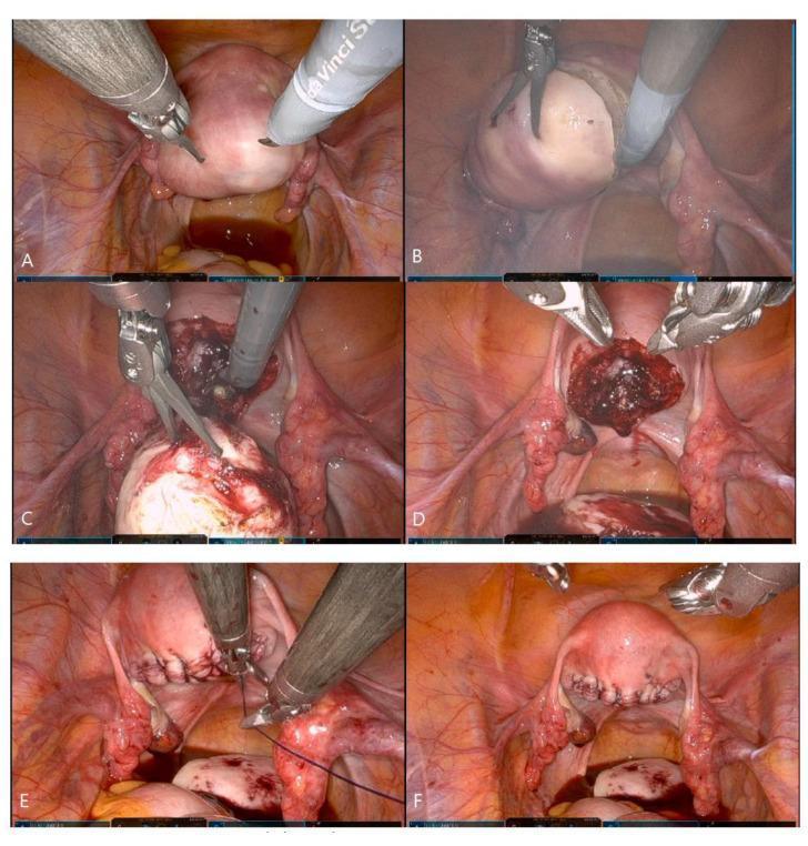
A series of six intraoperative endoscopic photographs (A-F) documenting a coaxial-robotic single-site myomectomy (C-RSSM). Panel (A) shows the initial robotic camera view of the uterus with surgical instruments positioned at the pelvic cavity. Panel (B) depicts the uterine serosal incision and the application of traction and countertraction to the intramural myoma. Panel (C) demonstrates the use of a robotic tenaculum grasper for powerful traction during the enucleation process. Panel (D) reveals the resulting uterine defect (cavity) after the myoma has been fully enucleated, showing visible hemorrhagic tissue within the bed. Panel (E) illustrates the reconstructive phase, where a mega-needle driver is used to perform layer-to-layer suturing of the uterine wall with barbed sutures. Panel (F) displays the final post-procedural state with the uterine defect successfully closed and anatomical integrity restored. The images highlight the limited range of motion and instrument positioning characteristic of single-site robotic gynecological surgery.
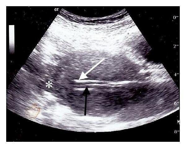
This diagnostic image is a transabdominal ultrasound in the sagittal plane, demonstrating ultrasound-guided intrauterine instrumentation. The scan shows two distinct, hyperechoic linear structures entering the uterine cavity. A white arrow identifies a suction curette, which is seen threaded over a secondary hyperechoic linear guide, indicated by a black arrow as a 10-French intubating stylet. A white asterisk (*) marks the uterine fundus, the intended anatomical destination within the uterine cavity. The surrounding myometrium and pelvic tissues exhibit heterogeneous echogenicity with characteristic speckled artifact. The image illustrates a clinical technique used to navigate difficult anatomy or false tracts (such as uterine perforation sites) by using a shaped stylet as a guide for the curette. This educational visual is relevant for obstetrics and gynecology, specifically regarding complications and management during surgical abortion or dilation and curettage (D&C) procedures.
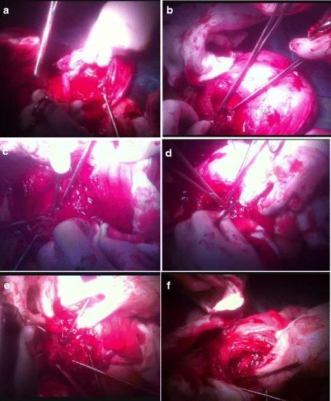
This composite of six intraoperative clinical photographs (a-f) illustrates the surgical steps of the cervical inversion technique used to manage massive hemorrhage from the placental bed. (a) Initial view of the surgical field showing active bleeding from the placental implantation site. (b) Traction and inversion of the cervical lips using two Allis forceps. (c) Insertion of a Hegar dilator into the cervical canal to maintain patency during reconstruction. (d) Suturing of the posterior cervical lip to the posterior wall of the lower uterine segment. (e) Suturing of the anterior cervical lip to the corresponding anterior wall of the lower uterine segment, effectively creating a surgical tamponade. (f) Final stage showing the removal of the Hegar dilator with the cervical inversion completed. This procedure is a conservative surgical intervention for placenta previa or accreta to achieve hemostasis while preserving the uterus. The images demonstrate the use of standard surgical instruments, including forceps, dilators, and needle drivers, in an acute obstetric surgical context.
Dilatation and Curettage (D&C)
What is D&C?
- Dilatation - the cervix (lower narrow part of the uterus) is mechanically opened (dilated) using progressively larger dilators
- Curettage - the endometrial lining of the uterus is scraped using a curette to remove tissue
Indications
- Abnormal uterine bleeding (AUB) - to rule out endometrial carcinoma or hyperplasia
- Postmenopausal bleeding
- Suspected endometrial polyp or submucosal fibroid
- Endometrial sampling when office biopsy is inconclusive
- Incomplete, missed, or inevitable abortion (evacuation of retained products of conception)
- Hydatidiform mole (suction D&C - first-line treatment)
- Menorrhagia not responding to medical therapy
- Gestational trophoblastic disease
- Elective or therapeutic termination of pregnancy (first trimester)
Instruments Required
1. Exposure and Visualization
| Instrument | Purpose |
|---|---|
| Sims speculum (posterior lip) + anterior vaginal retractor OR Bivalve (Cusco's) speculum | Retracts vaginal walls to expose the cervix |
| Deep weighted speculum (Auvard's) | Self-retaining posterior retractor used under general anesthesia |
| Narrow Deaver retractors | Additional lateral retraction if needed |
| Adequate light source / headlight | Illumination of the operative field |
2. Grasping the Cervix
| Instrument | Purpose |
|---|---|
| Vulsellum / Volsellum forceps (single-tooth or double-tooth tenaculum) | Grasps the anterior lip of the cervix at the 12 o'clock position for traction and to straighten the cervicouterine axis |
| Jacobs tenaculum or Ring forceps | Alternative to single-tooth tenaculum; less traumatic |
3. Sounding the Uterus
| Instrument | Purpose |
|---|---|
| Uterine sound | A long graduated probe inserted into the uterine cavity to measure depth (normally 6-8 cm) and assess the axis/direction of the uterus before dilating |
4. Cervical Dilation
| Instrument | Purpose |
|---|---|
| Hegar dilators (sizes 1-10 or beyond) | The most widely used set; double-ended, graduated metal dilators inserted progressively to dilate the cervical os |
| Pratt dilators | Tapered French-size dilators; an alternative to Hegar |
| Hank dilators | Another set of metal dilators; less commonly used |
5. Curettage
| Instrument | Purpose |
|---|---|
| Uterine curette (sharp) - small, medium, large sizes | The main working instrument; spoon-shaped end with sharp edges scrapes the endometrial lining systematically |
| Uterine curette (blunt/ovum forceps) | Used to remove larger fragments, especially products of conception |
| Suction curette / vacuum cannula | 5-12 mm plastic or metal cannulas connected to a manual (50 mL modified syringe) or electric vacuum pump for aspiration curettage - safer and more efficient than sharp curettage |
| Ovum forceps / sponge-holding forceps | Remove larger tissue fragments from the cavity |
6. General Surgical Support
| Item | Purpose |
|---|---|
| Sterile gloves, gown, drapes, face shield | Standard sterile precautions |
| Betadine (povidone-iodine) or Hibiclens solution | Antiseptic preparation of vulva, perineum, and vagina |
| Foley catheter | Bladder drainage before and during the procedure |
| Kidney tray, swabs, sponge-holding forceps | General surgical tray |
| Specimen jar with formalin | For tissue preservation and histopathology |
Patient Positioning on the Operating Table
Step-by-Step Positioning
-
Supine on the OT table - the patient lies flat on her back.
-
Buttocks at the table edge - the patient's buttocks are brought to the very edge (lower end) of the table so the perineum is accessible. The lower leaf/foot of the table is dropped or removed.
-
Legs raised into stirrups - both legs are raised simultaneously and symmetrically by two individuals. Legs are placed in Allen stirrups (preferred) or candy cane stirrups, supporting the legs at the popliteal fossa and heel.
-
Hip and knee position - hips are in slight flexion and abduction; knees are in slight flexion (not hyperflexed). Thighs should be in a near-neutral position.
-
Arms - positioned at the patient's sides (adducted and pronated) to reduce the risk of brachial plexus injury and to give the surgeon freedom of movement.
-
Bladder drained - a catheter is passed once the patient is positioned.
Key Points to Prevent Nerve Injury (per Medscape)
- Avoid excessive thigh abduction and external rotation - this can injure the femoral nerve (causing difficulty walking, numbness of the thigh)
- Avoid hyperflexion of the knee - this compresses the common peroneal nerve causing foot drop
- Both legs raised/lowered simultaneously - to prevent sacroiliac strain
- Lateral aspect of the knee padded - protects the common peroneal nerve
- Allen stirrups are preferred over candy cane stirrups for nerve safety
Step-by-Step Procedure Outline
- Anesthesia (general/spinal/paracervical block/IV sedation)
- Position in dorsal lithotomy; Foley catheter inserted
- Bimanual examination under anesthesia - assess uterine size, position (anteverted/retroverted), and cervical dilation
- Prep and drape in sterile fashion (Betadine to perineum and vagina)
- Speculum placed to expose cervix
- Vulsellum/tenaculum applied to anterior lip of cervix at 12 o'clock; gentle downward traction applied
- Uterine sound inserted to measure depth and confirm axis
- Serial cervical dilation with Hegar dilators starting from the smallest, progressing to No. 8 or 9
- Systematic curettage of all four walls, fundus, and cornua using the largest appropriate curette
- Tissue collected into specimen jar and sent for histopathology
- Check for completeness; assess for excessive bleeding