

photo-1789035772432.jpg
Act like my teacher teache me all of it with pictures
Reading File
analyse_media
Finding Sources
Searching PubMed
"anterior cruciate ligament" AND examination
Searching the Web
ACL PCL knee examination anatomy adductor canal reputable medical reference
Reading File
Finding Sources
Searching Images
labeled diagram of anterior cruciate ligament and posterior cruciate ligament in knee joint
Searching Images
labeled anatomy diagram adductor canal femoral artery vein saphenous nerve vastus medialis sartorius adductor longus
Reading File
Finding Sources
Reading Web Page
https://www.ncbi.nlm.nih.gov/books/NBK535416
Quick answer
Lower limb page: ACL, PCL and adductor canal
1. Start with the knee bones
The knee is mainly the joint between:
- Femur: thigh bone
- Tibia: main leg bone
- Patella: kneecap
Inside the knee, the ACL and PCL cross each other, hence the name cruciate meaning cross-shaped.
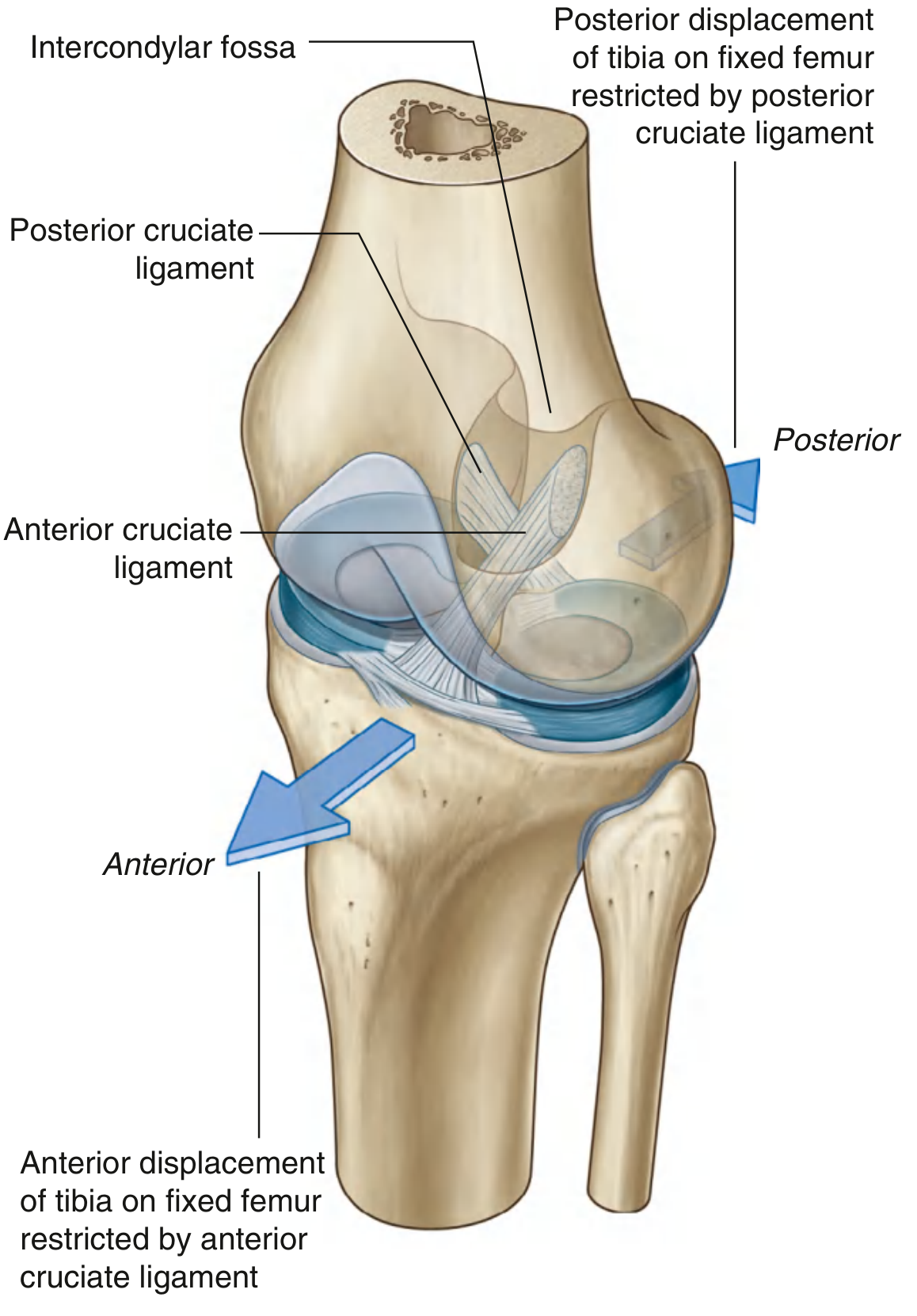
2. Cruciate ligaments: the core idea
A. ACL: Anterior Cruciate Ligament
Attachments
- Starts from the anterior intercondylar area of tibia
- Runs upward, backward, and laterally
- Attaches to the posterior part of the medial surface of the lateral femoral condyle
Main function
ACL prevents anterior displacement of the tibia on the femur.
In simple words:
ACL stops tibia from sliding FORWARD
If you keep the tibia fixed instead, the same fact can be said as:
ACL stops femur from sliding BACKWARD on tibia
Both statements mean the same thing.
Memory trick
ACL prevents Anterior movement of the tibia.
ACL injury
An ACL tear produces:
- Anterior instability of tibia on femur
- Often a twisting injury during sport, sudden deceleration, landing, or change in direction
- Possible “pop” sensation followed by rapid swelling
- Difficulty pivoting or changing direction
Tests for ACL
| Test | Meaning if positive |
|---|---|
| Lachman test | Best routine clinical test for ACL tear |
| Anterior drawer test | Tibia moves excessively forward |
| Pivot shift test | Suggests rotational instability, often ACL deficiency |
Lachman test: knee is held about 20-30° flexed and tibia is pulled forward. Excess forward movement or a soft end point suggests ACL injury.
For exams, say: Lachman is the most sensitive clinical test for ACL injury. It is generally preferred in an acute injury because hamstring spasm may make the anterior drawer test less reliable.
B. PCL: Posterior Cruciate Ligament
Attachments
- Starts from the posterior intercondylar area of tibia
- Runs upward, forward, and medially
- Attaches to the lateral surface of the medial femoral condyle
Main function
PCL prevents posterior displacement of the tibia on the femur.
PCL stops tibia from sliding BACKWARD
Equivalently:
PCL stops femur from sliding FORWARD on tibia
Memory trick
PCL prevents Posterior movement of tibia.
PCL injury
Classic cause: a force pushes the upper tibia backward while the knee is flexed.
Dashboard injury:
flexed knee hits dashboard
        ↓
tibia pushed posteriorly
        ↓
PCL injury
This is why a posterior cruciate injury can occur in road-traffic trauma.
Test for PCL
| Test | Meaning if positive |
|---|---|
| Posterior drawer test | Excess backward movement of tibia |
| Posterior sag sign | Tibia appears to sag backward when knee is flexed |
Posterior sag sign: with hip flexed to 45° and knee flexed to 90°, compare the tibial tuberosity with the other side. In a PCL tear, the tibia lies abnormally posterior.
3. The most important comparison
| Feature | ACL | PCL |
|---|---|---|
| Full name | Anterior cruciate ligament | Posterior cruciate ligament |
| Stops tibia moving | Anteriorly | Posteriorly |
| Equivalent femoral movement prevented | Posterior displacement of femur | Anterior displacement of femur |
| Important test | Lachman / anterior drawer | Posterior drawer / posterior sag |
| Classic injury | Pivoting, deceleration, sports | Dashboard injury |
| Tibial attachment | Anterior intercondylar area | Posterior intercondylar area |
| Femoral attachment | Lateral femoral condyle | Medial femoral condyle |
One-line exam answer
ACL prevents anterior displacement of tibia on femur.
PCL prevents posterior displacement of tibia on femur.
4. Correcting the page’s MCQs
“Anterior dislocation of tibia on femur is prevented by ACL”
✅ Correct.
“Anterior dislocation of femur on tibia is prevented by PCL”
✅ Correct.
This feels confusing because the same relative movement can be described from either bone:
Tibia forward = femur backward
Tibia backward = femur forward
“ACL is intracapsular”
✅ Correct, with an important refinement:
- ACL and PCL are intracapsular
- But they are extrasynovial, because synovial membrane reflects around them
So the best phrase is:
ACL and PCL are intracapsular but extrasynovial.
“ACL injury causes anterolateral rotatory instability”
✅ Correct. ACL is an important restraint to anterior tibial translation and contributes to rotational stability.
“Blow to lateral side of knee causes tibial collateral ligament injury”
✅ Correct.
A force from the lateral side opens the medial side of the knee, creating a valgus force, which injures the:
Medial collateral ligament = tibial collateral ligament
The unhappy triad
A valgus injury may damage:
MCL + medial meniscus + ACL
5. Collateral ligaments, quickly
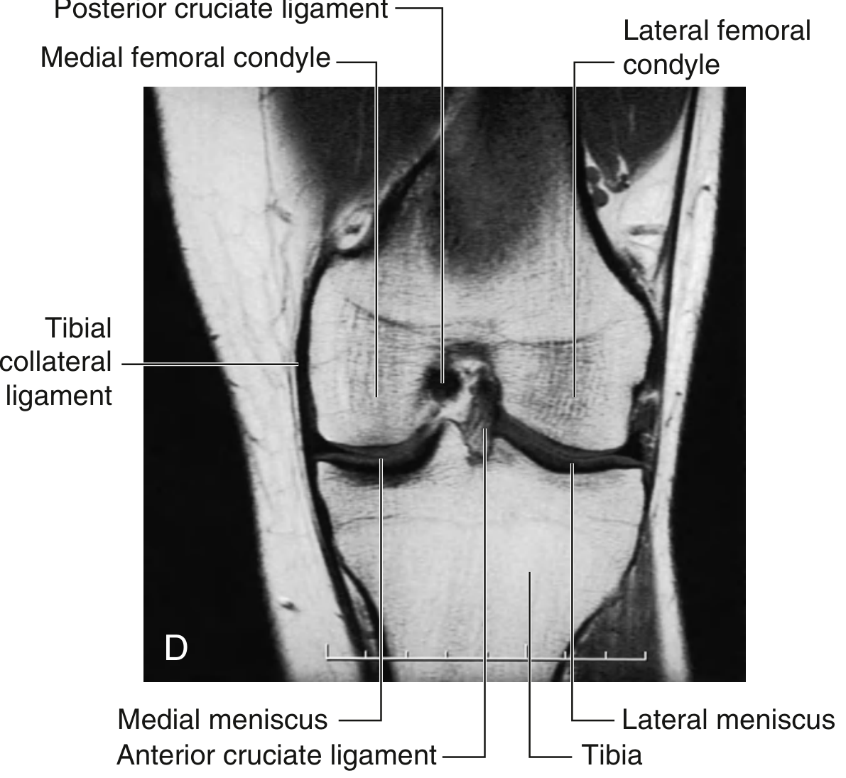
| Ligament | Other name | Main role | Typical force causing injury |
|---|---|---|---|
| Medial collateral ligament | Tibial collateral ligament | Resists valgus force | Blow to lateral side of knee |
| Lateral collateral ligament | Fibular collateral ligament | Resists varus force | Blow to medial side of knee |
6. Adductor canal
Also called:
Adductor canal = Hunter canal = Subsartorial canal
It is a tunnel in the middle third of the thigh through which major vessels and nerves travel toward the knee.
Boundaries
Think of a triangular tunnel:
              Roof
           Sartorius
              /\
             /  \
Vastus medialis   Adductor longus/magnus
   lateral             posteromedial
| Boundary | Structure |
|---|---|
| Anterolateral wall | Vastus medialis |
| Posteromedial wall | Adductor longus above, adductor magnus below |
| Roof | Sartorius and subsartorial fascia / vastoadductor membrane |
Easy mnemonic
SVA:
Sartorius = roof
Vastus medialis = lateral
Adductors = posteromedial
Contents of adductor canal
From anterior to posterior, important contents include:
- Femoral artery
- Femoral vein
- Saphenous nerve
- Nerve to vastus medialis
- Descending genicular vessels, near the lower end
Very important correction
The femoral nerve itself does not pass through the adductor canal.
Only its branches pass through, mainly:
- Saphenous nerve
- Nerve to vastus medialis
Adductor canal contents:
Femoral artery
Femoral vein
Saphenous nerve
Nerve to vastus medialis
The saphenous nerve is sensory and eventually supplies skin on the medial side of the leg and foot.
7. Clinical importance of adductor canal
Infection or pus in the canal
The structure most likely affected is the:
Femoral vein
This is because the vein lies close to the femoral artery within the confined canal.
Adductor canal block
An anesthetic injection around the saphenous nerve in this region can provide analgesia for the medial knee and leg, often with less quadriceps weakness than a femoral nerve block.
8. Final rapid revision
ACL: prevents anterior tibial translation
PCL: prevents posterior tibial translation

ACL tear: Lachman positive, anterior drawer positive
PCL tear: posterior drawer positive, posterior sag sign

ACL and PCL: intracapsular but extrasynovial

MCL injury: blow to lateral knee, valgus force
LCL injury: blow to medial knee, varus force

Adductor canal:
Roof - sartorius
Lateral wall - vastus medialis
Posteromedial wall - adductor longus and magnus

Contents:
Femoral artery, femoral vein,
saphenous nerve, nerve to vastus medialis
The anatomy of ACL and PCL on your page agrees with the textbook description: ACL limits anterior tibial displacement and PCL limits posterior tibial displacement, as described in Gray’s Anatomy for Students. The PCL posterior drawer and dashboard mechanism are also summarized in this NCBI clinical anatomy reference.
This is a shared conversation. Sign in to Orris to start your own chat.