
Pus filled something in underarm. Even after rupturing again growing and filling with pus
hidradenitis suppurativa axilla abscess
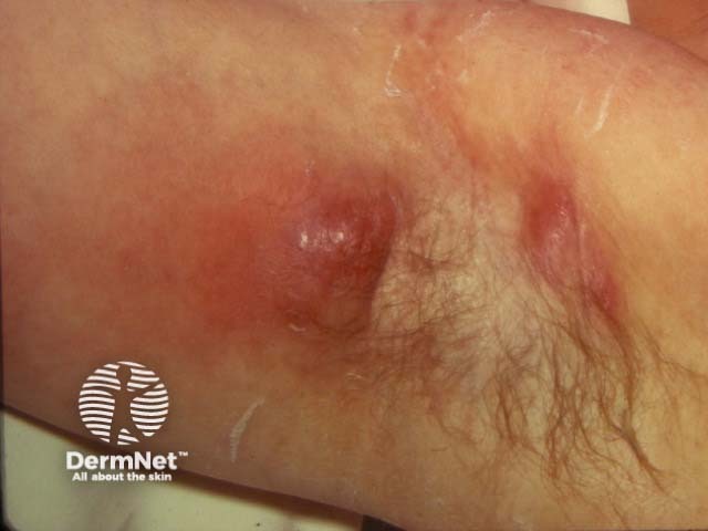
Clinical photograph of axillary skin demonstrating hidradenitis suppurativa (HS) in an apocrine-bearing region. The image shows a localized, inflamed erythematous nodular lesion with a raised, indurated border and a central fluctuant core suggestive of abscess formation. Surrounding skin exhibits mild edema and post-inflammatory changes, including hyperpigmentation and superficial crust. Hair follicles are visible at the periphery and there may be faint sinus tracts or scarring extending laterally toward surrounding axillary folds, consistent with chronic HS. The presentation is typical for HS of the axilla: recurrent, painful nodules that may ulcerate, discharge purulent material, and heal with scarring. The lesion site corresponds to the axillary region (apocrine gland-bearing area) within the integumentary system; this condition commonly involves both axillae but can be unilateral in early stages. Clinically, HS presents as a spectrum from tender papules and boils (furuncles) to draining sinus tracts and fibrotic plaques (Hurley stage II–III). Differential diagnoses include folliculitis, furunculosis, epidermal cyst, and simple abscess; risk factors include obesity, smoking, and metabolic syndrome. Management implications include anti-inflammatory and antibiotic therapy, surgical considerations for refractory disease, and potential biologic agents; this image is educational for recognizing HS features and differentiation from mimics in dermatology and medical education.
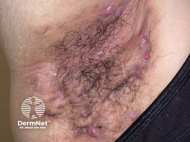
Clinical photograph of the axillary skin demonstrating hidradenitis suppurativa in an active and scarred phase. The image shows multiple inflamed nodules with surrounding erythema, coalescing abscess-like lesions, and visible sinus tracts with purulent drainage. Tender, firm-to-soft plaques with surrounding induration appear within the axillary crease, accompanied by scarring, sinus formation, and crusting. Hair follicles are involved with perifollicular inflammation; edema and erythema reflect ongoing inflammatory activity. Chronicity is suggested by extensive scar tissue and irregular, softened sinus openings. The modality is dermatologic photography, acquired as a close-up frontal/anterior view of the axilla under standard lighting. Notably, this presentation aligns with Hurley stage II-III disease, where recurrent inflamed nodules progress to sinus tracts and scarring. Clinically relevant features include pain, drainage, malodor, and restricted arm movement. This image is useful for educational discussions of HS morphology, differential diagnosis (folliculitis, abscess, epidermal cyst), and treatment planning including medical therapy (antibiotics, anti-inflammatory agents) and surgical considerations (excision, deroofing). It highlights the importance of early recognition to prevent progression and minimize scarring.
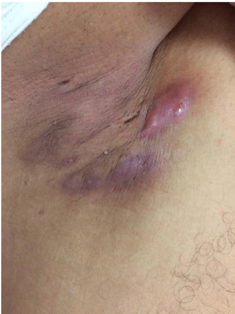
This clinical photograph displays a close-up view of the human axilla demonstrating signs of Hidradenitis Suppurativa (HS). The primary findings are multiple erythematous, inflamed lumps and deep-seated nodules. The largest lesion is a prominent, elongated, reddish-pink nodule with a shiny, tense surface, suggesting acute inflammation or early abscess formation. Below it, a linear, dusky-purple area indicates chronic inflammatory activity, possible sinus tract formation, and underlying fibrosis. The surrounding skin shows post-inflammatory hyperpigmentation, particularly within the skin folds, and textural irregularities consistent with scarring from previous flare-ups. A few scattered small, inflammatory papules are also visible in the periphery. This image illustrates the characteristic distribution of HS in intertriginous areas where apocrine glands are concentrated, highlighting the progression from acute nodules to chronic scarring and disfigurement, which is essential for clinical staging (Hurley criteria) and diagnostic classification in dermatology.
Pus-Filled Lump in the Underarm That Keeps Refilling
What Is It?
- The duct of a hair follicle or apocrine gland gets blocked by dead skin cells (keratin)
- This causes the gland to swell, rupture internally, and become infected
- Pus collects and forms an abscess (the painful lump)
- Even after it drains/ruptures, the underlying problem (blocked gland, inflammation) remains
- So it refills and recurs - often forming tunnels under the skin called sinus tracts
"Occlusion of gland ducts leads to bacterial proliferation, gland rupture and the spread of infection and epithelial components into the surrounding soft tissue and to adjacent glands." - Bailey and Love's Short Practice of Surgery, 28th Ed.
What It Looks Like
Who Gets It?
- 3x more common in women than men
- Usually starts after puberty, rarely after age 40
- Risk factors: obesity, smoking, possibly hormonal imbalance (androgen excess)
- Genetic predisposition plays a role
- Does NOT occur in children before puberty
Staging (Hurley Classification)
| Stage | Description |
|---|---|
| Stage I | Single or multiple abscesses, no sinus tracts or scarring |
| Stage II | Recurrent abscesses, sinus tracts, some scarring |
| Stage III | Multiple interconnected sinus tracts, extensive scarring |
Treatment
Mild / Early Stage:
- Antiseptic washes (chlorhexidine) daily on the area
- Weight loss if overweight - significantly helps
- Stop smoking - major trigger
- Topical clindamycin (antibiotic gel applied to skin)
- Oral antibiotics - tetracycline or doxycycline for 3+ months
- Avoid tight clothing, friction, and excessive sweating in that area
Moderate / Persistent:
- Combined oral antibiotics (clindamycin + rifampicin) for 10-12 weeks
- Hormonal therapy (anti-androgens like spironolactone in women)
- Intralesional steroid injections for individual nodules
Severe / Recurrent (Stage II-III):
- Biological agents: Adalimumab (Humira) - the only FDA-approved biologic for HS
- Surgical options: Deroofing (unroofing sinus tracts), wide local excision of affected skin
- Complete removal of all apocrine-gland-bearing skin in the axilla in severe cases
"Antibiotics may induce remission but often the disease relapses and progresses, at which point surgery is indicated." - Bailey and Love's Surgery
When to See a Doctor URGENTLY
- Rapidly enlarging, very painful lump with fever
- Red streaks spreading from the lump (sign of spreading infection/cellulitis)
- Multiple lumps appearing in different spots
- The lump has been present for more than 2 weeks without improvement
Important Points
- Do NOT keep squeezing or trying to burst it yourself - this pushes infection deeper and can worsen sinus tract formation
- Simple incision and drainage (I&D) is only a temporary fix - it relieves the acute abscess but does not address the underlying disease
- HS is a chronic medical condition, not just a one-time infection - it needs ongoing management with a dermatologist or surgeon
- The most common bacteria found are Staphylococcus, Streptococcus, and sometimes E. coli