
Explain ascending and descending tracts
ascending and descending tracts spinal cord diagram
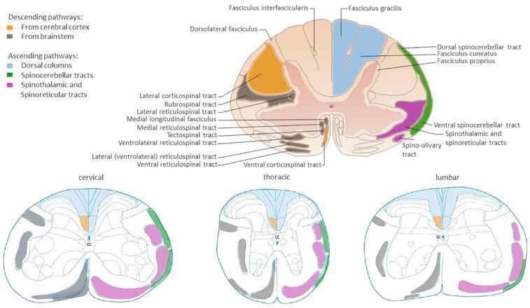
Anatomical diagrams illustrating the organization of major white matter tracts in the human spinal cord across cervical, thoracic, and lumbar cross-sections. A primary large-scale diagram provides detailed labeling of pathways, while three comparative smaller diagrams show regional variations. Descending (motor) pathways are color-coded in orange (corticospinal tracts from the cerebral cortex) and grey/black (brainstem-derived tracts including rubrospinal, reticulospinal, and tectospinal tracts). Ascending (sensory) pathways are highlighted in blue for dorsal columns (fasciculus gracilis and cuneatus), green for dorsal and ventral spinocerebellar tracts, and pink for the spinothalamic and spinoreticular tracts. The diagrams illustrate key neuroanatomical landmarks such as the central canal (CC), dorsal horns, and ventral horns. Educational focus is placed on the somatotopic arrangement and the relative change in white matter volume versus grey matter across different spinal levels, highlighting the clinical relevance for localized spinal cord injury and neurological deficit mapping.
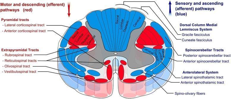
This medical anatomical diagram illustrates a transverse section of the human spinal cord, specifically detailing the organization of white matter tracts. The visual uses a color-coded schema to differentiate between motor and sensory pathways: descending (efferent) motor tracts are highlighted in red, while ascending (afferent) sensory tracts are shown in blue. Key anatomical structures identified include the Pyramidal tracts (lateral and anterior corticospinal tracts), Extrapyramidal tracts (rubrospinal, reticulospinal, olivospinal, and vestibulospinal), the Dorsal Column-Medial Lemniscus system (gracile and cuneate fasciculi), Spinocerebellar tracts, and the Anterolateral system (lateral and anterior spinothalamic tracts). A significant educational feature of the diagram is the depiction of somatotopic organization, showing the concentric arrangement of sacral, lumbar, thoracic, and cervical fibers within the lateral corticospinal and dorsal column tracts. The image provides a comprehensive overview of spinal cord neuroanatomy, useful for understanding clinical localization of spinal cord lesions and the functional topography of the central nervous system.
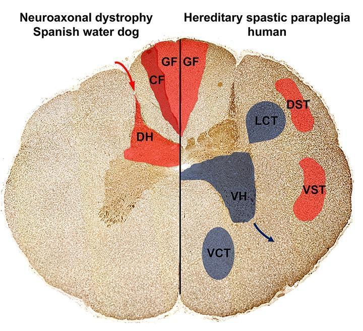
This Comparison Diagram presents a cross-sectional illustration of the spinal cord, contrasting the localization of axonal spheroids and neuronal loss in Neuroaxonal Dystrophy (NAD) and Hereditary Spastic Paraplegia (HSP). The diagram is split into two halves: the left represents a Spanish Water Dog with NAD, and the right represents human HSP. Ascending sensory pathways are highlighted in red, including the Gracile Fasciculus (GF), Cuneate Fasciculus (CF), and Dorsal Horn (DH) on the NAD side, and GF, Dorsal Spinocerebellar Tract (DST), and Ventral Spinocerebellar Tract (VST) on the HSP side. Descending motor pathways, affected only in the HSP model, are highlighted in blue, depicting the Lateral Corticospinal Tract (LCT), Ventral Corticospinal Tract (VCT), and Ventral Horn (VH). The visual demonstrates that in NAD, pathology is restricted to sensory, ascending tracts in the dorsal funiculus and dorsal horn. In contrast, human HSP shows involvement of both sensory pathways and descending motor pathways (pyramidal tracts and ventral horns). This diagram illustrates the pathophysiology of upper motor neuron and sensory system degeneration relevant to neurology and neurohistology.
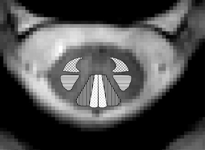
This diagnostic image is an axial cross-sectional Magnetic Resonance (MR) image of the human spinal cord at the C2/C3 cervical level. The image serves as an anatomical diagram by overlaying specific patterns on the white matter to identify major ascending and descending tracts. Centrally located within the spinal cord's white matter, four bilateral regions are highlighted: 1) The lateral corticospinal tracts are indicated by horizontal lines, showing their posterior-lateral position. 2) The cuneate fasciculi are marked with vertical lines in the dorsal column. 3) The gracile fasciculi are represented by dotted patterns, located medially in the dorsal column adjacent to the posterior median sulcus. 4) The combined spinothalamic and spinoreticular tracts are shown with crosshatched patterns in the anterolateral region. This visual aid is designed for advanced medical education to illustrate neuroanatomical localization and the functional organization of spinal cord pathways, providing clinical relevance for assessing regional white matter integrity in spinal cord pathologies or injuries.
Ascending and Descending Tracts of the Spinal Cord
ASCENDING TRACTS (Sensory)
1. Posterolateral (Anterolateral) System
a) Lateral Spinothalamic Tract (Neospinothalamic Tract)
- Modalities: Pain (sharp, localized) and temperature
- 1st order neuron: Cell body in dorsal root ganglion; enters via lateral bundle of dorsal root, travels briefly in Lissauer's tract (tract of Lissauer), synapses in laminae I and V of the dorsal horn
- 2nd order neuron: Crosses the midline via the ventral white commissure over 2-3 segments, then ascends in the anterolateral quadrant (contralateral side)
- 3rd order neuron: Synapses in the ventral posterolateral (VPL) nucleus of the thalamus; projects to primary somatosensory cortex
- Somatotopic arrangement: Sacral fibers are ventrolateral, cervical fibers are dorsomedial (clinically important in cordotomy)
- Localization in Clinical Neurology, 8e, p. 245
b) Spinoreticular Tract (Paleospinothalamic System)
- Modalities: Poorly localized, dull/burning pain; emotional and arousal aspects of pain
- 2nd order axons have short axons synapsing in the brainstem reticular formation, ultimately ending in the intralaminar nuclei of the thalamus
- Localization in Clinical Neurology, 8e, p. 245
c) Spinomesencephalic Tract
- Projects to the periaqueductal gray matter and superior colliculi in the midbrain
- Role in central modulation (descending inhibition) of pain
- Gray's Anatomy for Students, p. 1328
d) Ventral Spinothalamic Tract
- Modality: Light (crude) touch
- Crosses in the ventral white commissure, ascends to VPL nucleus of thalamus
- Light touch is also duplicated in the dorsal column system, which is why it is relatively preserved in pure anterolateral lesions
2. Dorsal Column-Medial Lemniscal Pathway
- Modalities: Fine/discriminative touch, vibration, conscious proprioception (joint position sense), two-point discrimination, stereognosis, weight perception
- 1st order neuron: Enters via the medial bundle of the dorsal root; ascends ipsilaterally in the dorsal funiculus without synapsing first
- Fasciculus gracilis (Goll's column) - medial position - carries sacral, lumbar, and lower 6 thoracic segments (lower limb and trunk)
- Fasciculus cuneatus (Burdach's column) - lateral position - carries upper 6 thoracic and all cervical segments (upper limb and neck)
- 2nd order neuron: Synapses in nucleus gracilis and nucleus cuneatus in the caudal medulla; axons then decussate in the lower medulla (sensory decussation) and ascend as the medial lemniscus to the VPL nucleus of the thalamus
- 3rd order neuron: Projects through the posterior limb of the internal capsule to the primary somatosensory cortex
- Gray's Anatomy for Students, p. 1329; Localization in Clinical Neurology, 8e, p. 245
Key difference from spinothalamic tract: The dorsal columns cross in the medulla (ipsilateral ascent), while the spinothalamic tract crosses within the spinal cord (at the level of entry). This explains why in a Brown-Sequard hemisection, ipsilateral fine touch is lost but contralateral pain/temperature is lost.
3. Spinocerebellar Tracts (Subconscious Proprioception)
| Tract | Source | Pathway | Function |
|---|---|---|---|
| Posterior (Dorsal) Spinocerebellar Tract | Nucleus dorsalis (Clarke's column) | Ipsilateral; enters cerebellum via inferior cerebellar peduncle | Proprioception from lower limb & lower trunk |
| Anterior (Ventral) Spinocerebellar Tract | Laminae V-VII | Crosses twice (at cord + superior cerebellar peduncle); enters via superior cerebellar peduncle | Proprioception from lower limb |
| Cuneocerebellar Tract | Accessory cuneate nucleus | Ipsilateral; inferior cerebellar peduncle | Proprioception from upper limb & upper trunk |
| Rostrocerebellar Tract | - | Superior cerebellar peduncle | Upper body proprioception |
- Localization in Clinical Neurology, 8e, p. 245
DESCENDING TRACTS (Motor)
LATERAL MOTOR SYSTEM
1. Lateral Corticospinal Tract (Pyramidal Tract)
- Origin: Primary motor cortex (Brodmann area 4, precentral gyrus), premotor cortex (area 6), supplementary motor cortex, plus contributions from sensory cortex (areas 3,1,2) and parietal areas
- Course: Corona radiata → posterior limb of internal capsule → crus cerebri (midbrain) → anterior pons (as small bundles) → medullary pyramids → pyramidal decussation at the caudal medulla (~90% of fibers cross) → lateral funiculus of the spinal cord
- Termination: Synapses on LMN cell bodies in the lateral part of the anterior horn (laminae IV-VII and IX)
- Function: Voluntary, skilled movements of the distal extremities (especially fine finger movements)
- Somatotopy: Sacral fibers are most lateral, cervical fibers most medial within the tract
- Localization in Clinical Neurology, 8e, p. 246
2. Rubrospinal Tract
- Origin: Red nucleus of the midbrain
- Course: Crosses at the ventral tegmental decussation → lateral column of spinal cord (alongside lateral corticospinal tract)
- Extent: Only reaches cervical levels of the spinal cord
- Function: Facilitates flexor muscles and inhibits extensor muscles of the upper limb; relatively minor in humans
- Gray's Anatomy for Students, p. 1330
MEDIAL MOTOR SYSTEM
3. Anterior (Ventral) Corticospinal Tract
- The ~10% of corticospinal fibers that do NOT decussate at the medulla
- Descends ipsilaterally in the anterior funiculus; most fibers eventually cross in the ventral white commissure at cervical/upper thoracic levels
- Terminates on LMN cell bodies in the medial anterior horn (axial muscles, neck muscles)
- Localization in Clinical Neurology, 8e, p. 246; Gray's Anatomy for Students, p. 1330
4. Tectospinal Tract
- Origin: Superior colliculus of the dorsal midbrain (receives visual input)
- Course: Crosses in the dorsal tegmental decussation → descends near midline close to the medial longitudinal fasciculus (MLF) → cervical spinal cord only
- Function: Reflex postural movements of the head and neck in response to visual stimuli
- Gray's Anatomy for Students, p. 1331
5. Vestibulospinal Tracts
| Component | Origin | Laterality | Extent | Function |
|---|---|---|---|---|
| Lateral vestibulospinal tract | Lateral vestibular nucleus (Deiter's) | Ipsilateral | Entire spinal cord | Facilitates extensor (antigravity) muscles; important for standing balance |
| Medial vestibulospinal tract | Medial vestibular nucleus | Bilateral | Cervical/thoracic | Head and neck orientation in response to vestibular input |
- Localization in Clinical Neurology, 8e, p. 245; Gray's Anatomy for Students, p. 1332
6. Reticulospinal Tracts
| Component | Origin | Function |
|---|---|---|
| Medial (pontine) reticulospinal tract | Pontine reticular formation | Facilitates antigravity muscles (extensors); descends ipsilaterally |
| Lateral (medullary) reticulospinal tract | Medullary reticular formation | Inhibits antigravity muscles; descends bilaterally |
Summary Table
| Tract | Column | Modality | Crosses? | Key Relay |
|---|---|---|---|---|
| Lateral spinothalamic | Anterolateral | Pain, temperature | Cord (2-3 segments above entry) | VPL thalamus |
| Anterior spinothalamic | Anterior | Light touch | Cord | VPL thalamus |
| Spinoreticular | Anterolateral | Dull/poorly localized pain | Cord | Reticular formation → intralaminar thalamus |
| Spinomesencephalic | Anterolateral | Pain modulation | Cord | Periaqueductal gray |
| Fasciculus gracilis | Posterior (medial) | Fine touch, vibration, proprioception (lower body) | Medulla | Nucleus gracilis → VPL |
| Fasciculus cuneatus | Posterior (lateral) | Fine touch, vibration, proprioception (upper body) | Medulla | Nucleus cuneatus → VPL |
| Post. spinocerebellar | Lateral | Unconscious proprioception (lower) | Does NOT cross | Cerebellum (inf. peduncle) |
| Ant. spinocerebellar | Lateral | Unconscious proprioception (lower) | Crosses twice | Cerebellum (sup. peduncle) |
| Lateral corticospinal | Lateral | Voluntary motor (limbs) | Medulla (pyramidal decussation) | Anterior horn LMN |
| Anterior corticospinal | Anterior | Voluntary motor (axial) | Cord (ventral commissure) | Medial anterior horn |
| Rubrospinal | Lateral | Flexor facilitation (upper limb) | Midbrain (ventral tegmental) | Cervical anterior horn |
| Vestibulospinal (lateral) | Anterior | Extensor facilitation / balance | Ipsilateral (does NOT cross) | Anterior horn |
| Reticulospinal | Anterior/lateral | Tone, posture, pain modulation | Variable | Anterior horn via interneurons |
| Tectospinal | Anterior | Visual-guided head/neck reflexes | Midbrain (dorsal tegmental) | Cervical anterior horn |
Clinical Relevance
- Brown-Sequard Syndrome (hemisection): Ipsilateral loss of fine touch/proprioception (dorsal columns) + ipsilateral UMN weakness (corticospinal) + contralateral loss of pain/temperature (spinothalamic crosses in cord)
- Central cord syndrome: Preferential damage to cervical fibers (most medial in lateral corticospinal tract) → greater upper limb weakness than lower limb
- Syringomyelia: Expands from the central canal outward, first damaging crossing spinothalamic fibers in the anterior commissure → "cape-like" suspended bilateral loss of pain/temperature
- Subacute combined degeneration (B12 deficiency): Damages dorsal columns and lateral corticospinal tracts simultaneously → loss of proprioception + UMN signs