I now have rich, comprehensive material from multiple authoritative textbooks. Let me compile the full response.
Atrial Fibrillation (AF)
Definition and Overview
Atrial fibrillation is the most common sustained cardiac arrhythmia. It is characterized by disorganized, rapid, irregular atrial electrical activity (350-700 impulses/min) that results in loss of effective atrial contraction. The ventricles respond irregularly through the AV node, producing the hallmark "irregularly irregular" pulse.
ECG Findings
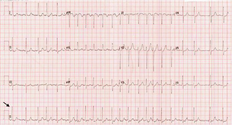
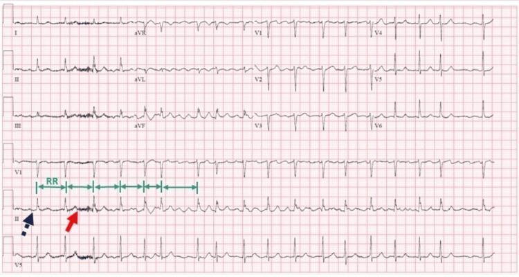
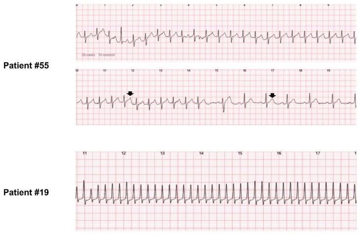
The two pathognomonic ECG features are:
- Absence of distinct P waves - replaced by irregular fibrillatory (f) waves, best seen in V1 and II
- Irregularly irregular QRS complexes - varying R-R intervals with usually narrow QRS (unless aberrant conduction or pre-excitation)
Classification
| Type | Definition |
|---|
| Paroxysmal | Terminates spontaneously or with intervention within 7 days |
| Persistent | Lasts >7 days or requires cardioversion |
| Long-standing persistent | Continuous AF >12 months |
| Permanent | Accepted by patient and clinician; no further rhythm control |
| Lone AF | No identifiable structural heart disease, hypertension, or other cause (historical term) |
Epidemiology
AF is extremely common, affecting approximately 12% of patients ≥75 years and 18% of those ≥85 years. The 2019 AHA/ACC/HRS guideline estimates that about one-third of all AF patients are ≥80 years. Global prevalence is rising due to aging populations and increasing rates of hypertension, obesity, and heart failure. - Braunwald's Heart Disease, p. 2808
Pathophysiology
AF requires both a trigger and a substrate:
Electrophysiological Mechanisms
- The classical model is Moe's multiple-wavelet hypothesis: multiple disorganized reentrant wavelets perpetuate the arrhythmia
- A competing theory involves focal discharge or rotor sources - single or small numbers of drivers that sustain AF. Evidence strongly supports the pulmonary veins (PVs) as the dominant source of both focal triggers and reentrant activity; PV isolation is thus the cornerstone of ablation therapy
- Underlying organization is reflected in dominant frequencies detectable by spectral analysis - Braunwald's Heart Disease, p. 721
Structural Remodeling
- Atrial fibrosis and conduction heterogeneity provide the substrate for reentry
- Age-related atrial changes, LA enlargement (e.g., from mitral stenosis or hypertension), and inflammation all promote remodeling
- AF begets AF ("electrical remodeling"): sustained AF shortens atrial effective refractory periods, stabilizing the arrhythmia
Ion Channel Abnormalities
- Familial (monogenic) AF: mutations in genes encoding K+ (IK) and Na+ (INa) channel subunits
- Gain-of-function IK mutations → shortened APD and atrial refractoriness → facilitates reentry
- Augmented inward INa → increased excitability and triggered activity
- Mutations in KCNJ2, KCNA5, and GJA5 (connexin 40) have been identified
- Polygenic forms: GWAS studies have identified multiple susceptibility loci - Braunwald's Heart Disease, pp. 733-762
Risk Factors / Causes
Cardiac:
- Hypertension (most common modifiable RF)
- Heart failure (HFpEF, HFrEF)
- Coronary artery disease
- Valvular disease (especially mitral stenosis - carries particularly high embolic risk)
- Cardiomyopathy, pericarditis, myocarditis
- Congenital heart disease
Non-cardiac:
- Obesity, obstructive sleep apnea
- Hyperthyroidism
- Diabetes mellitus
- Chronic kidney disease
- Excessive alcohol ("holiday heart")
- Pulmonary disease (COPD, PE)
- Post-surgery (especially cardiac surgery)
Clinical Features
Symptoms:
- Palpitations (may be less prominent in older adults)
- Dyspnea, reduced exercise tolerance
- Fatigue, light-headedness
- Chest discomfort
- Acute pulmonary edema (with rapid ventricular response, especially in HFpEF/stiff LV)
- Syncope, falls, or stroke as initial presentation (particularly in elderly)
- Many patients are asymptomatic - Braunwald's Heart Disease, p. 2814
Hemodynamic consequences:
- Loss of atrial kick reduces cardiac output by ~15-25% (more in diastolic dysfunction)
- Rapid ventricular rate further impairs diastolic filling
- Tachycardia-mediated cardiomyopathy if rate is uncontrolled chronically
Diagnosis
- ECG: Hallmark test - irregular rhythm without P waves
- Holter monitor / event recorder: For paroxysmal AF
- Echocardiogram: Assess structural disease, LA size, LV function, valvular disease, LAA thrombus
- TEE: Gold standard for excluding LAA thrombus before cardioversion
- Blood tests: TFTs (thyroid), CBC, electrolytes, renal function, BNP
- Wearable devices: Apple Watch and similar devices show utility for screening; ESC recommends AF screening at age ≥65 by pulse palpation or ECG rhythm strip (Class I)
Stroke Risk: CHA₂DS₂-VASc Score
Nonvalvular AF confers a fivefold increase in stroke risk. Stroke risk is quantified using the CHA₂DS₂-VASc score: - Tintinalli's Emergency Medicine, p. 1723
| Factor | Points |
|---|
| C - Congestive Heart Failure | 1 |
| H - Hypertension | 1 |
| A₂ - Age ≥75 years | 2 |
| D - Diabetes mellitus | 1 |
| S₂ - Prior Stroke/TIA/thromboembolism | 2 |
| V - Vascular disease (prior MI, PAD) | 1 |
| A - Age 65-74 years | 1 |
| Sc - Sex category (female) | 1 |
Anticoagulation thresholds:
- Score 0: anticoagulation not recommended
- Score 1: individualize (consider anticoagulation in men; generally treat in women)
- Score ≥2: oral anticoagulation strongly recommended
- All patients ≥75 years automatically score ≥2 and are candidates for anticoagulation - Braunwald's Heart Disease, p. 2821
Special case - Valvular AF (rheumatic mitral stenosis or mechanical heart valve): DOACs are contraindicated; warfarin (target INR 2-3) is mandated regardless of CHA₂DS₂-VASc score.
Bleeding Risk: HAS-BLED Score
Before initiating anticoagulation, assess bleeding risk:
| Factor | Points |
|---|
| H - Uncontrolled Hypertension (SBP >160) | 1 |
| A - Abnormal renal/liver function | 1-2 |
| S - Stroke history | 1 |
| B - Bleeding history or predisposition | 1 |
| L - Labile INR | 1 |
| E - Elderly (>65 years) | 1 |
| D - Drugs (antiplatelets, NSAIDs) or alcohol | 1-2 |
Score ≥3 = high bleeding risk. Importantly, a high HAS-BLED score should prompt modification of reversible bleeding risk factors, not automatic withholding of anticoagulation (in most patients, stroke risk still outweighs bleed risk). - Braunwald's Heart Disease, p. 2824
Anticoagulation Therapy
Direct Oral Anticoagulants (DOACs) - Preferred for Nonvalvular AF
| Drug | Mechanism | Dose |
|---|
| Apixaban (Eliquis) | Factor Xa inhibitor | 5 mg BID (2.5 mg BID if ≥2 of: age ≥80, wt ≤60 kg, Cr ≥1.5) |
| Rivaroxaban (Xarelto) | Factor Xa inhibitor | 20 mg OD with evening meal |
| Dabigatran (Pradaxa) | Direct thrombin inhibitor | 150 mg BID (110 mg BID if high bleed risk) |
| Edoxaban (Savaysa) | Factor Xa inhibitor | 60 mg OD (30 mg OD if CrCl 15-50 or wt ≤60 kg) |
DOACs offer similar or better stroke prevention versus warfarin, no dietary restrictions, no INR monitoring, and in elderly patients (≥75 years) demonstrate similar or less bleeding. - Braunwald's Heart Disease, p. 2826
Warfarin
- Target INR: 2.0-3.0 (2.0-2.5 in elderly); 2.5-3.5 for mechanical heart valves
- Required for valvular AF (rheumatic MS, mechanical valves) where DOACs are contraindicated
- Challenges: dietary interactions, multiple drug interactions, regular monitoring, increased osteoporosis risk
- Estimated maintenance dose in elderly: typically 2-5 mg/day - Braunwald's Heart Disease, p. 2826
Rate Control vs. Rhythm Control
Rate Control
Goal: Resting heart rate <80-100 bpm (lenient: <110 bpm acceptable in asymptomatic patients)
| Drug Class | Examples | Notes |
|---|
| Beta-blockers | Metoprolol, bisoprolol, carvedilol | First-line; preferred in CAD, HFrEF |
| Non-DHP CCBs | Diltiazem, verapamil | Avoid in HFrEF or accessory pathways |
| Digoxin | Digoxin | Useful in HF; less effective at exercise |
| Amiodarone | Amiodarone | Reserved for refractory cases |
Rhythm Control
Goal: Restore and maintain sinus rhythm
Indications:
- Persistent symptoms despite rate control
- Young patients with first-episode AF
- AF precipitating heart failure
- Patient preference
Cardioversion:
- Electrical cardioversion (DC cardioversion): 200 J biphasic; effective for acute restoration
- Pre-cardioversion anticoagulation ≥3 weeks if AF >48 hours (or TEE to exclude LAA thrombus)
- Post-cardioversion anticoagulation ≥4 weeks regardless of CHA₂DS₂-VASc score (due to "atrial stunning")
Antiarrhythmic Drugs (AADs):
| Drug | Use Case | Cautions |
|---|
| Flecainide / Propafenone | No structural disease | Contraindicated in CAD/HF ("pill-in-pocket" strategy) |
| Amiodarone | Any structural disease; HF | Pulmonary/thyroid/hepatic toxicity with long-term use |
| Dronedarone | Non-permanent AF without severe HF | Reduced mortality in ATHENA; avoid in HFrEF |
| Sotalol | Mild-to-moderate structural disease | QTc prolongation; requires renal monitoring |
| Dofetilide | HF, post-MI | Requires in-hospital initiation |
Ablation (Catheter Ablation / PVI)
- Pulmonary vein isolation (PVI) is the cornerstone procedure
- Superior to AADs for maintaining sinus rhythm in paroxysmal and persistent AF
- A 2024 meta-analysis (PMID 38040282) found ablation superior to AADs as first-line therapy in treatment-naive AF for maintaining sinus rhythm, with lower AF recurrence and comparable safety
- A separate 2024 meta-analysis (PMID 37937825) confirmed ablation improves LV function and quality of life vs. medical therapy
- Surgical maze procedure (Cox-Maze): used during concomitant cardiac surgery; high efficacy for permanent rhythm restoration
AF with Special Conditions
AF + Heart Failure with Preserved EF (HFpEF)
- AF frequently complicates HFpEF; loss of atrial kick and fast rate can precipitate acute decompensation
- Anticoagulation mandatory if coexisting AF
- Whether rhythm control reduces HF hospitalizations remains under investigation; rhythm control trials in HFrEF (e.g., CASTLE-AF) showed benefit - Braunwald's Heart Disease, p. 329
AF + Mitral Stenosis (Valvular AF)
- MS + AF carries embolic risk as high as mechanical prosthetic valves
- DOACs are contraindicated; warfarin is mandatory irrespective of CHA₂DS₂-VASc score
- Even transient episodes (<30 sec) detected on ambulatory monitoring significantly raise thromboembolic risk
- Rate control is difficult; ivabrading has been tried
- Restoring sinus rhythm improves QoL, especially with small LA and short AF duration - Braunwald's Heart Disease, p. 760
AF in the Elderly (≥75 years)
- All patients ≥75 automatically qualify for anticoagulation (CHA₂DS₂-VASc ≥2)
- DOACs preferred over warfarin (similar/better efficacy, similar/less bleeding)
- Symptoms are often atypical or minimal; syncope, falls, or stroke may be first presentation
- Screening: ESC recommends pulse palpation or ECG strip at age ≥65 (Class I); systematic ECG screening at ≥75 is Class IIb - Braunwald's Heart Disease, p. 2817
Emergency Management (Acute AF)
Hemodynamically unstable (hypotension, acute pulmonary edema, angina):
- Immediate synchronized DC cardioversion regardless of anticoagulation status
Hemodynamically stable:
- Rate control (IV metoprolol, diltiazem, digoxin)
- Consider rhythm control if AF onset <48 hours and patient appropriate
- Anticoagulation: heparin bridge if cardioversion planned; initiate long-term OAC based on CHA₂DS₂-VASc
- Discharge criteria per Tintinalli's: SpO2 >95%, BP at baseline, resting HR <100 bpm; 80%+ of ED patients can be safely discharged - Tintinalli's Emergency Medicine, p. 1718-1720
Complications
| Complication | Notes |
|---|
| Stroke / thromboembolism | 5× increased risk; main driver of anticoagulation decisions |
| Heart failure | Loss of atrial kick + uncontrolled rate |
| Tachycardia-induced cardiomyopathy | Reversible with rate/rhythm control |
| Cognitive decline / dementia | Independent association |
| Reduced quality of life and physical performance | Even with rate control |
| Increased mortality | HR ~1.5-2× vs. age-matched controls |
Recent Evidence (2024)
- Ablation vs. AADs as first-line therapy (PMID 38040282) - Systematic review/meta-analysis: catheter ablation superior to AADs in treatment-naive AF for maintaining sinus rhythm with comparable safety profile
- Ablation vs. medical therapy - updated meta-analysis (PMID 37937825) - Ablation improves LVEF and reduces recurrence vs. medical therapy
- Emerging therapies in AF management (PMID 37716839) - Reviews novel antiarrhythmics, left atrial appendage occlusion (LAAO), and pulsed-field ablation as emerging strategies
Key Take-Home Points
- AF = irregularly irregular rhythm with no P waves on ECG
- Anticoagulate all patients with CHA₂DS₂-VASc ≥2 (DOACs preferred over warfarin for nonvalvular AF)
- Valvular AF (rheumatic MS, mechanical valve) = warfarin only, no DOACs
- Rate control (HR <100-110) is adequate for asymptomatic patients; rhythm control for symptomatic patients
- PVI catheter ablation is the most effective rhythm control strategy, increasingly used as first-line
- Assess both stroke risk (CHA₂DS₂-VASc) AND bleeding risk (HAS-BLED) before anticoagulation
- All patients ≥75 years qualify for anticoagulation regardless of other risk factors
Sources: Braunwald's Heart Disease (10th ed.), Tintinalli's Emergency Medicine, Harrison's Principles (22nd ed., 2025), Miller's Anesthesia (10th ed.), Fuster and Hurst's The Heart (15th ed.)