
How to read x ray
Reading File
"How to read x-ray" covers a lot of ground since the approach differs by body region. Here's a breakdown:
Topics covered under X-ray interpretation:
- General principles - patient/technical check (name, date, projection, exposure, rotation), then a systematic scan pattern
- Chest X-ray (CXR) - most commonly taught; ABCDE approach (Airway, Bones, Cardiac, Diaphragm, Effusion/lung fields)
- Abdominal X-ray (AXR) - bowel gas pattern, free air, calcifications, bones
- Musculoskeletal/bone X-ray - fracture description (site, pattern, displacement, angulation), joint alignment
- Basic radiographic physics - density differences (air, fat, soft tissue, bone, metal) that let you interpret shadows
A quick overview: every X-ray read starts the same way - confirm it's the right patient/date/projection and check technical adequacy (penetration, rotation, inspiration for CXR). Then you scan systematically rather than jumping straight to the "obvious" finding, comparing left-to-right and, if available, to prior films. Each region has its own checklist (e.g., CXR uses ABCDE; bone films use the "4 A's" - Alignment, Adequacy of bone density, Articular surfaces, Soft tissues).
Which one do you want me to go into detail on?
Asking for Preferences
Which type of X-ray do you want a detailed reading guide for?
"How to read x-ray" covers a lot of ground since the approach differs by body region. Here's a breakdown:
Topics covered under X-ray interpretation:
- General principles - patient/technical check (name, date, projection, exposure, rotation), then a systematic scan pattern
- Chest X-ray (CXR) - most commonly taught; ABCDE approach (Airway, Bones, Cardiac, Diaphragm, Effusion/lung fields)
- Abdominal X-ray (AXR) - bowel gas pattern, free air, calcifications, bones
- Musculoskeletal/bone X-ray - fracture description (site, pattern, displacement, angulation), joint alignment
- Basic radiographic physics - density differences (air, fat, soft tissue, bone, metal) that let you interpret shadows
A quick overview: every X-ray read starts the same way - confirm it's the right patient/date/projection and check technical adequacy (penetration, rotation, inspiration for CXR). Then you scan systematically rather than jumping straight to the "obvious" finding, comparing left-to-right and, if available, to prior films. Each region has its own checklist (e.g., CXR uses ABCDE; bone films use the "4 A's" - Alignment, Adequacy of bone density, Articular surfaces, Soft tissues).
Which one do you want me to go into detail on?
How to Read an X-ray
The core method is the same for any X-ray: check the technical quality first, then scan systematically rather than jumping to the obvious finding. Below is the standard approach used in most radiology teaching, broken down by region.
1. Before You Even Look at the Pathology - Check the Basics (RIPE)
- R - Right patient/film: name, date, comparison with prior films if available
- I - Image orientation/projection: PA vs AP, erect vs supine, left/right markers
- P - Penetration/exposure: on a chest film you should just be able to see the thoracic vertebrae through the heart shadow. Too dark = overpenetrated, too white = underpenetrated
- E - Everything included: make sure the whole area of interest is in the field, and check for rotation (on CXR, the medial ends of both clavicles should be equidistant from the spinous process)
2. Chest X-ray (CXR) - the "ABCDE" system
This is the most widely taught systematic approach:
- A - Airway: trachea central (not deviated), carina, main bronchi
- B - Bones/soft tissues: ribs, clavicles, scapulae, spine - look for fractures, lytic/sclerotic lesions; check soft tissues for surgical emphysema
- C - Cardiac: heart size (cardiothoracic ratio should be <50% on a PA film), heart borders, mediastinal contours
- D - Diaphragm: both hemidiaphragms should be domed and sharp; look under them for free air (suggests perforation) and check costophrenic angles for blunting (effusion)
- E - Effusions/Everything else (lung fields): compare left and right lung fields for symmetry, look for consolidation (fluffy white opacity, air bronchograms), pneumothorax (absent lung markings peripherally, visible pleural line), masses, and check all the tubes/lines if it's an ICU film
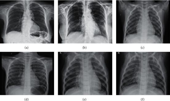
Here is an example comparing a normal chest X-ray to abnormal patterns, useful for practicing the scan:
3. Abdominal X-ray (AXR)
- Bowel gas pattern: distinguish small bowel (central, valvulae conniventes crossing the full width) from large bowel (peripheral, haustra that don't cross the full width) and look for dilation (>3 cm small bowel, >6 cm large bowel, >9 cm caecum)
- Free air (suggests perforation - better seen on erect chest X-ray as air under the diaphragm)
- Calcifications: gallstones, renal stones, appendicoliths, vascular calcification
- Bones and soft tissue as with any film
4. Bone/Skeletal X-ray - the "4 A's" (or ABCS)
- A - Alignment: bones in normal anatomical position, joint congruity
- A/B - Bone density: cortex intact, no lucency (lytic lesion) or sclerosis, look specifically along the cortical margin for a fracture line
- A - Articular surfaces: joint space width, effusion
- S - Soft tissues: swelling, foreign bodies, fat pad displacement (e.g., elbow fat pad sign suggesting occult fracture)
If a fracture is found, describe it fully: which bone, location (proximal/mid/distal shaft), pattern (transverse, oblique, spiral, comminuted), displacement, angulation, and whether it's intra-articular or open.
5. General Tip
Always compare left to right, and to a previous film if one exists - subtle asymmetry is often the first clue to pathology that's easy to miss when scanning a single image in isolation.
If you want, tell me which specific type of X-ray (chest, abdominal, a particular bone/joint) or a specific clinical scenario you're working through, and I can go deeper with example findings and how to phrase a formal report.
This is a shared conversation. Sign in to Orris to start your own chat.