
Could you please explain medula oblongata from grays anatomy textbook in deatil Dont miss out on any topic Prepare me to answer usmle questions please
Reading File
Now let me search the library for Gray's Anatomy and medulla oblongata content simultaneously.
Finding Sources
Finding Sources
Gray's Anatomy for Students has the medulla oblongata content in block 6. Let me also search for related headings and read all content simultaneously.
Finding Sources
Reading File
Excellent content found. Now let me read more of this block to get the internal medulla section (around line 698+) and also look for any preceding context:
Reading File
Reading File
Excellent! I now have rich content. Let me fetch the key anatomical images and also read the vascular supply section:
Reading File
Loading Image
Loading Image
Now let me fetch the cross-section images of the medulla (rostral and mid-medulla diagrams):
Loading Image
Loading Image
Loading Image
I now have all the images and content I need. Let me also check if there are additional paragraphs about the medulla in Gray's, specifically about clinical correlates and cranial nerve nuclei details:
Finding Sources
Finding Sources
I now have comprehensive content to write a detailed, USMLE-focused explanation. Let me also quickly get the Wallenberg syndrome details from the Localization in Clinical Neurology textbook:
Reading File
Reading File
I now have extremely comprehensive content. Let me also get the Wallenberg syndrome clinical features section from the same book:
Reading File
Reading File
I now have everything I need for a comprehensive, USMLE-focused answer. Let me compile it:# Medulla Oblongata - Complete USMLE Guide
Source: Gray's Anatomy for Students + Localization in Clinical Neurology
1. OVERVIEW & POSITION
The medulla oblongata (also called the myelencephalon) is the most caudal part of the brainstem, measuring approximately 3 cm in length. It connects the pons rostrally to the spinal cord caudally, with the junction at the foramen magnum. Of the three brainstem segments, the medulla is the longest.
USMLE Memory Hook: Brainstem from rostral to caudal = Midbrain - Pons - Medulla (mnemonic: "My Pretty Medulla")
2. EXTERNAL ANATOMY (What You See on the Surface)
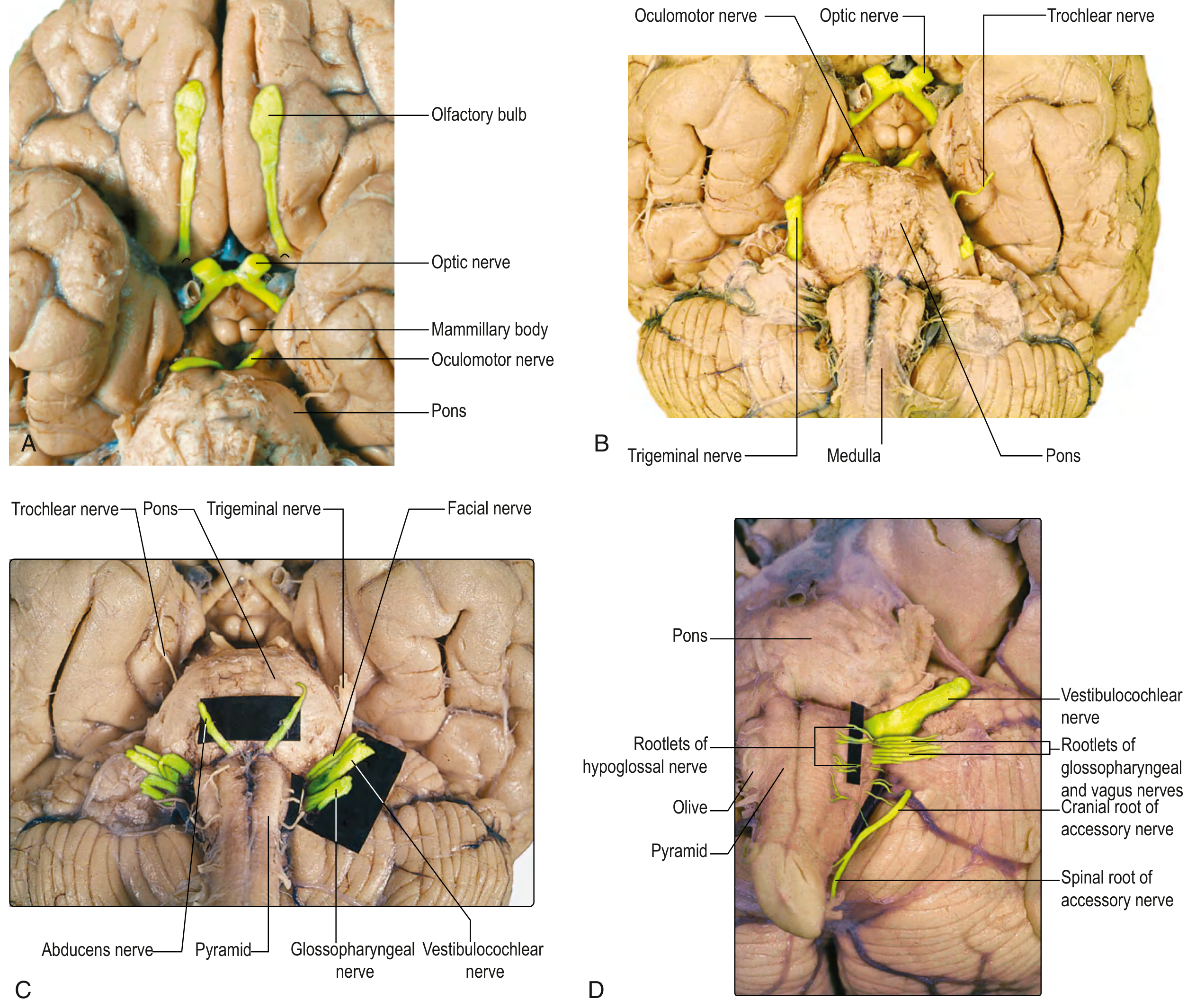
Anterior (Ventral) Surface
| Structure | Location | What It Contains / Represents |
|---|---|---|
| Anterior median fissure | Midline | Continuous with anterior fissure of spinal cord |
| Medullary pyramids | Lateral to fissure | Corticospinal (motor) fibers descending from cerebral cortex |
| Decussation of pyramids | Caudal end | ~90% of corticospinal fibers cross here to contralateral side |
| Olives | Lateral to pyramids | Surface bulges representing the inferior olivary nuclei |
| CN XII rootlets | Junction of pyramid & olive | Hypoglossal nerve exits here |
| CN IX, X, XI rootlets | Junction of olive & inferior cerebellar peduncle (rostral to caudal) | Glossopharyngeal, Vagus, Spinal Accessory nerve rootlets |
Posterior (Dorsal) Surface
- Forms the floor of the fourth ventricle (upper medulla)
- Posterior median sulcus runs down the midline
- Gracile tubercles (medial) and Cuneate tubercles (lateral) - surface bumps formed by the underlying gracile and cuneate nuclei
- The obex is the V-shaped inferior tip of the fourth ventricle at the posterior medulla
USMLE Pearl: CN XII exits between the pyramid and the olive (pre-olivary sulcus). CN IX, X, XI exit in the post-olivary sulcus (between olive and inferior cerebellar peduncle). This localization is tested frequently.
3. INTERNAL ANATOMY - THREE LEVELS (Most Tested on USMLE)
The medulla is best understood at three cross-sectional levels from rostral to caudal:
LEVEL 1: Rostral Medulla (Level of the Inferior Olivary Nucleus)
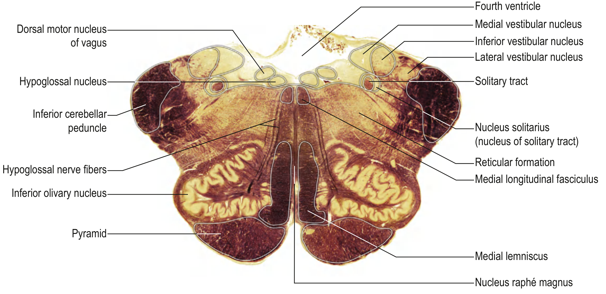
Key structures visible in this section:
| Structure | Location | Function |
|---|---|---|
| Inferior olivary nucleus | Ventrolateral (large, convoluted) | Relay to cerebellum; involved in motor learning |
| Pyramid | Anteromedial | Corticospinal tract |
| Medial lemniscus | Vertical, adjacent to midline | Carries proprioception, vibration, 2-point touch (already decussated) |
| Medial longitudinal fasciculus (MLF) | Dorsal to medial lemniscus | Coordinates eye movements (CN III, IV, VI), also connects to CN VIII |
| Hypoglossal nucleus (CN XII) | Dorsomedial | Motor to tongue |
| Dorsal motor nucleus of vagus (CN X) | Dorsolateral to hypoglossal nucleus | Parasympathetic output to thoracic/abdominal viscera |
| Nucleus solitarius (NTS) | Ventrolateral to dorsal vagal nucleus | Taste (CN VII, IX, X) + cardiovascular/respiratory reflexes |
| Vestibular nuclei (CN VIII) | Dorsal, near ICP | Balance/coordination |
| Inferior cerebellar peduncle (ICP) | Large lateral structure | Connects medulla to cerebellum (spinocerebellar tract, etc.) |
| Nucleus ambiguus | Reticular formation (medial to spinal V nucleus) | Motor to pharynx, larynx, palate (CN IX, X, XI) |
| Reticular formation | Central core | Autonomic control, arousal, respiratory/cardiovascular centers |
| Nucleus raphé magnus | Midline | Serotonin production; descending pain modulation |
LEVEL 2: Mid-Medulla (Level of Internal Arcuate Fiber Decussation / Sensory Decussation)
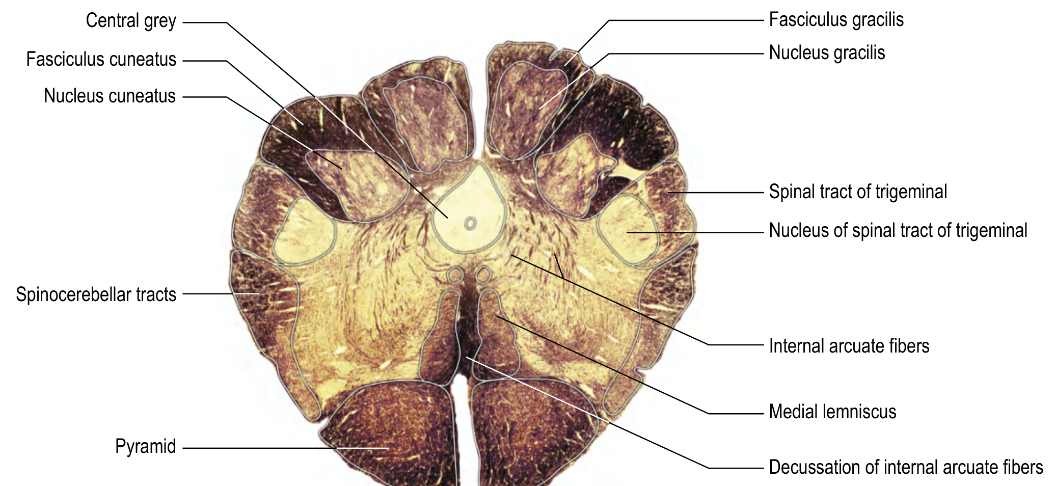
Key structures:
| Structure | Location | Function |
|---|---|---|
| Gracile nucleus | Dorsomedial | Receives fibers from lower limbs/trunk (fasciculus gracilis) |
| Cuneate nucleus | Dorsolateral | Receives fibers from upper limbs/trunk (fasciculus cuneatus) |
| Internal arcuate fibers | Sweep ventrally from dorsal columns | Axons of gracile and cuneate nuclei curving to midline to decussate |
| Decussation of internal arcuate fibers | Midline crossing | Forms the medial lemniscus after crossing = SENSORY decussation |
| Spinal trigeminal nucleus | Lateral | Pain/temp from ipsilateral face (CN V pars caudalis) |
| Spinal trigeminal tract | Lateral to nucleus | Descending fibers of CN V |
| Spinocerebellar tracts | Lateral perimeter | Unconscious proprioception to cerebellum |
| Anterolateral (spinothalamic) tract | Lateral perimeter | Pain and temperature from contralateral body (already crossed in spinal cord) |
| Pyramid | Ventral | Corticospinal fibers |
LEVEL 3: Caudal Medulla (Level of Pyramidal Decussation / MOTOR Decussation)
- Pyramidal decussation occurs at the midline of the caudal medulla
- ~90% of corticospinal fibers cross to the contralateral lateral funiculus of the spinal cord to become the lateral corticospinal tract
- ~10% remain uncrossed, forming the anterior corticospinal tract
- Gracile and cuneate nuclei begin to emerge here from their fasciculi
- CN XI nucleus (spinal accessory) is in the cervical spinal cord, NOT the medulla
USMLE Memory Tool - Two Decussations in the Medulla:
- Motor decussation (pyramidal) = CAUDAL medulla, corticospinal fibers cross
- Sensory decussation (internal arcuate fibers / lemniscal) = MID-medulla, dorsal column-medial lemniscal fibers cross
4. CRANIAL NERVE NUCLEI IN THE MEDULLA
The medulla houses nuclei for CN VIII, IX, X, XI, and XII:
| Cranial Nerve | Nucleus in Medulla | Function |
|---|---|---|
| CN VIII (Vestibulocochlear) | Vestibular nuclei (medial, inferior, lateral, superior) + Cochlear nuclei | Hearing & balance |
| CN IX (Glossopharyngeal) | Nucleus ambiguus (motor), NTS (taste/visceral afferents), Inferior salivatory nucleus (parasympathetic) | Stylopharyngeus, taste posterior 1/3 tongue, parotid gland, carotid body |
| CN X (Vagus) | Nucleus ambiguus (motor), Dorsal motor nucleus (parasympathetic), NTS (visceral afferents) | Pharynx/larynx, parasympathetic to thorax/abdomen |
| CN XI (Spinal Accessory) | Nucleus in cervical spinal cord (C1-C5), cranial root joins vagus | Sternocleidomastoid, trapezius |
| CN XII (Hypoglossal) | Hypoglossal nucleus (dorsomedial) | All intrinsic + most extrinsic tongue muscles |
USMLE Pearl on CN XII: Upper motor neuron (UMN) lesion above the level of CN XII nucleus = tongue deviates AWAY from lesion. Lower motor neuron (LMN) lesion of CN XII nucleus or nerve = tongue deviates TOWARD the side of lesion (fibers of genioglossus push tongue to opposite side, so when one side is weak, the other pushes tongue ipsilaterally to the lesion).
5. KEY TRACTS PASSING THROUGH THE MEDULLA
| Tract | Origin | Destination | What it Carries | Where it Crosses |
|---|---|---|---|---|
| Corticospinal tract (pyramids) | Motor cortex | Spinal cord anterior horn | Voluntary motor | Caudal medulla (pyramidal decussation) |
| Medial lemniscus | Gracile + cuneate nuclei | Thalamus (VPL) | Proprioception, vibration, 2-point touch | Mid-medulla (sensory decussation) |
| Spinothalamic (anterolateral) | Spinal cord | Thalamus (VPL) | Pain, temperature | Already crossed in spinal cord |
| Spinocerebellar tracts | Spinal cord | Cerebellum via ICP | Unconscious proprioception | Varies (dorsal uncrossed, ventral crosses twice) |
| MLF | Vestibular nuclei / PPRF | CN III, IV, VI nuclei | Conjugate eye movement coordination | Bilateral |
| Descending sympathetic fibers | Hypothalamus | Ciliospinal center (C8-T2) | Sympathetic control of eye / face | Runs through lateral tegmentum |
6. VASCULAR SUPPLY (VERY HIGH-YIELD for USMLE)
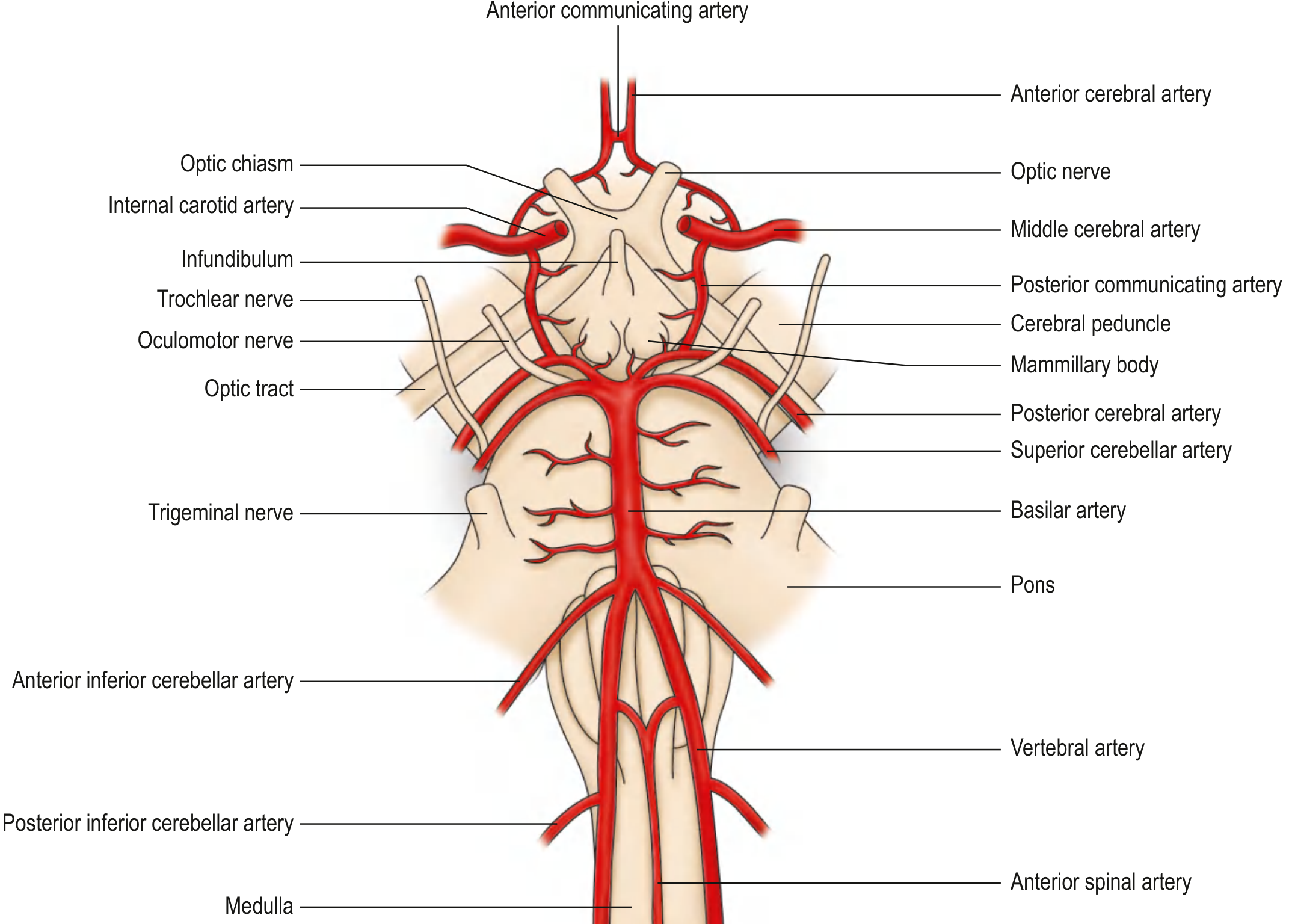
The medulla is supplied by the vertebrobasilar system:
| Region | Artery | Clinical Syndrome if Occluded |
|---|---|---|
| Lateral medulla | PICA (Posterior Inferior Cerebellar Artery) from vertebral artery | Wallenberg Syndrome (Lateral Medullary Syndrome) |
| Medial/anterior medulla | Paramedian branches of vertebral + anterior spinal artery | Dejerine Syndrome (Medial Medullary Syndrome) |
| Inferior cerebellum | PICA | Cerebellar infarction |
USMLE Fact: The vertebral arteries arise from the subclavian arteries, ascend through foramina transversaria of C6-C2, then fuse at the pontomedullary junction to form the single basilar artery.
7. THE TWO MAJOR MEDULLARY STROKE SYNDROMES (HIGH-YIELD)
A. LATERAL MEDULLARY SYNDROME (Wallenberg Syndrome)
Cause: PICA or vertebral artery occlusion - most common
Structure damaged = lateral wedge of medulla
| Structure Damaged | Clinical Finding |
|---|---|
| Spinal trigeminal nucleus/tract | Ipsilateral facial pain/temperature loss |
| Spinothalamic tract | Contralateral body pain/temperature loss |
| Nucleus ambiguus (CN IX, X) | Ipsilateral dysphagia, dysarthria, hoarseness, palatal palsy |
| Descending sympathetic fibers | Ipsilateral Horner syndrome (ptosis, miosis, anhidrosis) |
| Vestibular nuclei | Vertigo, nausea, vomiting, nystagmus |
| Inferior cerebellar peduncle | Ipsilateral cerebellar ataxia (limb + gait) |
Classic USMLE Pattern: Crossed sensory findings - ipsilateral face loss + contralateral body loss (crossed sensory dissociation)
Memory Trick: PICA = "People I Can't Agree with":
- Palatal palsy, Ipsilateral face sensory loss, Contralateral body sensory loss, Ataxia + Horner + Vertigo
B. MEDIAL MEDULLARY SYNDROME (Dejerine / Anterior Bulbar Syndrome)
Cause: Vertebral artery or anterior spinal artery occlusion
Structure damaged = paramedian medulla (pyramid, medial lemniscus, CN XII)
| Structure Damaged | Clinical Finding |
|---|---|
| Pyramid (corticospinal) | Contralateral hemiplegia (body, face spared) |
| Medial lemniscus | Contralateral loss of vibration, proprioception, 2-point touch |
| CN XII nucleus/fibers | Ipsilateral tongue weakness (LMN), tongue deviates toward lesion |
Classic Triad for USMLE:
- Contralateral hemiplegia (UMN, face spared)
- Contralateral loss of dorsal column sensation
- Ipsilateral tongue deviation toward lesion (LMN CN XII)
8. COMPARISON TABLE: WALLENBERG vs. DEJERINE
| Feature | Lateral Medullary (Wallenberg / PICA) | Medial Medullary (Dejerine / Ant. Spinal A.) |
|---|---|---|
| Artery occluded | PICA / vertebral | Vertebral / anterior spinal |
| Motor | No hemiplegia (corticospinal spared) | Contralateral hemiplegia |
| Sensation - body | Crossed (ipsi face, contra body pain/temp) | Contra loss of vibration/proprioception |
| CN XII (tongue) | Usually spared | Ipsilateral LMN weakness |
| Horner syndrome | YES (ipsilateral) | NO |
| Dysphagia / dysarthria | YES (nucleus ambiguus) | Usually NO |
| Vertigo | YES | Usually NO |
| Cerebellar signs | YES (ipsilateral) | Usually NO |
9. OTHER IMPORTANT MEDULLARY STRUCTURES FOR USMLE
Reticular Formation
- Located in the central core of the medulla
- Contains the respiratory centers (dorsal respiratory group, ventral respiratory group)
- Contains cardiovascular control centers (vasomotor, cardiac centers)
- Contains the vomiting center (area postrema - chemoreceptor trigger zone is just lateral)
Area Postrema
- Located at the caudal end of the fourth ventricle
- A circumventricular organ - outside the blood-brain barrier
- Functions as the chemoreceptor trigger zone (CTZ) for vomiting
- Responds to emetic drugs, toxins, uremia
Inferior Olivary Nucleus
- Large convoluted structure in the ventrolateral medulla
- Sends climbing fibers to contralateral cerebellar cortex (Purkinje cells)
- Involved in motor learning and coordination
- Undergoes hypertrophic olivary degeneration after dentatorubroolivary pathway (triangle of Guillain-Mollaret) damage - seen as T2 hyperintensity on MRI
Nucleus Solitarius (NTS - Nucleus Tractus Solitarius)
- Receives taste from CN VII (anterior 2/3 tongue), CN IX (posterior 1/3 tongue), CN X (epiglottis)
- Receives visceral sensory input (baroreceptors, chemoreceptors)
- Integrates cardiovascular and respiratory reflexes
- Projects to hypothalamus, thalamus, reticular formation
Nucleus Ambiguus
- Located in the reticular formation, medial to the spinal trigeminal nucleus
- Motor nucleus for CN IX, X, and cranial root of CN XI
- Controls pharynx, larynx, soft palate
- Damage = dysphagia, hoarseness, nasal regurgitation
10. QUICK USMLE QUESTION BANK SCENARIOS
Q1: A patient develops sudden vertigo, ipsilateral facial numbness, contralateral limb pain/temperature loss, ipsilateral Horner syndrome, and dysphagia. Where is the lesion and what artery?
A: Lateral medulla - PICA occlusion (Wallenberg syndrome)
Q2: A patient has contralateral hemiplegia (face spared), loss of vibration/proprioception on the opposite side, and ipsilateral tongue deviation with atrophy. What syndrome?
A: Medial medullary syndrome (Dejerine) - anterior spinal artery or vertebral artery occlusion
Q3: Where does 90% of the corticospinal tract cross?
A: Caudal medulla - pyramidal decussation
Q4: The gracile and cuneate nuclei second-order neurons cross via what fibers to form what tract?
A: Internal arcuate fibers decussate at the mid-medulla to form the medial lemniscus
Q5: A patient with multiple sclerosis develops upbeat nystagmus and his MRI shows a lesion in the caudal medulla near the midline. Which structure is involved?
A: Medial longitudinal fasciculus (MLF) - or nucleus intercalatus in the medullary tegmentum
Q6: A patient vomits after IV morphine administration. Which area lacks a blood-brain barrier and is the chemoreceptor trigger zone?
A: Area postrema (floor of fourth ventricle, caudal medulla)
Q7: Which nucleus contains the cell bodies that give rise to climbing fibers to the cerebellum?
A: Inferior olivary nucleus
Q8: A patient with a bullet injury to the left medulla develops atrophy of the right half of the tongue with rightward deviation. Where is the lesion relative to the pyramidal decussation?
A: Below the pyramidal decussation (CN XII is LMN lesion ipsilateral to lesion; hemiplegia if any would be ipsilateral too)
11. SUMMARY DIAGRAM - MEDULLA AT A GLANCE
CAUDAL MEDULLA MID-MEDULLA ROSTRAL MEDULLA
----------------------- ----------------------- -------------------------
Pyramidal decussation Gracile nucleus (medial) Inferior olivary nucleus
(motor fibers cross) Cuneate nucleus Pyramid
Internal arcuate fibers Medial lemniscus (vertical)
Sensory decussation MLF
Spinal V nucleus Hypoglossal nucleus
Spinocerebellar tracts Dorsal motor vagal nucleus
Anterolateral tract Nucleus solitarius
Vestibular nuclei
Inferior cerebellar peduncle
Nucleus ambiguus
Reticular formation
Sources:
- Gray's Anatomy for Students, External Medulla Oblongata & Internal Medulla Oblongata sections, pp. 1317-1321
- Localization in Clinical Neurology, 8e, Chapter 15: Medullary Anatomy and Medullary Syndromes, pp. 915-925
- Gray's Anatomy for Students, Vascular Supply to the Brainstem, p. 1322
Thats all??
You're right - there's a lot more to cover! Let me go deeper into the medulla, pulling from additional textbooks in the library.
Finding Sources
Finding Sources
Finding Sources
Finding Sources
Finding Sources
Reading File
Finding Sources
Finding Sources
Finding Sources
Reading File
Reading File
Reading File
Reading File
Reading File
Loading Image
Loading Image
Loading Image
Excellent - I now have the clinical cross-section showing both medullary syndromes, plus MRI images of real medullary infarcts. Let me now compile the full, massively expanded answer:Fair point - here is the full, deeply expanded edition covering everything the previous answer missed:
MEDULLA OBLONGATA - COMPLETE DEEP DIVE (USMLE Edition)
Sources: Gray's Anatomy for Students | Localization in Clinical Neurology 8e | Adams & Victor's Principles of Neurology | Neuroanatomy through Clinical Cases 3e | Guyton & Hall | Harrison's
PART 1 - THE DORSAL COLUMN - MEDIAL LEMNISCAL PATHWAY (Deep Dive)
This is the most tested sensory pathway and its medullary relay is critical.
The Full 3-Neuron Arc
| Neuron | Cell Body | Axon Travels | Ends At |
|---|---|---|---|
| 1st order | Dorsal root ganglion (DRG) | Ipsilateral dorsal column of spinal cord (fasciculus gracilis / cuneatus) | Nucleus gracilis or nucleus cuneatus in caudal medulla |
| 2nd order | Nucleus gracilis (lower body) or nucleus cuneatus (upper body) in medulla | Internal arcuate fibers cross in mid-medulla sensory decussation → medial lemniscus | VPL nucleus of thalamus |
| 3rd order | VPL thalamus | Thalamocortical radiation | Primary somatosensory cortex (S1), postcentral gyrus |
Somatotopic Organization
- Fasciculus gracilis (medial in dorsal column) = lower limbs + lower trunk (T6 and below)
- Fasciculus cuneatus (lateral in dorsal column) = upper limbs + upper trunk (T6 and above)
- After decussation in the medial lemniscus, the somatotopy flips: lower limb fibers shift lateral, upper limb shift medial
- Modalities carried: fine touch, 2-point discrimination, vibration, conscious proprioception, pressure
What It Does NOT Carry
Pain and temperature are carried by the spinothalamic (anterolateral) tract, which crosses in the spinal cord (not the medulla). This is the core of "dissociated sensory loss" questions on USMLE.
USMLE Trap: A patient with a medullary lesion will have contralateral loss of vibration/proprioception (medial lemniscus already crossed) but the spinothalamic deficits from a medullary lesion depend on the specific location. In Wallenberg, the spinothalamic tract is damaged BEFORE it reaches the thalamus, causing contralateral body pain/temp loss.
PART 2 - THE SPINOTHALAMIC (ANTEROLATERAL) SYSTEM
| Feature | Detail |
|---|---|
| 1st order neuron | DRG → enters spinal cord, synapses in dorsal horn (Rexed laminae I, II, V) |
| Crossing | 2nd order crosses within 1-2 spinal cord levels (via anterior white commissure) |
| Location in medulla | Lateral perimeter of medulla (anterolateral position) |
| Destination | VPL of thalamus |
| Modalities | Pain, temperature, crude touch, tickle, itch, sexual sensation |
Key USMLE Point: Because it crosses in the spinal cord, a lesion in the medulla damages fibers that have already crossed - so symptoms appear contralateral to the lesion, on the same side of the body as the contralateral vibration/proprioception loss (both cross, just at different levels). This is why Wallenberg causes contralateral body pain/temp loss AND contralateral body vibration loss but ipsilateral face sensory loss (because the spinal trigeminal nucleus descends ipsilaterally through the medulla).
PART 3 - ALL CRANIAL NERVE NUCLEI - DETAILED BREAKDOWN
CN VIII - Vestibulocochlear Nerve
The vestibular nuclei are a complex of four nuclei at the pontomedullary junction, extending into the medulla:
| Nucleus | Location | Main Function |
|---|---|---|
| Medial vestibular nucleus | Dorsomedial, near ICP | Mediates MLF connections → eye movements |
| Lateral vestibular nucleus (Deiters) | Lateral vestibulospinal tract → extensor tone | |
| Superior vestibular nucleus | Eye movements | |
| Inferior vestibular nucleus | Medullary | Cerebellar connections |
Cochlear nuclei (dorsal and ventral) are at the lateral border of the inferior cerebellar peduncle - they receive CN VIII and their axons form the acoustic striae heading to the superior olivary complex and lateral lemniscus.
USMLE: Vestibular nuclei damage in Wallenberg → vertigo, nystagmus, nausea, vomiting. Nystagmus is almost invariable and often direction-changing. Skew deviation (hypotropia on side of lesion) is also common.
CN IX - Glossopharyngeal Nerve (exits post-olivary sulcus, most rostral)
| Component | Nucleus | Function |
|---|---|---|
| Motor (SVE) | Nucleus ambiguus | Stylopharyngeus muscle |
| Parasympathetic (GVE) | Inferior salivatory nucleus | Parotid gland (via lesser petrosal nerve → auriculotemporal nerve) |
| Visceral afferent (GVA) | Nucleus solitarius (caudal part) | Carotid body/sinus (baroreflex, chemoreceptor) |
| Taste (SVA) | Nucleus solitarius (rostral part) | Taste from posterior 1/3 tongue |
| General sensation (GSA) | Spinal trigeminal nucleus | Posterior tongue, tonsil, pharynx, middle ear |
Clinical: CN IX lesion → loss of gag reflex (along with CN X), loss of taste on posterior tongue, dysphagia, ipsilateral parotid gland hyposecretion.
CN X - Vagus Nerve (exits post-olivary sulcus, multiple rootlets)
| Component | Nucleus | Function |
|---|---|---|
| Motor to pharynx/larynx (SVE) | Nucleus ambiguus | Soft palate, pharynx, larynx (vocal cord) |
| Parasympathetic (GVE) | Dorsal motor nucleus of vagus | Heart (slows rate), lungs (bronchoconstriction), GI (peristalsis, secretion) - up to splenic flexure |
| Visceral afferent (GVA) | Nucleus solitarius | Gut distension, cardiac/pulmonary info |
| Taste from epiglottis (SVA) | Nucleus solitarius (rostral) | Taste from epiglottis/pharynx |
| Somatic sensation (GSA) | Spinal trigeminal nucleus | External ear (Arnold's nerve reflex - triggers cough when ear is touched) |
USMLE Fact on Dorsal Motor Nucleus of Vagus: It is located dorsolateral to the hypoglossal nucleus and is the preganglionic parasympathetic source for all thoracic and abdominal viscera up to the splenic flexure. Lesion → tachycardia, GI hypomotility.
USMLE Fact on Nucleus Ambiguus: Located in the reticular formation, medial to spinal trigeminal nucleus. It controls the vocal cord via the recurrent laryngeal nerve (branch of vagus). Unilateral nucleus ambiguus lesion → hoarse voice, dysarthria, dysphagia, uvula deviation AWAY from lesion.
CN XI - Spinal Accessory Nerve
- Spinal nucleus is in the anterior horn of C1-C5 of the cervical spinal cord - NOT the medulla
- Fibers ascend through foramen magnum, join cranial root briefly, then exit jugular foramen
- Cranial root briefly joins CN X and supplies laryngeal muscles via recurrent laryngeal nerve
- Spinal root supplies sternocleidomastoid and trapezius
USMLE Pearl: CN XI spinal nucleus is in the cervical cord. A medullary lesion alone does NOT typically destroy CN XI. This is frequently tested - "which cranial nerve nucleus is NOT in the medulla?" - answer: CN XI (spinal accessory).
CN XII - Hypoglossal Nerve (exits pre-olivary sulcus)
| Detail | Fact |
|---|---|
| Nucleus location | Dorsomedial medulla, floor of fourth ventricle (hypoglossal triangle) |
| Fibers course | Sweep ventrally between pyramid and inferior olivary nucleus |
| Exit | Pre-olivary sulcus (between pyramid and olive) |
| Supplies | All intrinsic muscles + most extrinsic tongue muscles (except palatoglossus = CN X) |
| Main action tested | Genioglossus = protrudes tongue (toward opposite side) |
Tongue Deviation Rules:
- UMN lesion (above nucleus): Tongue deviates AWAY from lesion (toward weak side) - because UMN is from contralateral cortex, the ipsilateral genioglossus still pushes
- LMN lesion (nucleus or nerve): Tongue deviates TOWARD the lesion side (ipsilateral genioglossus paralyzed, contralateral pushes tongue to the lesion side) + fasciculations + atrophy
PART 4 - NUCLEUS SOLITARIUS (NTS) - DEEP DIVE
The nucleus tractus solitarius is one of the most clinically important structures in the medulla and often overlooked.
Location: Dorsal medulla, ventrolateral to the dorsal motor nucleus of vagus. Runs the entire length of the medulla.
Inputs (afferents) via CN VII, IX, X:
| Input | Via | From |
|---|---|---|
| Taste | CN VII (chorda tympani) | Anterior 2/3 tongue |
| Taste | CN IX | Posterior 1/3 tongue |
| Taste | CN X | Epiglottis, pharynx |
| Baroreceptors | CN IX (from carotid sinus) | BP changes |
| Chemoreceptors | CN IX (from carotid body) | O2/CO2/pH |
| Visceral sensation | CN X | Gut distension, lung stretch |
Functions of NTS:
- Cardiovascular control - receives baroreceptor input, regulates heart rate and BP; projects to RVLM (rostral ventrolateral medulla) and nucleus ambiguus
- Respiratory control - dorsal respiratory group (DRG) = the main inspiration-driving nucleus
- Vomiting reflex - integrates emetic signals
- Cough reflex, gag reflex, carotid sinus reflex
- Taste relay - projects to VPM of thalamus → somatosensory cortex insula for taste perception
USMLE Pearl: Bilateral NTS lesions can cause acute neurogenic hypertension (baroreceptor afferents are lost, so the reflex that dampens BP is gone). This is tested in the context of neck surgery and medullary infarction.
PART 5 - NUCLEUS AMBIGUUS - DEEP DIVE
Location: Reticular formation of the medulla, ventromedial to the spinal trigeminal nucleus, extending from the level of the inferior olivary nucleus caudally to the pyramidal decussation.
Rostral part = more dysphagia (pharyngeal muscles affected)
Caudal part = more laryngeal involvement (vocal cords)
Participates in:
- Control of automatic respiration (along with NTS and dorsal motor vagal nucleus)
- Swallowing reflex
- Phonation
Clinical significance in Wallenberg syndrome: Ipsilateral nucleus ambiguus damage → hoarseness, dysphagia, nasal regurgitation, absent gag reflex ipsilaterally. The uvula deviates AWAY from the lesion (toward the normal side).
PART 6 - INFERIOR OLIVARY NUCLEUS - DEEP DIVE
Location: The large, convoluted, bag-like nucleus in the ventrolateral medulla, which creates the surface olive bulge.
Connections:
- Receives input from: cerebral cortex, red nucleus, spinal cord (spino-olivary tract)
- Projects via: olivocerebellar tract → crosses midline → enters cerebellum via inferior cerebellar peduncle (restiform body) → climbing fibers to Purkinje cells of contralateral cerebellar cortex
Function:
- Motor learning and timing
- Provides instructive signals ("error signals") to the cerebellum for motor adaptation
- Part of the triangle of Guillain-Mollaret (dentate nucleus → red nucleus → inferior olivary nucleus → back to dentate via cerebellum)
Clinical - Hypertrophic Olivary Degeneration (HOD):
When the dentatorubroolivary pathway (triangle of Guillain-Mollaret) is damaged (e.g., by stroke in the tegmentum, cavernous malformation), the inferior olive loses its inhibitory input and undergoes a unique hypertrophy (not atrophy!), with enlarged cells and increased connexins that synchronize their firing.
- Seen as T2/FLAIR hyperintensity of the inferior olivary nucleus on MRI, weeks to months after the brainstem lesion
- Clinical result: palatal myoclonus (palatal tremor) - rhythmic elevation of the soft palate at 2-3 Hz
- Can also cause pendular nystagmus (oscillopsia)
PART 7 - THE RETICULAR FORMATION - DEEP DIVE
The reticular formation is the central core of the medulla (and entire brainstem). It is not a single structure but a diffuse network. In the medulla it has several named functional zones:
Respiratory Centers
| Center | Location | Function |
|---|---|---|
| Dorsal Respiratory Group (DRG) | NTS (nucleus solitarius) | Main inspiratory rhythm generator; fires in bursts to drive the diaphragm via CN X → phrenic nerve |
| Ventral Respiratory Group (VRG) | Nucleus ambiguus region | Active during heavy breathing; drives expiratory and accessory muscles |
| Pre-Bötzinger Complex | Just rostral to VRG | Pacemaker/rhythm generator for breathing (intrinsic automaticity) |
USMLE: Bilateral medullary lesions can cause central hypoventilation (Ondine's curse) - the patient cannot breathe automatically during sleep. This is Lateral Medullary Syndrome complication. Also seen in Arnold-Chiari malformation.
Cardiovascular Centers
| Center | Function |
|---|---|
| Rostral Ventrolateral Medulla (RVLM) | Tonic sympathetic output to heart and vasculature; "the pacemaker of sympathetic tone" |
| Caudal Ventrolateral Medulla (CVLM) | Inhibits RVLM; mediates baroreceptor reflex (receives NTS input) |
| Dorsal vagal nucleus + nucleus ambiguus | Parasympathetic cardiac control (slowing) |
USMLE: Sudden death from cardiac/respiratory arrest can occur even after apparent recovery from Wallenberg syndrome - due to involvement of reticular cardiovascular/respiratory centers.
Vomiting Center
The vomiting center (lateral reticular formation, medullary) receives inputs from:
- Area postrema (CTZ) - bloodborne toxins, drugs, uremia, ketoacidosis
- Vestibular apparatus - motion sickness (via histamine H1 and muscarinic M1)
- Nucleus solitarius (NTS) - vagal afferents from GI tract
- Cortex and limbic system - anticipatory/psychological nausea
Neurotransmitters at CTZ: Dopamine (D2), serotonin (5-HT3), neurokinin-1
Neurotransmitters at vestibular system: H1, M1
USMLE Drug Applications:
- Ondansetron - 5-HT3 antagonist at CTZ and vagal afferents → chemo-induced nausea
- Metoclopramide/prochlorperazine - D2 antagonist at CTZ
- Scopolamine - M1 antagonist → motion sickness
- Promethazine - H1 antagonist → motion sickness
- Aprepitant - NK1 antagonist → delayed chemo nausea
PART 8 - AREA POSTREMA
| Feature | Detail |
|---|---|
| Location | Floor of fourth ventricle, caudal medulla (lateral walls of the obex region) |
| BBB status | Outside the blood-brain barrier (circumventricular organ) |
| Function | Chemoreceptor trigger zone (CTZ) for emesis |
| Responds to | Drugs (opioids, cytotoxics, digitalis), bacterial toxins, uremia, metabolic acidosis, radiation |
| Projects to | Vomiting center in lateral reticular formation, NTS |
USMLE Buzz: The area postrema is why IV morphine, chemotherapy, and uremia cause vomiting - the drug circulates in blood and directly triggers the area postrema without needing to cross the BBB.
PART 9 - DESCENDING SYMPATHETIC TRACT (Critical for USMLE)
Course:
Hypothalamus → descends through tegmentum of midbrain → pons → lateral tegmentum of medulla → cervical spinal cord → ciliospinal center (C8-T2) → preganglionic sympathetic fibers → superior cervical ganglion → postganglionic fibers to the eye and face
Horner Syndrome = disruption of this pathway anywhere along its course:
| Feature | Finding |
|---|---|
| Ptosis | Partial (1-2 mm) - Müller's muscle paralysis |
| Miosis | Dilator pupillae paralysis |
| Anhidrosis | Face ipsilateral (if preganglionic or hypothalamic lesion) |
| Enophthalmos | Apparent (not true) |
Localizing Horner in Medullary Lesions:
- Wallenberg syndrome → descending sympathetic fibers are in the lateral medullary tegmentum
- Horner is ipsilateral to the medullary lesion
- Anhidrosis of the body may occur (sweating pathway also descends here)
Central vs. Peripheral Horner - Pharmacologic Testing:
| Test | Central (1st/2nd order) | Peripheral (3rd order) |
|---|---|---|
| Cocaine (blocks NE reuptake) | No dilation | No dilation |
| Hydroxyamphetamine (releases NE) | Dilation | NO dilation (depleted NE) |
| Apraclonidine (alpha-2 agonist, denervation supersensitivity) | No/little response | Dilation (reverse Horner) |
PART 10 - INFERIOR CEREBELLAR PEDUNCLE (RESTIFORM BODY)
One of the most prominent structures in the rostral medullary cross-section.
Carries fibers:
| Tract | Direction | From → To | Carries |
|---|---|---|---|
| Posterior spinocerebellar | Afferent | Spinal cord → cerebellum | Unconscious proprioception, lower limbs |
| Cuneocerebellar | Afferent | Cuneate nucleus → cerebellum | Unconscious proprioception, upper limbs |
| Olivocerebellar | Afferent | Inferior olivary nucleus → cerebellum | Motor error signals (climbing fibers) |
| Reticulocerebellar | Afferent | Lateral reticular nucleus → cerebellum | Motor coordination |
| Vestibulocerebellar | Both | Vestibular nuclei ↔ flocculonodular lobe | Balance |
| Cerebellovestibular | Efferent | Cerebellum → vestibular nuclei | Output from cerebellar cortex |
USMLE: ICP damage in Wallenberg → ipsilateral cerebellar signs (ataxia, dysmetria, past-pointing). The patient falls or has lateropulsion toward the IPSILATERAL side (the same side as the lesion).
PART 11 - MEDIAL LONGITUDINAL FASCICULUS (MLF)
Location in medulla: Dorsomedial, adjacent to midline, dorsal to medial lemniscus
Connects:
- Vestibular nuclei (CN VIII) → CN III, IV, VI nuclei (for coordinated eye movements with head movement = VOR - vestibulo-ocular reflex)
- PPRF (paramedian pontine reticular formation) → CN III nucleus (horizontal gaze coordination)
- Interstitial nucleus of Cajal and riMLF → torsional and vertical eye movements
MLF damage in medulla:
- At medullary level, MLF damage causes disruption of vestibulo-ocular reflex
- Nystagmus (especially in Wallenberg)
- Internuclear ophthalmoplegia (INO) = more of a pontine lesion (MLF between CN VI nucleus and CN III nucleus) but MLF runs through medulla too
PART 12 - MEDULLARY STROKE MRI FINDINGS
Lateral Medullary Infarct (Wallenberg) on MRI
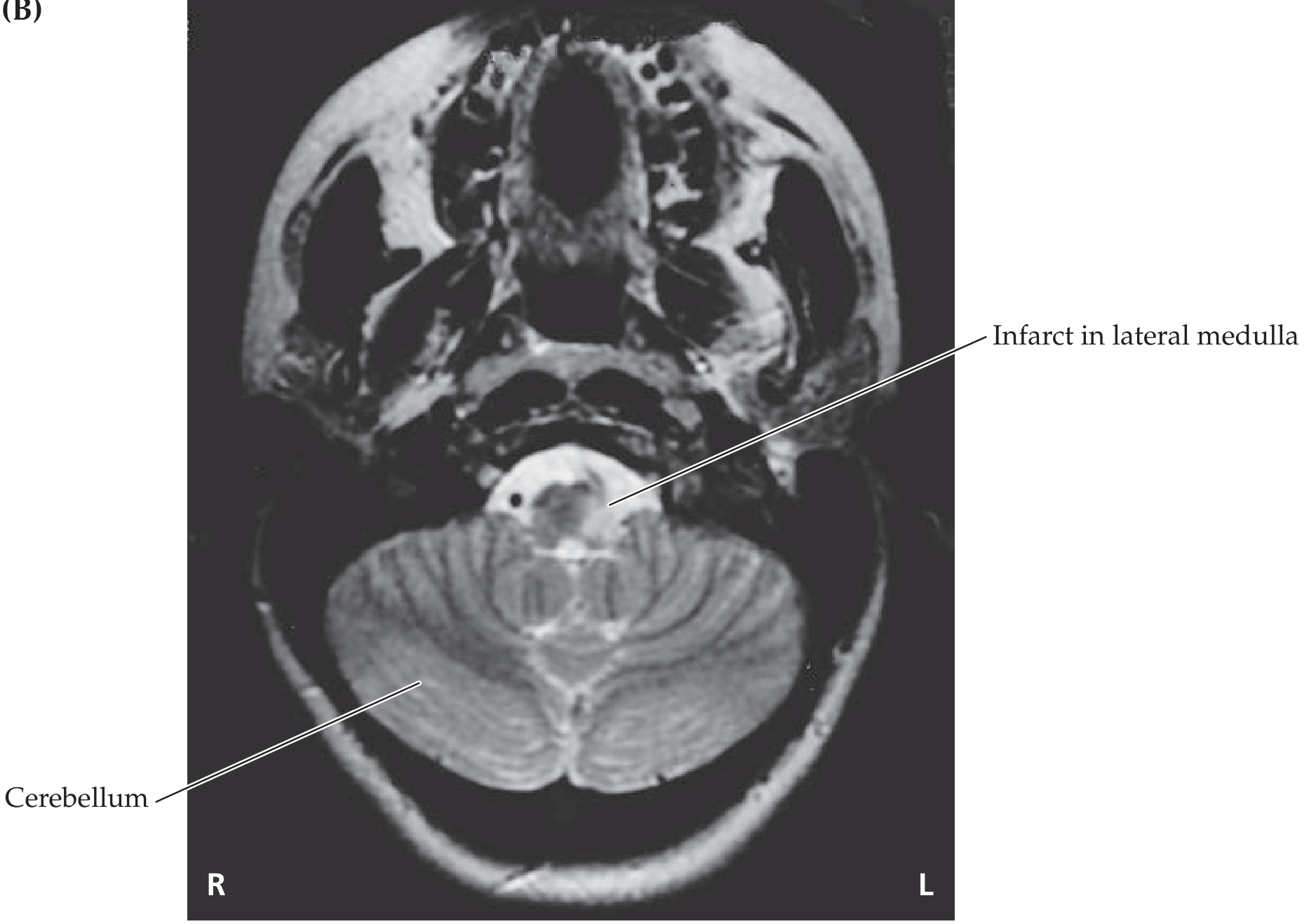
- DWI (diffusion-weighted imaging): Bright signal in lateral medullary wedge within hours of onset
- T2/FLAIR: Hyperintense signal in lateral medulla
- MRA: May show absent flow void in vertebral artery (dissection or thrombosis)
- T1 fat-sat (neck): Thickened, bright vertebral artery wall = dissection with intramural hematoma
Medial Medullary Infarct (Dejerine) on MRI
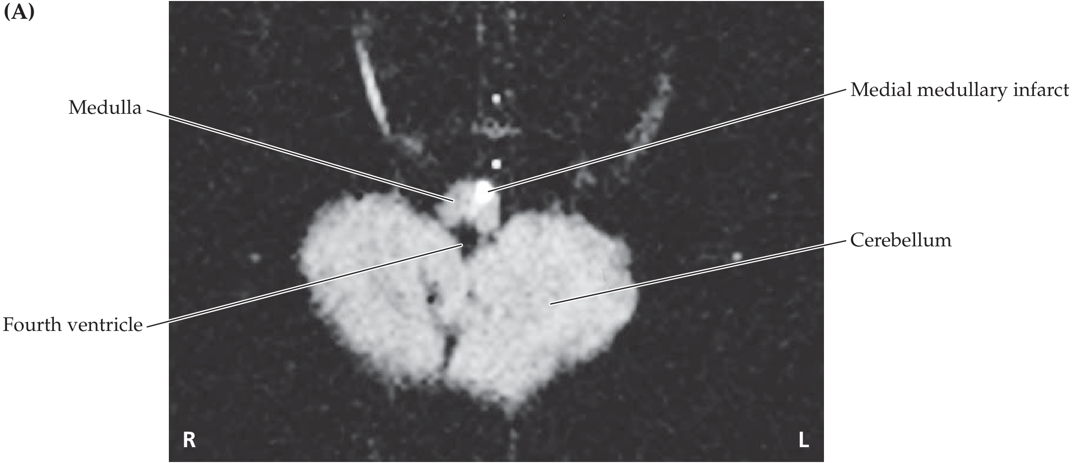
- DWI: Bright paramedian signal (often resembles a "V" or "heart" shape on axial cuts)
- Can be missed on initial MRI - follow-up imaging at 5 days may be needed
- Associated with: Occlusion of paramedian branches of vertebral artery or anterior spinal artery
PART 13 - CROSS-SECTION SHOWING BOTH SYNDROMES SIMULTANEOUSLY
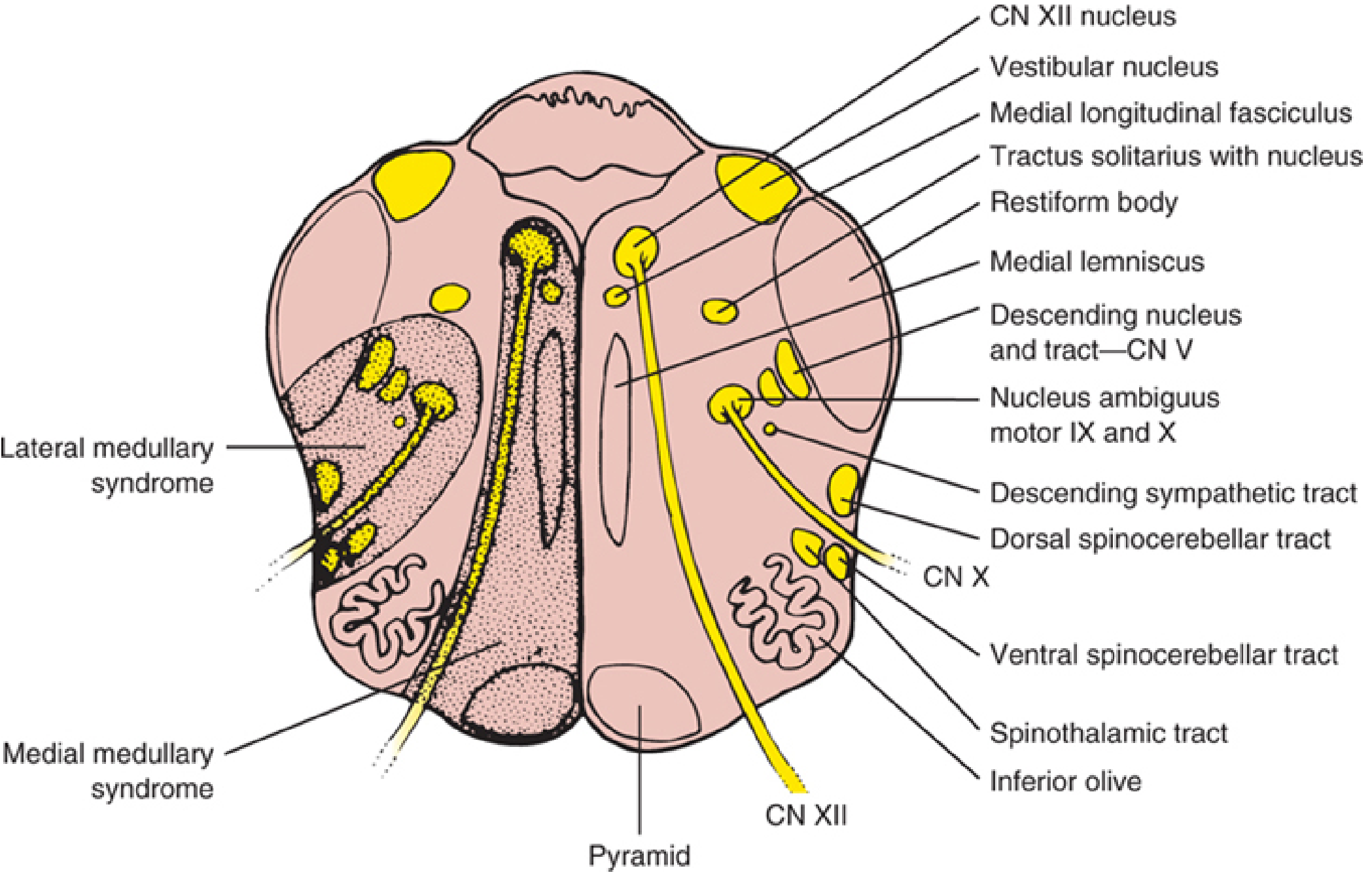
This diagram is the most useful visual for USMLE - memorize which structures fall in the lateral wedge (Wallenberg) vs. the medial/paramedian zone (Dejerine).
PART 14 - ADDITIONAL MEDULLARY SYNDROMES (Less Common but Tested)
Hemimedullary Syndrome (Babinski-Nageotte)
- Combines lateral medullary + medial medullary features on the SAME side
- Results from infarction of the entire lateral + medial medullary territory on one side
- Signs: ipsilateral Horner + ipsilateral tongue weakness + ipsilateral cerebellar signs + contralateral hemiplegia + crossed sensory findings
Bilateral Medial Medullary Syndrome
- Both anterior spinal arteries occluded or bilateral vertebral artery disease
- Flaccid quadriplegia (face spared) + bilateral tongue atrophy and weakness + complete loss of vibration/proprioception all four limbs + respiratory failure (life-threatening)
Opalski Syndrome
- Ipsilateral hemiparesis/hemiplegia + lateral medullary syndrome features
- Due to extension of lateral medullary infarction below the pyramidal decussation
- The corticospinal lesion is now BELOW the decussation → ipsilateral hemiplegia (instead of contralateral)
PART 15 - DORSAL COLUMN PATHWAY vs. SPINOTHALAMIC - COMPLETE COMPARISON
| Feature | Dorsal Column-Medial Lemniscal | Anterolateral (Spinothalamic) |
|---|---|---|
| Modalities | Fine touch, vibration, proprioception, 2-point discrimination, pressure | Pain, temperature, crude touch, tickle, itch |
| 1st order | DRG → dorsal column (ipsilateral) | DRG → dorsal horn (synapse immediately) |
| Where it crosses | Mid-medulla (internal arcuate fibers) | Spinal cord (anterior white commissure, 1-2 levels above entry) |
| 2nd order | Nucleus gracilis/cuneatus → internal arcuate → medial lemniscus | Wide dynamic range neurons in dorsal horn → spinothalamic tract |
| 3rd order | VPL thalamus → S1 cortex | VPL thalamus → S1 cortex |
| Fiber type | Large myelinated (A-alpha, A-beta) | Small myelinated/unmyelinated (A-delta, C) |
| Speed | Fast (well myelinated) | Slow (C fibers) |
| Cortical representation | Excellent spatial localization | Poor localization (crude) |
PART 16 - EMBRYOLOGY
| Structure | Embryologic Origin |
|---|---|
| Medulla oblongata | Myelencephalon (most caudal part of the rhombencephalon) |
| Neurons, glia | Neuroectoderm from neural tube |
| Alar plate | Dorsal - gives rise to sensory nuclei (gracile, cuneate, spinal trigeminal, vestibular, cochlear, NTS) |
| Basal plate | Ventral - gives rise to motor nuclei (hypoglossal, nucleus ambiguus, dorsal motor vagal nucleus) |
| Neural crest | Contributes to peripheral ganglia (inferior ganglion of IX, inferior ganglion of X) |
USMLE: Alar plate = sensory, Basal plate = motor. This holds throughout the brainstem and spinal cord. In the medulla, the alar plate actually flips laterally (because the fourth ventricle opens up dorsally), so the sensory nuclei end up LATERAL and motor nuclei MEDIAL - exactly opposite to the spinal cord arrangement.
PART 17 - GLOSSARY OF KEY MEDULLARY TERMS FOR USMLE
| Term | Definition |
|---|---|
| Obex | V-shaped caudal tip of the fourth ventricle at posterior medulla |
| Restiform body | Inferior cerebellar peduncle (ICP) |
| Internal arcuate fibers | Axons of gracile/cuneate nuclei sweeping to decussate = form medial lemniscus |
| Fasciculus gracilis | Dorsal column tract carrying lower limb dorsal column inputs |
| Fasciculus cuneatus | Dorsal column tract carrying upper limb dorsal column inputs |
| Anterior median fissure | Midline groove on ventral medulla |
| Pre-olivary sulcus | Between pyramid and olive; CN XII exits here |
| Post-olivary sulcus | Between olive and ICP; CN IX, X, XI exit here |
| Medullary pyramids | Bilateral ventral swellings containing corticospinal tract |
| Gracile tubercle | Posterior medullary bump over nucleus gracilis |
| Cuneate tubercle | Posterior medullary bump over nucleus cuneatus |
| Olivocerebellar decussation | Inferior olivary axons cross before entering ICP |
| Lateral reticular nucleus | Part of reticular formation; relay for spinocerebellar inputs |
| Nucleus raphé magnus | Midline serotonergic nucleus; descending pain modulation (inhibits pain) |
| Nucleus raphé obscurus | Medullary midline; serotonin; cardiovascular/respiratory roles |
| Pre-Bötzinger complex | Rhythmic breathing pacemaker in VRG |
| RVLM | Rostral ventrolateral medulla; tonic sympathetic output |
| Ondine's curse | Central hypoventilation from medullary damage (automatic breathing lost) |
PART 18 - MASTER USMLE QUESTION PATTERNS
Pattern 1 - Crossed Findings = Brainstem (almost always)
If a patient has cranial nerve signs on ONE side and motor/sensory signs on the OTHER side of the body → brainstem lesion. The level is determined by which CN is involved.
Pattern 2 - "Tongue deviates to..."
- Toward the lesion = LMN (CN XII nucleus/nerve damage = ipsilateral)
- Away from the lesion = UMN (contralateral cortex lesion)
Pattern 3 - Horner + brainstem signs
= Lateral medullary lesion (Wallenberg) - the descending sympathetic runs in the lateral tegmentum
Pattern 4 - Dysphagia + hoarseness + cerebellar ataxia + Horner + crossed sensory
= Wallenberg. PICA or vertebral artery.
Pattern 5 - Hemiplegia + tongue deviation + no facial involvement
= Medial medullary infarct (Dejerine). Face is SPARED (CN VII nucleus is in the pons, above the medulla).
Pattern 6 - Vomiting triggered by IV drug
= Area postrema (CTZ) outside BBB responds to bloodborne emetic agents
Pattern 7 - Palatal tremor + oscillopsia months after pontine/midbrain hemorrhage
= Hypertrophic olivary degeneration from triangle of Guillain-Mollaret disruption
Pattern 8 - "Which decussation is lower?"
= Motor decussation (pyramidal) is at the caudal medulla; sensory decussation (internal arcuate) is at the mid medulla. Motor decussation is caudal to sensory decussation.
Pattern 9 - Central hypoventilation during sleep
= Ondine's curse - reticular respiratory centers of medulla damaged (often bilateral Wallenberg)
Pattern 10 - Bilateral tongue atrophy + quadriplegia + no facial involvement + respiratory failure
= Bilateral medial medullary syndrome - both hypoglossal nuclei and both pyramids affected
PART 19 - PHARMACOLOGY TIED TO MEDULLARY FUNCTION
| Drug | Target in Medulla | Clinical Use |
|---|---|---|
| Ondansetron | 5-HT3 receptor at area postrema + vagal afferents (NTS) | Chemo/post-op nausea |
| Metoclopramide | D2 receptor at area postrema | Nausea, gastroparesis |
| Apomorphine | D2/D3 agonist at area postrema | Emetic agent (toxin antidote); also used in Parkinson's |
| Atropine | M2 receptors (cardiac = nucleus ambiguus/dorsal vagal effects) | Bradycardia reversal |
| Scopolamine | M1 at vestibular nuclei | Motion sickness |
| Meclizine | H1 + M1 at vestibular system | Vertigo, motion sickness |
| Morphine/opioids | μ receptors at area postrema (emetic) + NTS | Cause nausea via area postrema |
| Dextromethorphan | NMDA receptors in medulla | Cough suppression (acts on NTS cough center) |
| Codeine | μ-opioid at NTS cough center | Cough suppression |
PART 20 - HIGH-YIELD SUMMARY TABLE (Memorize This)
| Medullary Zone | Key Structures | Artery | Syndrome |
|---|---|---|---|
| Lateral wedge | Spinal V nucleus/tract, spinothalamic tract, nucleus ambiguus, descending sympathetic, ICP, vestibular nuclei | PICA (or vertebral a.) | Wallenberg (Lateral Medullary) |
| Paramedian zone | Pyramid (corticospinal), medial lemniscus, CN XII nucleus/fibers | Anterior spinal a. or vertebral a. paramedian branches | Dejerine (Medial Medullary) |
| Dorsomedial | Hypoglossal nucleus, dorsal motor vagal nucleus, MLF | Perforating vertebral branches | Isolated CN XII palsy, autonomic dysfunction |
| Entire medulla | All of above | Vertebral artery occlusion | Hemimedullary (Babinski-Nageotte) |
Sources: Gray's Anatomy for Students, pp. 1317-1323 | Localization in Clinical Neurology, 8e, pp. 915-925 | Adams and Victor's Principles of Neurology, 12e, p. 816 | Neuroanatomy through Clinical Cases, 3e, pp. 691-695 | Guyton and Hall Textbook of Medical Physiology, p. 597 | Katzung's Basic and Clinical Pharmacology, 16e
This is a shared conversation. Sign in to Orris to start your own chat.