
Detailed management of melesma
melasma treatment management
melasma facial hyperpigmentation clinical photo
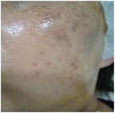
This clinical photograph displays the facial skin of a patient with melasma, specifically focusing on the lateral cheek and temple area. The image reveals numerous hyperpigmented macules and patches of varying sizes and shapes. These pigmented lesions range in color from light tan to dark brown, demonstrating a patchy, heterogeneous distribution typical of dermal or mixed melasma. The skin surface appears smooth and hydrated with a visible reflective sheen, and there are no signs of scaling, erythema, or post-inflammatory hyperpigmentation (PIH). The visual evidence represents a follow-up state after multiple treatment modalities, including Q-switched 1064 nm Nd:YAG laser sessions and adjuvant vitamin C therapy. The educational focus is on identifying clinical patterns of facial hyperpigmentation and evaluating treatment response in dermatologic practice for disorders of pigmentation.
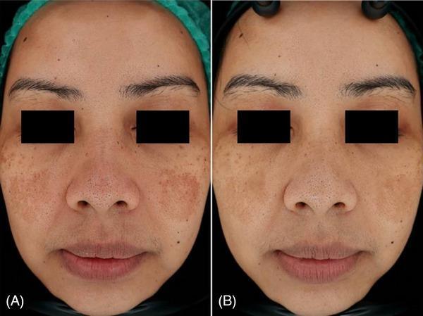
This clinical photograph comparison shows the face of a 39-year-old female patient diagnosed with facial melasma, highlighting a before-and-after treatment progression over three months. Image (A) displays baseline hyperpigmentation characterized by confluent, brownish patches with irregular borders, symmetrically distributed across the malar regions (cheeks) and the nasal bridge. The pigmentation appears dense with a mottled texture. Image (B), taken three months after laser therapy, demonstrates a significant clinical improvement. There is a visible reduction in the size, color intensity, and overall density of the melasma patches. The skin tone appears more uniform and brightened, with less prominent follicular or pore visibility in the affected areas. This comparison illustrates the effectiveness of dermatological laser intervention for managing epidermal and dermal hyperpigmentation disorders in adult patients.
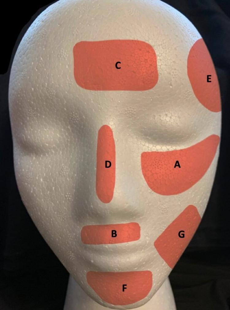
This anatomical diagram uses a white mannequin head to illustrate the most frequent facial regions affected by melasma. Seven orange shaded areas, labeled A through G, represent localized zones of hyperpigmentation based on clinical prevalence. The regions identified include: (A) zygomatic (cheek) area, the most frequent site; (B) supralabial (upper lip) area; (C) frontal (forehead) area; (D) nasal (nose) bridge; (E) temporal area; (F) mental (chin) area; and (G) mandibular (jawline) area. The diagram demonstrates the distribution of centrofacial, malar, and mandibular melasma patterns. These zones correspond to facial convexities most susceptible to ultraviolet (UV) radiation and visible light exposure, which are key triggers for melanocyte activation and exacerbation of melasma. The visual serves as an educational tool for dermatologists and medical students to recognize common clinical presentations and diagnostic patterns of this acquired pigmentary disorder.
melasma management guidelines 2024 2025 tranexamic acid cysteamine treatment
Detailed Management of Melasma
Overview
- (A) Zygomatic (cheek) - most frequent
- (B) Supralabial (upper lip)
- (C) Frontal (forehead)
- (D) Nasal bridge
- (E) Temporal
- (F) Mental (chin)
- (G) Mandibular (jawline)
Classification
| Type | Melanin Location | Wood's Lamp | Response to Topicals |
|---|---|---|---|
| Epidermal | Basal/suprabasal epidermis | Accentuated | Good |
| Dermal | Dermal melanophages | Not accentuated | Poor |
| Mixed | Both layers | Variable | Partial |
Note: Most cases show an admixture of both epidermal and dermal types on histology/confocal microscopy, regardless of Wood's lamp findings. A therapeutic trial should be offered independent of subtype classification. - Dermatology 2-Volume Set 5e
Step 1 - Trigger Avoidance (Foundational)
- Discontinue oral contraceptives and hormone replacement therapy if feasible
- Avoid phototoxic/melanogenic drugs: phenytoin, finasteride, doxycycline, amiodarone
- Avoid pregnancy-related exacerbation counseling (melasma of pregnancy usually resolves months after delivery; OCP-related melasma often persists even after discontinuation)
- Avoid irritant topical agents that can cause postinflammatory hyperpigmentation (PIH)
Step 2 - Photoprotection (Non-Negotiable, Ongoing)
| Measure | Details |
|---|---|
| Sunscreen | Broad-spectrum SPF ≥30 daily; physical blockers (zinc oxide, titanium dioxide) preferred |
| Visible light protection | Iron oxide-containing formulations for Fitzpatrick types IV-VI (visible light is a significant trigger in darker skin) |
| Protective clothing | Wide-brimmed hats, UPF-rated clothing |
| Sun avoidance | Avoid peak UV hours (10 AM - 4 PM); avoid tanning beds |
| Camouflage makeup | With iron oxide to block visible light |
Step 3 - Topical Therapies (First-Line Active Treatment)
A. Hydroquinone (HQ) - Gold Standard
- Mechanism: Tyrosinase inhibitor; inhibits conversion of DOPA to melanin
- 2% HQ: Available OTC, modestly effective
- 4% HQ: Prescription strength, first-line active treatment
- Higher concentrations (>4%): For refractory cases - risk of satellite pigmentation and exogenous ochronosis increases
- Use at bedtime for 2-4 months, then taper to 1-2x/week for maintenance
- Side effects: Irritant/allergic contact dermatitis, exogenous ochronosis (with prolonged overuse), perioral dermatitis
B. Kligman's Formula - Most Effective Topical Regimen
5% Hydroquinone + 0.1% Tretinoin + Mild topical corticosteroid (applied at bedtime)
4% Hydroquinone + 0.05% Tretinoin + 0.01% Fluocinolone acetonide
C. Retinoids (Monotherapy or Adjunct)
- Tretinoin (retinoic acid): Stimulates keratinocyte turnover, reduces melanosome transfer, inhibits tyrosinase. Approved in the US for melasma treatment.
- Less effective than HQ monotherapy but synergistic in combination
- Also: adapalene, tazarotene (approved for acne, used off-label)
- Used long-term as maintenance therapy
D. Azelaic Acid (15-20%)
- Tyrosinase inhibitor; selectively toxic to hyperactive melanocytes
- Useful as HQ alternative or adjunct
- Safe in pregnancy (Category B)
- Less irritating than HQ; good tolerability profile
E. Kojic Acid (1-4%)
- Inhibits tyrosinase by chelating copper at the active site
- Used as adjunct; may cause contact dermatitis
- Often combined with glycolic acid or HQ
F. Topical Tranexamic Acid (2-5%)
- Mechanism: Plasmin inhibition reduces UV-induced prostaglandin synthesis, thereby reducing melanocyte stimulation; antioxidant
- Effective as adjunct for both epidermal and dermal melasma
- Good safety profile; useful in HQ-intolerant patients
G. Other Adjunctive Agents
| Agent | Concentration | Mechanism |
|---|---|---|
| L-ascorbic acid (Vitamin C) | 10-15% | Antioxidant; reduces dopaquinone |
| Niacinamide | 4% | Inhibits melanosome transfer to keratinocytes |
| Arbutin | Variable | Tyrosinase inhibitor; inhibits melanosome maturation |
| Methimazole | Variable | Antithyroid agent; peroxidase inhibitor |
| Cysteamine 5% | 5% | Potent antioxidant; inhibits tyrosinase and peroxidase |
| Licorice extract | Variable | Glabridin - tyrosinase inhibitor |
2024-2025 evidence: Cysteamine 5% cream has strong recent evidence as an HQ-alternative. A 2024 systematic review and meta-analysis (PMID: 39673630) confirmed its efficacy and safety for melasma. A nano-formulated cysteamine + tranexamic acid combination cream has also shown significant MASI score reduction with excellent tolerability.
Step 4 - Oral Systemic Therapies (Adjunctive)
Oral Tranexamic Acid (OTA)
- Dose: 250 mg BID for 8-12 weeks (low dose; some protocols use 500-700 mg/day)
- Indication: Treatment-resistant, moderate-to-severe, or dermal melasma
- Mechanism: Inhibits the plasminogen-keratinocyte interaction, reducing UV-stimulated prostaglandin E2 and alpha-MSH release, ultimately reducing melanogenesis
- Proven effective for both epidermal and dermal components - Fitzpatrick's Dermatology, p. 1410
- A 2024 meta-analysis (PMID: 38843906) of RCTs confirmed OTA as an effective therapeutic option
- Intradermal tranexamic acid injection also shows meta-analytic support (PMID: 39574359, 2025)
- Contraindications/screening: Screen for thromboembolic risk, hypercoagulable states, history of DVT/PE, renal impairment; avoid in pregnancy
Step 5 - Procedural Therapies (Second and Third Line)
Chemical Peels (Second-Line)
| Peel Agent | Concentration | Notes |
|---|---|---|
| Glycolic acid | Start at 30%, increase as tolerated | Every 4-6 weeks; adjunct to topicals |
| Salicylic acid | 20-30% | Safer in darker skin types |
| TCA | Low-concentration | Risk of PIH; use cautiously |
| Tretinoin 1% | 1% | Mild improvement |
Laser and Light-Based Therapies (Third-Line)
| Modality | Notes |
|---|---|
| Low-fluence Q-switched Nd:YAG (1064 nm) ("laser toning") | Most popular; safest in skin of color; efficacious when few treatments used in carefully selected cases |
| Non-ablative fractional lasers (1550 nm, 1927 nm) | Moderate evidence; may improve HQ absorption via microchannels; PIH risk |
| Intense Pulsed Light (IPL) | Improves melasma but high relapse rate; PIH risk |
| Erbium:YAG (Er:YAG) | Temporary improvement; risk of PIH |
| Picosecond lasers | Emerging evidence; potentially safer |
| Standard Q-switched lasers | Can paradoxically increase pigmentation; use cautiously |
Always perform a test spot before widespread laser/light therapy. The inflammatory response from laser treatment can perpetuate or aggravate dermal melasma via PIH. - Dermatology 2-Volume Set 5e, p. 2859
A 2025 meta-analysis (PMID: 40555739) supports microneedling combined with tranexamic acid as an effective procedural approach.
Step 6 - Long-Term Maintenance
- Daily broad-spectrum sunscreen (non-negotiable)
- Sun-protective measures (hats, clothing, shade-seeking)
- Topical retinoid (nightly; low-concentration)
- Topical alpha-hydroxy acid (e.g., glycolic acid cream)
- Rotating adjuncts: Vitamin C 10-15%, azelaic acid 15-20%, kojic acid 1-4%, tranexamic acid 2-5%
- HQ or triple combination 1-2x/week as needed (avoid prolonged daily HQ use)
Special Populations
| Population | Considerations |
|---|---|
| Pregnancy | Sunscreen + azelaic acid (safe); avoid HQ, retinoids, kojic acid; melasma often resolves postpartum |
| Darker skin types (IV-VI) | Iron oxide sunscreen for visible light; avoid aggressive peels/lasers; lower PIH threshold; prefer gentler agents |
| Dermal melasma | Topicals less effective; oral tranexamic acid, laser toning are preferred options |
| HQ-intolerant | Use azelaic acid, cysteamine, or tranexamic acid-based formulations |
Summary Algorithm
ALL PATIENTS: Trigger avoidance + Photoprotection (SPF ≥30 + iron oxide)
        ↓
FIRST-LINE: Triple combination (HQ 4% + tretinoin + topical steroid) OR HQ 4% alone
        ↓ (insufficient response at 3-6 months)
ADD ADJUNCTS: Topical tranexamic acid, vitamin C, niacinamide, azelaic acid, kojic acid
        ↓ (still refractory)
SYSTEMIC: Oral tranexamic acid 250 mg BID x 8-12 weeks
        ↓ (persistent/dermal melasma)
PROCEDURAL: Chemical peels → Laser toning (low-fluence QS Nd:YAG) → Fractional lasers / IPL
        ↓
MAINTENANCE: Sunscreen + retinoid ± rotating topical agents indefinitely
Key Points
- Hydroquinone 4% remains the gold standard topical agent - Fitzpatrick's Dermatology, p. 1409
- Kligman's formula (triple combination) is the most effective single topical regimen
- Dermal melasma responds poorly to topicals; oral tranexamic acid and low-fluence laser toning offer the best options
- Cysteamine 5% is a well-evidenced, safe HQ-alternative with strong 2024-2025 meta-analytic support
- Laser therapy must be approached cautiously; PIH is a real and common complication
- Melasma is chronic - patients must understand that no treatment is curative and maintenance is lifelong
- Quality of life impact (depression, anxiety) should be explicitly assessed and addressed