
Mefenamic acid vs tolfenamic acid
Both are fenamate NSAIDs (anthranilic acid derivatives) that inhibit COX-1 and COX-2, but they have different clinical profiles and primary uses.
Searching sources...
Reading File
Finding Sources
Finding Sources
Searching PubMed
mefenamic acid tolfenamic acid comparison
Reading File
Finding Sources
Searching PubMed
tolfenamic acid migraine dysmenorrhea
Finding Sources
Searching the Web
tolfenamic acid vs mefenamic acid pharmacology clinical uses comparison
Finding Sources
Searching PubMed
tolfenamic acid migraine treatment clinical trial
Reading File
Now I have enough data to give a thorough comparison. Let me compile the full answer.
Mefenamic Acid vs Tolfenamic Acid
Both belong to the fenamate (anthranilic acid derivative) subclass of NSAIDs. They share a core diphenylamine backbone but differ in their chlorine substituent position and, more importantly, in their clinical niches.
1. Chemical Structure
| Feature | Mefenamic Acid | Tolfenamic Acid |
|---|---|---|
| IUPAC name | N-(2,3-dimethylphenyl)anthranilic acid | N-(2-methyl-3-chlorophenyl)anthranilic acid |
| Key substituent | 2,3-dimethyl on the phenyl ring | 2-methyl-3-chloro on the phenyl ring |
| Halogenation | None | Chlorine at position 3 |
2. Mechanism of Action
Both drugs are non-selective COX-1 and COX-2 inhibitors that reduce prostaglandin and thromboxane synthesis. They share the additional fenamate property of antagonizing prostaglandin receptors (not just blocking synthesis), which is a distinguishing feature of the fenamate class not shared by most other NSAIDs. This dual action - reduced prostaglandin production + receptor blockade - may partly explain their efficacy in dysmenorrhea and migraine.
Tolfenamic acid also has documented anti-endotoxic and mild antibacterial properties, and at least one in vitro signal for anticancer activity, though these have limited clinical translation.
3. Pharmacokinetics
| Parameter | Mefenamic Acid | Tolfenamic Acid |
|---|---|---|
| Peak plasma concentration | 2-4 hours | ~2-3 hours |
| Half-life | 2-4 hours (short) | ~1-2 hours (even shorter; rapid redistribution) |
| Protein binding | ~90% | >99% |
| Metabolism | CYP2C9 oxidation + glucuronidation | Hydroxylation to less active metabolites |
| Excretion | Renal (urine) | Renal (urine) |
| Duration of action | 4-6 hours (requires dosing every 6 h) | 4-6 hours |
Both have short half-lives requiring multiple daily doses. The short t1/2 actually makes tolfenamic acid suitable for acute migraine (single-dose or short-course therapy) rather than chronic use. - Goodman & Gilman's Pharmacological Basis of Therapeutics
4. Clinical Uses
| Indication | Mefenamic Acid | Tolfenamic Acid |
|---|---|---|
| Primary dysmenorrhea | ✅ First-line; well established | ✅ Effective; less commonly used |
| Acute migraine | Used off-label | ✅ Primary indication in many countries (Scandinavia, Finland); RCT-supported |
| Migraine prophylaxis | Rarely used | ✅ Studied in RCTs vs propranolol |
| Short-term pain (dental, soft tissue) | ✅ Approved | ✅ Approved |
| Rheumatoid arthritis / OA | ✅ Short-term use | ✅ Inflammatory/rheumatic disorders |
| Antipyretic | Yes | Yes (notably studied in children: 0.5 mg/kg effective) |
Tolfenamic acid has the strongest NSAID evidence base for acute migraine treatment - RCTs have shown it effective alone and in combination with metoclopramide and caffeine (Tokola et al., 1984, PMID: 6394143), and it has been compared against propranolol for migraine prophylaxis (Rasmussen et al., 1994, PMID: 7976233).
Mefenamic acid's dominant niche is dysmenorrhea, where it is among the best-studied NSAIDs due to its dual prostaglandin synthesis inhibition + receptor antagonism. Treatment is typically limited to 2-3 days per cycle.
5. Dosing
| Parameter | Mefenamic Acid | Tolfenamic Acid |
|---|---|---|
| Standard adult dose | 500 mg loading dose, then 250 mg every 6 hours | 100-200 mg up to 3x daily |
| Migraine (acute) | Off-label ~500 mg | 200 mg single dose (may repeat once) |
| Max duration | 7 days (pain); 2-3 days (dysmenorrhea) | Short-term (acute/episodic use) |
| Pediatric use | Not recommended | Antipyretic use studied (0.5 mg/kg) |
6. Adverse Effects
Both share the standard NSAID adverse effect profile:
| Adverse Effect | Mefenamic Acid | Tolfenamic Acid |
|---|---|---|
| GI toxicity | Common; diarrhea can be severe with steatorrhea, bowel inflammation | Common (~1.7% in large epidemiological study of 16,521 patients) |
| Hepatotoxicity | ~15% get elevated liver enzymes; 5% reversible transaminase elevation | Less emphasized |
| CNS toxicity | ⚠️ Notably higher risk of seizures in overdose vs other NSAIDs (UK NPIS data) | Seizure risk not specifically highlighted |
| Autoimmune hemolytic anemia | Rare but serious | Not specifically reported |
| Pseudoporphyria (skin) | Reported | Not highlighted |
| Thyroid hormone displacement | Yes (displaces T3/T4 from TBG) | Not reported |
| Pregnancy | Not recommended | Not recommended |
| Overall adverse event rate | ~5-10% | ~3% (from 16,521-patient study) |
Mefenamic acid carries a specific warning about CNS toxicity and seizures in overdose, which is higher than most NSAIDs. It is also listed as a cause of pseudoporphyria and T3/T4 displacement from thyroid-binding globulin.
7. Contraindications & Precautions
Both share standard NSAID contraindications:
- Active peptic ulcer disease
- Severe renal/hepatic impairment
- Third trimester pregnancy (risk of premature closure of ductus arteriosus)
- Aspirin-exacerbated respiratory disease
- Children/adolescents for mefenamic acid specifically (not recommended)
8. Key Distinguishing Points
| Feature | Mefenamic Acid | Tolfenamic Acid |
|---|---|---|
| Primary clinical niche | Dysmenorrhea | Acute migraine |
| Seizure risk in overdose | Higher (notable warning) | Not specifically highlighted |
| Liver enzyme elevation | ~15% rate | Less documented |
| Pediatric use | Avoided | Antipyretic use studied |
| Veterinary use | No | Widely used (cattle, pigs, dogs, cats) |
| Regulatory availability | USA (brand: Ponstel), UK, widespread | Primarily Europe/Scandinavia; not FDA-approved |
Summary
Both are short-acting fenamate NSAIDs with similar mechanisms and comparable efficacy. The main practical difference is indication focus: mefenamic acid is the go-to fenamate for dysmenorrhea (2-3 day cycles), while tolfenamic acid has the strongest evidence as an acute migraine abortive agent and has been studied for migraine prophylaxis. Mefenamic acid carries a higher signal for seizures in overdose and liver enzyme elevation. Tolfenamic acid appears to have a somewhat better overall tolerability profile based on large-scale post-marketing data.
Sources: Goodman & Gilman's Pharmacological Basis of Therapeutics (fenamate pharmacology and PK table); EMA tolfenamic acid summary report; clinical trial data from PubMed.
Imaging to rule out prolactinoma
MRI of the pituitary gland with gadolinium contrast is the gold standard imaging for prolactinoma. CT is a fallback when MRI is contraindicated.
Searching sources...
Reading File
Finding Sources
Finding Sources
Searching PubMed
prolactinoma pituitary MRI diagnosis guidelines
Reading File
Reading File
Searching PubMed
prolactinoma diagnosis management Endocrine Society guideline
Finding Sources
Finding Sources
Searching PubMed
hyperprolactinemia pituitary MRI diagnosis workup
Reading File
Finding Sources
Finding Sources
Reading File
Finding Sources
Loading Image
Excellent - I now have a thorough set of data including the clinical flowchart. Let me compile the full answer.
Imaging to Rule Out Prolactinoma
The Modality: MRI of the Pituitary with Gadolinium
MRI with gadolinium (contrast) enhancement of the sella turcica and pituitary gland is the imaging modality of choice. It provides the best anatomic detail for evaluating the hypothalamic-pituitary region. CT scanning is generally avoided because:
- Cumulative radiation dose from repeated scans can cause cataracts
- "Coned-down" views and tomograms of the sella are very insensitive
- MRI far outperforms CT in soft tissue resolution for small tumors
CT is only used when MRI is contraindicated (e.g., pacemaker, severe claustrophobia).
- Berek & Novak's Gynecology; Textbook of Family Medicine 9e
When to Image
Imaging is not required in every case of elevated prolactin. The indication depends on clinical context:
| Situation | Image? |
|---|---|
| Unexplained, persistent prolactin elevation after drugs/hypothyroidism excluded | Yes - MRI |
| Prolactin >100 ng/mL (strong suspicion of adenoma) | Yes - MRI |
| Symptoms suggesting mass lesion (headache, visual field defects) | Yes - MRI urgently |
| Clearly drug-induced hyperprolactinemia, no mass symptoms | Not necessarily required |
| Patient desires future fertility (even modest elevation) | Yes - MRI to distinguish micro from macroadenoma |
| Macroadenoma confirmed on MRI | Formal visual field testing also required |
What MRI Shows: Micro vs Macroadenoma
The key anatomical distinction is tumor size, which drives management:
| Feature | Microadenoma | Macroadenoma |
|---|---|---|
| Size | < 10 mm | > 10 mm |
| Prolactin level correlation | Usually < 100-200 ng/mL (though overlap exists) | Often > 200-250 ng/mL; very high levels (>1000 ng/mL) essentially diagnostic |
| MRI appearance | Hypointense focus on dynamic contrast T1; may be subtle | Obvious sellar mass, often with suprasellar extension |
| Risk of progression | Low (~7% progress to macro) | Already large; may compress optic chiasm |
| Visual field testing | Rarely needed unless imaging suggests optic compression | Mandatory (risk of bitemporal hemianopsia) |
| Neurosurgical consult | Not typically needed | Required |
Important caveat: up to half of all prolactin-secreting microadenomas are too small to be detected by any imaging method. A normal MRI does not exclude a microadenoma - it may represent a very small adenoma, prolactin-cell hyperplasia, or idiopathic hyperprolactinemia. - Tietz Textbook of Laboratory Medicine, 7th ed.
Key Diagnostic Pitfalls
1. The "Stalk Effect" (Pseudoprolactinoma)
Any large pituitary or suprasellar mass (craniopharyngioma, non-functioning adenoma, Rathke's cleft cyst) can compress the hypothalamic-pituitary portal system, impairing dopamine delivery to lactotrophs, causing mild-moderate prolactin elevation (50-200 ng/mL) - this is not a true prolactinoma. If a macroadenoma is on MRI but prolactin is only modestly elevated, suspect stalk effect rather than a true prolactinoma.
- A dopamine agonist (bromocriptine/cabergoline) will reduce prolactin in a true prolactinoma but not in stalk effect.
2. The Hook Effect
Very high prolactin concentrations can saturate the immunoassay, causing a falsely low reading. If MRI shows a large macroadenoma but prolactin seems only modestly elevated, request a 1:10 and 1:100 dilution of the sample to unmask the true (much higher) level.
3. Macroprolactinemia
Large-molecular-weight prolactin aggregates with IgG can register as elevated prolactin on immunoassay despite no pathology. Nearly 20% of patients referred for prolactinoma workup may have this. The lab can rule it out with polyethylene glycol (PEG) precipitation before ordering an MRI. A normal pituitary MRI in a patient with high prolactin and no symptoms should prompt consideration of macroprolactinemia.
4. Pituitary Incidentaloma
Incidental pituitary adenomas occur in 10-20% of the general population on imaging. A lesion on MRI is not automatically the cause of elevated prolactin - it may be an incidentaloma. Clinical correlation between prolactin level, tumor size, and symptoms is essential.
Prolactin Level and Its Correlation with Imaging Findings
| Prolactin Level | Likely Interpretation |
|---|---|
| Mildly elevated (< 50 ng/mL) | Drug-induced, physiological, macroprolactinemia, small microadenoma, idiopathic |
| 50-200 ng/mL | Microadenoma, stalk effect from non-functioning adenoma, hypothyroidism |
| > 200 ng/mL | Strongly suggests prolactinoma |
| > 300 ng/mL | Almost always a pituitary adenoma (in the absence of pregnancy) |
| > 1000 ng/mL | Essentially diagnostic of macroprolactinoma - consider hook effect if MRI doesn't match |
- Swanson's Family Medicine Review; Smith & Tanagho's General Urology
Clinical Workup Flowchart
This is the standard diagnostic pathway from Berek & Novak's Gynecology:
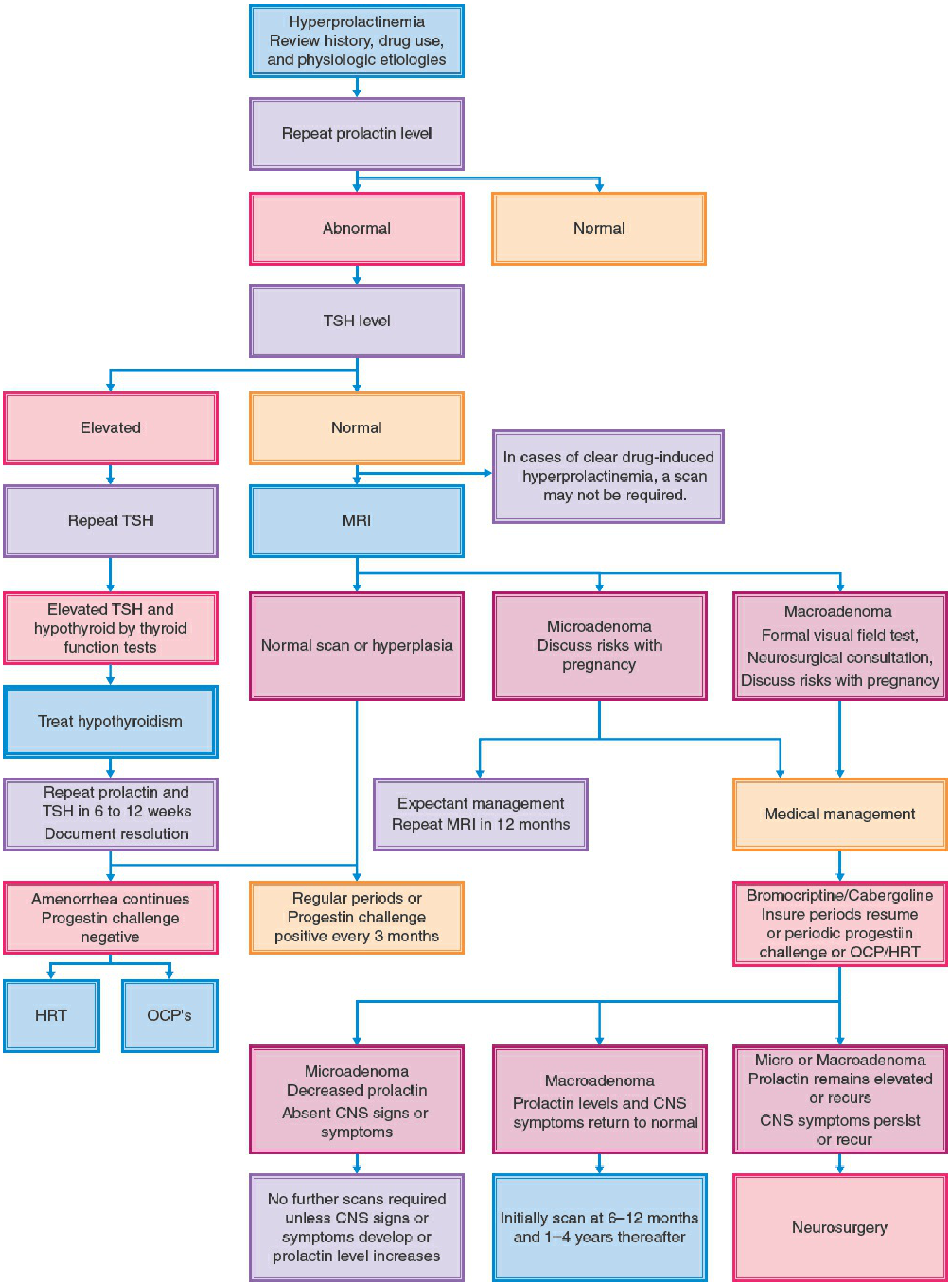
Workup for hyperprolactinemia - Berek & Novak's Gynecology, Fig. 35-9
Follow-up Imaging After Diagnosis
| Scenario | Follow-up MRI |
|---|---|
| Microadenoma, treated, prolactin normalized, no CNS symptoms | Repeat at 12 months; if stable, no further scans unless prolactin rises or CNS symptoms develop |
| Macroadenoma, medical treatment, prolactin and CNS symptoms normalize | Rescan at 6-12 months, then every 1-4 years |
| Symptoms persist or recur despite treatment | Neurosurgery referral |
Summary
- MRI pituitary with gadolinium = gold standard for prolactinoma
- Image when prolactin is persistently elevated without a clear drug/physiological cause, or when mass symptoms exist
- A normal MRI does not exclude a small microadenoma
- Always rule out macroprolactinemia (PEG test) and the hook effect before concluding imaging findings
- Distinguish true prolactinoma from stalk effect (the latter won't respond to dopamine agonists)
- Macroadenoma always requires formal visual field testing and neurosurgical consultation
Tests for hyperthyrpidism
The core tests are TSH (suppressed), free T4 (elevated), and free T3. Additional tests include thyroid antibodies, radioiodine uptake scan, and thyroid ultrasound depending on the suspected cause.
Searching sources...
Finding Sources
Finding Sources
Searching PubMed
hyperthyroidism diagnosis laboratory tests guidelines
Reading File
Reading File
Finding Sources
Reading File
Finding Sources
Finding Sources
Reading File
Finding Sources
Reading File
Finding Sources
I now have comprehensive data. Here is the full structured answer:
Tests for Hyperthyroidism
Step-by-Step Diagnostic Approach
Step 1 - TSH (First-Line / Best Initial Test)
Serum TSH is the single best initial test for suspected hyperthyroidism.
- Normal range: ~0.5-4.5 mIU/L
- In hyperthyroidism: TSH is suppressed (typically < 0.1 μU/mL in overt disease)
- A normal TSH virtually excludes clinical hyperthyroidism - no further thyroid testing is needed
- TSH is sensitive because even small rises in free T4 suppress TSH through negative feedback
Important caveat: TSH can be falsely suppressed by severe non-thyroidal illness, dopamine infusions, and high-dose glucocorticoids. TSH also remains suppressed for 6-8 weeks after hyperthyroidism is corrected, so it is not useful for monitoring early treatment response. - Washington Manual of Medical Therapeutics
Step 2 - Free T4 (If TSH is Low)
If TSH is suppressed, measure free T4 (fT4):
| Result | Interpretation |
|---|---|
| TSH low + fT4 elevated | Overt hyperthyroidism confirmed |
| TSH low + fT4 normal | Measure free T3 (possible T3 toxicosis or subclinical disease) |
| TSH low + fT4 and fT3 both normal | Subclinical hyperthyroidism |
Free T4 measured by equilibrium dialysis is the gold standard but rarely available rapidly. Standard immunoassay fT4 is reliable in most settings. - Goldman-Cecil Medicine
Step 3 - Free T3 (Selected Cases)
Measure free T3 when TSH is suppressed but fT4 is normal:
- T3 toxicosis occurs in ~5% of hyperthyroid patients - elevated free T3 with normal free T4. Seen early in hyperthyroidism and in iodine-deficient states combined with Graves disease.
- Free T3 also confirms severity of hyperthyroidism when TSH is low.
- Total T3 is sometimes used instead because accurate fT3 measurement is technically challenging at low concentrations.
- Free T3 is not useful for diagnosing hypothyroidism. - Rosen's Emergency Medicine; Harrison's Principles of Internal Medicine 22e
Reverse pattern - elevated fT4 with normal T3 (T4 toxicosis): suggests thyroiditis, exogenous levothyroxine ingestion, or hyperthyroidism in elderly patients with suppressed T4-to-T3 conversion.
Summary: Pattern Recognition
| TSH | Free T4 | Free T3 | Diagnosis |
|---|---|---|---|
| Low | High | High | Overt hyperthyroidism |
| Low | Normal | High | T3 toxicosis (~5% of cases) |
| Low | Normal | Normal | Subclinical hyperthyroidism |
| Normal/High | High | High | Secondary hyperthyroidism (TSH-secreting pituitary adenoma - rare) |
Additional Tests to Establish the Cause
Once hyperthyroidism is biochemically confirmed, additional tests identify the etiology, which drives treatment:
4. TSH Receptor Antibodies (TRAb / TSI)
- Highly sensitive and specific for Graves disease
- TRAb (TSH receptor antibodies) act as TSH agonists, stimulating uncontrolled thyroid hormone production
- Elevated TRAb essentially confirms Graves disease and has largely replaced radioiodine scanning for this purpose in most centres
- Also called TSI (thyroid-stimulating immunoglobulin) or LATS (long-acting thyroid stimulator)
- A 2024 Practice Guideline (Kalra et al., PMID: 39707289) highlights TRAb as the best laboratory marker for Graves disease diagnosis, prognosis, and monitoring
5. Anti-TPO and Anti-Thyroglobulin Antibodies
- Anti-thyroid peroxidase (anti-TPO) and anti-thyroglobulin antibodies are elevated in autoimmune thyroid disease (both Graves and Hashimoto's)
- They are not specific to Graves disease - more strongly associated with Hashimoto's thyroiditis
- Useful to confirm autoimmune aetiology but not to distinguish Graves from thyroiditis in isolation
6. ESR (Erythrocyte Sedimentation Rate)
- Used in the diagnosis of subacute (de Quervain's) thyroiditis - typically very elevated (often > 50 mm/hr)
- Thyroiditis causes transient hyperthyroidism (release of stored hormone) followed by hypothyroidism
7. Radioactive Iodine Uptake (RAIU) Scan with I-123
Used when the cause of hyperthyroidism is unclear after antibody testing, particularly to distinguish:
| Cause | RAIU Pattern |
|---|---|
| Graves disease | Diffusely increased uptake |
| Toxic multinodular goiter | Patchy/focal increased uptake |
| Toxic adenoma (Plummer disease) | Single hot nodule, suppressed background |
| Subacute/postpartum thyroiditis | Very low/absent (hormone release, not overproduction) |
| Iodine-induced hyperthyroidism | Low (gland saturated with non-radioactive iodine) |
| Factitious hyperthyroidism | Very low |
Note: RAIU is contraindicated in pregnancy. If a patient had recent iodinated contrast exposure, the gland is iodine-saturated - use pertechnetate scanning as an alternative. - Goldman-Cecil Medicine; Washington Manual
8. Thyroid Ultrasound with Doppler
- Best modality for thyroid structure
- In Graves disease: diffuse enlargement, heterogeneous echotexture, markedly increased vascularity on Doppler ("thyroid inferno")
- In thyroiditis: decreased or absent vascularity on Doppler (helpful to distinguish from Graves when RAIU is not done)
- Characterises nodules in toxic multinodular goiter or toxic adenoma
- Useful when TRAb is negative but Graves is still suspected, or when RAIU is contraindicated
9. Other Associated Tests
| Test | Indication |
|---|---|
| Serum calcium | Hyperthyroidism can cause hypercalcaemia |
| Liver enzymes / ALP | Often elevated in hyperthyroidism |
| CBC | May show mild anaemia, lymphocytosis (Graves) |
| Cholesterol | Low total cholesterol suggests hyperthyroidism |
| Blood glucose | Worsening glycaemic control in diabetics |
| Serum SHBG | Elevated in hyperthyroidism; useful marker |
| Urine/serum hCG | Rule out gestational trophoblastic disease causing hyperthyroidism (hCG cross-reacts with TSH receptor) |
Common Causes and Their Diagnostic Fingerprint
| Cause | TSH | fT4/fT3 | TRAb | RAIU | Ultrasound |
|---|---|---|---|---|---|
| Graves disease | ↓ | ↑ | Positive | Diffuse ↑ | ↑ vascularity |
| Toxic multinodular goiter | ↓ | ↑ | Negative | Patchy ↑ | Multiple nodules |
| Toxic adenoma | ↓ | ↑ | Negative | Single hot nodule | Single nodule |
| Subacute thyroiditis | ↓ | ↑ | Negative | Very low | ↓ vascularity, tender |
| Postpartum thyroiditis | ↓ | ↑ | Sometimes + | Low | Variable |
| Exogenous T4/T3 | ↓ | ↑ fT4 | Negative | Very low | Normal/small gland |
| TSH-secreting pituitary adenoma | Normal/↑ | ↑ | Negative | ↑ | Normal thyroid |
Pitfalls: False Thyroid Function Tests
Be aware of conditions that give spurious thyroid results (Goldman-Cecil Medicine, Table 207-1):
- Biotin supplements (>100 mg/day): falsely decreased TSH, increased fT4/fT3 - stop biotin for 2-3 days before testing
- Heterophile antibodies: artificially increased TSH
- Thyroid hormone autoantibodies: falsely increased total and free T4/T3
- Macro-TSH: artificially elevated TSH (can be ruled out with PEG precipitation)
- Severe non-thyroidal illness (euthyroid sick syndrome): low T3 (low conversion), and in severe illness low T4 too - TSH may be transiently suppressed mimicking hyperthyroidism
Sources: Washington Manual of Medical Therapeutics; Goldman-Cecil Medicine; Quick Compendium of Clinical Pathology 5th ed; Rosen's Emergency Medicine; Harrison's Principles of Internal Medicine 22e (2025); Sabiston Textbook of Surgery
This is a shared conversation. Sign in to Orris to start your own chat.