
Pt female came to emergency with unconscious drowsiness fiex and pin point pupil taken 5 mg morphine single dose hgt came out to be 30 mg /dl and Na 126 What investigation and what management to be given and if naloxe dose to be given what dose to be given and examination should be done before giving nalaxone
Reading File
Now let me search the medical library in parallel for the key topics in this case.
Finding Sources
Finding Sources
Finding Sources
Now let me read the key sections on opioid overdose management and naloxone dosing:
Reading File
Reading File
Reading File
Now let me get the hyponatremia and hypoglycemia management sections:
Finding Sources
Finding Sources
Finding Sources
I now have all the information needed for a comprehensive answer. Let me compile the full clinical response.
Clinical Assessment: Unconscious Female - Morphine Overdose + Hypoglycemia + Hyponatremia
This patient presents with the classic opioid toxidrome plus two metabolic emergencies. This is a multi-problem emergency requiring simultaneous management.
Problem Summary
| Finding | Value | Significance |
|---|---|---|
| Consciousness | Drowsy / unconscious | Opioid CNS depression + metabolic |
| Pupils | Fixed, pinpoint (miosis) | Opioid toxidrome |
| HGT (blood glucose) | 30 mg/dL | Severe hypoglycemia |
| Serum Na | 126 mEq/L | Symptomatic hyponatremia |
| Morphine | 5 mg single dose | Therapeutic dose - suggests heightened sensitivity |
IMMEDIATE MANAGEMENT (First 5 minutes - simultaneous)
1. Airway, Breathing, Circulation (ABC)
- Position airway - jaw thrust, head tilt
- High-flow O2 via non-rebreather mask
- Attach pulse oximetry + continuous cardiac monitor
- IV access x2 (large bore)
- If respiratory rate <8/min or SpO2 <90% despite O2 - prepare for bag-mask ventilation or intubation
2. Correct Hypoglycemia FIRST (HGT 30 mg/dL)
- Give 50% Dextrose (D50W) 50 mL IV bolus (= 25 g glucose)
- Follow immediately with 10% dextrose infusion to maintain glucose
- Recheck HGT at 15 and 30 minutes
- If IV access fails: Glucagon 1 mg IM
- This must happen simultaneously with/before naloxone - hypoglycemia alone can cause unconsciousness and mimic opioid toxicity
- (Goldman-Cecil Medicine; Miller's Anesthesia 10e)
3. Naloxone for Opioid Reversal
Before giving naloxone - examination to perform:
| Examination | What to assess |
|---|---|
| Respiratory rate + effort | Is it apnea/near-apnea (<8/min) or just depressed? |
| GCS / mental status | Depth of coma |
| Pupil size + reactivity | Confirm miosis |
| Skin | Track marks (IV drug use history), needle sites |
| Signs of opioid dependence | Withdrawal signs (sweating, tremor, tachycardia) in background |
| SpO2 / cyanosis | Guides urgency and dose of naloxone |
| Drug use history | Opioid-naive vs. opioid-dependent - this changes the starting dose |
Naloxone Dosing (Titrate to clinical response)
This patient took a single 5 mg morphine dose - likely opioid-naive (e.g., post-operative or prescribed analgesic). This is a relatively low dose, suggesting she has heightened sensitivity, possibly due to the concomitant hyponatremia or an underlying condition.
Route: IV preferred (fastest onset ~1-2 min)
| Clinical state | Starting dose | Repeat |
|---|---|---|
| Apnea / near-apnea / cyanosis | 2 mg IV immediately | 2 mg IV every 3 min; up to 10 mg maximum |
| Depressed mental status + moderate respiratory depression (RR 8-12) - opioid-naive | 0.4 mg IV | 0.4 mg IV every 2-3 min; titrate to effect |
| Opioid-dependent patient (to avoid precipitating withdrawal) | 0.04 mg IV | 0.04-0.4 mg IV every 2-3 min |
For this patient (likely non-dependent, moderate-to-deep sedation): start with 0.4 mg IV, repeating every 2-3 minutes. If no response to 10 mg total - reconsider pure opioid etiology.
If IV not available:
- IM: 0.4-2 mg
- Intranasal: 2 mg (as effective as IM/IV)
After reversal - Naloxone infusion: Because morphine's duration of action exceeds naloxone's half-life (60-90 min):
- Calculate "wake-up dose" (e.g., 0.8 mg), then infuse 2/3 of that per hour (= 0.5 mg/hr in this case) in normal saline
- Monitor for re-sedation (naloxone wears off before morphine does)
- (Tintinalli's Emergency Medicine; Roberts & Hedges' Clinical Procedures)
If no IV access and severe respiratory depression: Give naloxone via endotracheal tube.
Investigations to Order
Urgent (bedside / stat):
- Serial HGT (every 15 min initially)
- ECG (Na 126 can cause arrhythmias)
- SpO2 / ABG (assess respiratory failure, CO2 retention)
- Urine dipstick
Blood tests (stat):
- Serum electrolytes (Na, K, Cl, bicarbonate) - confirm Na 126 and check K
- RFT (renal function - creatinine, BUN) - for cause of hyponatremia
- LFT (hepatic cause of altered sensorium)
- CBC (infection, bleeding)
- Serum osmolality + urine osmolality + urine Na (to classify hyponatremia: SIADH vs. hypovolemic vs. dilutional)
- Serum cortisol (adrenal insufficiency can cause both hyponatremia and hypoglycemia - important cause to rule out)
- Blood glucose (formal lab value)
- Urine/serum toxicology screen (confirm opioids; rule out poly-drug ingestion)
- Thyroid function (TSH) (hypothyroidism causes hyponatremia + hypoglycemia + CNS depression)
- Serum lactate (if shocked)
- Coagulation (if bleeding suspected)
Imaging (as needed):
- CT brain (if no response to glucose + naloxone - rule out structural cause of coma)
- CXR (aspiration pneumonia, pulmonary edema)
Management of Hyponatremia (Na 126 mEq/L)
Na 126 is symptomatic (unconsciousness, lethargy). Treat, but correct slowly.
- Target: raise Na by no more than 8-10 mEq/L per 24 hours to prevent osmotic demyelination syndrome (ODS/central pontine myelinolysis)
- If severely symptomatic (seizures, coma): 3% NaCl infusion - 150 mL bolus over 20 min; can repeat once; recheck Na hourly
- For less urgent: fluid restriction + treat underlying cause
- Identify and treat cause: SIADH, adrenal insufficiency, hypothyroidism, diuretics, polydipsia
- (Goldman-Cecil Medicine; Bradley & Daroff's Neurology)
Key Diagnostic Point
This patient's altered consciousness likely has 3 contributing factors:
- Opioid effect (morphine - miosis, CNS depression)
- Severe hypoglycemia (HGT 30 mg/dL - can cause coma independently)
- Symptomatic hyponatremia (Na 126 - CNS edema, encephalopathy)
Why 5 mg morphine caused this: The hyponatremia and/or hypoglycemia markedly amplified CNS sensitivity to opioids. Even a therapeutic dose caused profound toxicity.
Priority: Dextrose + Naloxone together, then manage Na cautiously and investigate the root cause (adrenal insufficiency or hypothyroidism topping the differential for the triple combination of hyponatremia + hypoglycemia + CNS depression).
Sources: Tintinalli's Emergency Medicine; Harrison's Principles of Internal Medicine 22E; Roberts & Hedges' Clinical Procedures in Emergency Medicine; Goldman-Cecil Medicine; Morgan & Mikhail's Clinical Anesthesiology 7e; Miller's Anesthesia 10e
MRI BRAIN and CT brain can be done
Great question. Here's the clinical guidance on when and how to use CT and MRI brain in this specific patient.
Searching sources...
Finding Sources
Finding Sources
Finding Sources
Reading File
Finding Sources
Finding Sources
Finding Sources
Reading File
Finding Sources
Excellent - I have all the information needed. Here is the full structured answer:
CT Brain vs MRI Brain - When and What to Look For in This Patient
The Core Rule: Stabilise FIRST, Scan SECOND
This patient cannot go to CT or MRI while unstable. Both scans require a patient who:
- Has a protected airway (or is intubated)
- Has been given glucose (HGT 30 - treat before scan)
- Has received naloxone trial with reassessment
- Has blood pressure and SpO2 monitored
Only send for imaging if she has NOT improved after:
- IV dextrose (glucose correction)
- Naloxone 0.4-2 mg IV with repeat doses
- Supportive airway management
If she wakes up after glucose + naloxone - imaging may not be urgently needed and can be done electively.
CT Brain (Non-Contrast) - Do FIRST
When to do it in this patient:
- No response to naloxone up to 10 mg total + glucose correction
- Focal neurological signs appear (asymmetric pupils, hemiplegia, extensor posturing)
- Suspected head injury (fall at time of collapse)
- History unclear - need to rule out structural cause of coma
- New onset seizure
- Evidence of raised ICP (hypertension + bradycardia + irregular breathing = Cushing's triad)
Why CT first (not MRI):
- Faster (minutes vs. 20-40 min for MRI)
- Available at almost all emergency departments
- Does not require patient cooperation or stillness for long periods
- Better for detecting acute bleeds, skull fractures, mass effect
- Safer for unstable patients
What CT Brain will show in this patient's context:
| Finding on CT | What it means |
|---|---|
| Normal CT (most likely here) | Opioid + metabolic cause - no structural lesion - proceed with medical management |
| Hyperdense bleed (subarachnoid, subdural, intracerebral) | Haemorrhagic cause of coma - neurosurgery consult |
| Midline shift / herniation | Space-occupying lesion - urgent neurosurgery |
| Cerebral oedema (sulcal effacement, compressed ventricles) | Can be seen with acute severe hyponatremia (Na 126 with rapid onset) |
| Hypodense areas (ischaemia) | Ischaemic stroke - but usually not the primary cause here |
| Normal CT + opioid toxidrome | Confirms drug-related cause; opioid intoxication shows no structural abnormality on CT |
In pure opioid toxidrome: CT brain is NORMAL. CT is done to rule out concurrent structural pathology.
MRI Brain - Do AFTER CT or When CT is Normal but Patient Doesn't Improve
When to do MRI in this patient:
| Scenario | What MRI adds |
|---|---|
| Na rapidly over-corrected (>10-12 mEq/L in 24 hrs) | Detect Osmotic Demyelination Syndrome (ODS) / Central Pontine Myelinolysis |
| No response to treatment and CT is normal | More sensitive for posterior fossa lesions, brainstem lesions, early ischaemia |
| Suspected Wernicke's encephalopathy (alcoholic, malnourished) | MRI shows hyperintensity in mammillary bodies, periaqueductal grey, medial thalami |
| Suspected encephalitis | FLAIR/DWI sequences detect early cortical/temporal lobe changes |
| Delayed neurological deterioration after initial recovery | Osmotic demyelination presents 2-7 days after sodium correction |
The most important MRI scenario in THIS patient - Osmotic Demyelination Syndrome (ODS):
If sodium was rapidly corrected (either by you aggressively, or if the hyponatremia was chronic), the patient can develop ODS:
- Timing: Symptoms appear 2-7 days after sodium correction
- Clinical: Initially improves, then deteriorates again with: dysarthria, dysphagia, spastic quadriparesis, locked-in syndrome, coma
- MRI findings:
- T2/FLAIR hyperintensity in the central pons (trident or bat-wing pattern)
- May also involve thalami, basal ganglia, corpus callosum
- DWI (diffusion-weighted imaging) shows restricted diffusion early
- CT is often normal in early ODS - this is exactly why MRI is needed
(Goldman-Cecil Medicine; Adams & Victor's Principles of Neurology 12e)
MRI Sequences - What to Request
If ordering MRI, specify:
| Sequence | Purpose in this patient |
|---|---|
| DWI (Diffusion Weighted) | Early ischaemia, ODS (most sensitive) |
| T2/FLAIR | Demyelination, oedema, pontine lesions |
| T1 with/without contrast | Mass lesions, abscess, enhancement |
| T2 / SWI (susceptibility)* | Microbleeds, old haemorrhage |
| MR Angiography (MRA) | If vascular cause suspected |
Practical Decision Flow for This Patient
Glucose + Naloxone given
        |
        ↓
    Patient wakes up?
   /              \
 YES               NO
  |                 |
Elective CT      URGENT CT Brain (non-contrast)
brain if          (rule out bleed, mass, herniation)
clinically              |
indicated         CT Normal?
                 /        \
              YES           NO → Act on finding
               |             (neurosurgery, etc.)
        MRI Brain
        (if still not
        responding or
        Na overcorrected)
Key Points Summary
| Imaging | Role | Priority |
|---|---|---|
| CT Brain (non-contrast) | Rule out bleed, mass, herniation, acute oedema | First line - do if no response to treatment |
| MRI Brain (DWI + FLAIR) | Detect ODS if Na over-corrected; Wernicke's; brainstem lesions; early ischaemia | After CT; or 2-7 days later if neurological deterioration occurs |
Important: In pure opioid toxidrome, both CT and MRI are normal. Imaging confirms nothing structural is contributing - diagnosis remains clinical. (Adams & Victor's Principles of Neurology 12e)
Dysplasia vs metaplasia
Reading File
Searching sources...
Finding Sources
Finding Sources
Finding Sources
Reading File
Finding Sources
Finding Sources
Now let me get the cellular adaptation section for metaplasia from the earlier chapters of Robbins:
Finding Sources
Finding Sources
Reading File
Reading File
I now have excellent authoritative content from Robbins. Let me also grab the histology images:
Loading Image
Loading Image
Excellent - great images. Now I have everything needed for a thorough, well-illustrated comparison. Here is the full answer:
Dysplasia vs Metaplasia
Head-to-Head Comparison Table
| Feature | Metaplasia | Dysplasia |
|---|---|---|
| Definition | Replacement of one differentiated adult cell type by another | Disordered growth - abnormal differentiation and proliferation of cells |
| Literal meaning | "Change in form" | "Disordered growth" |
| Nature of change | Adaptive - a protective response | Pre-neoplastic - potentially dangerous |
| Reversibility | Fully reversible if stimulus removed | May be reversible (mild-moderate); severe dysplasia often irreversible |
| Cell architecture | Normal architecture maintained - just a different cell type | Disorganised architecture - loss of polarity, abnormal layering |
| Nuclear changes | Nuclei appear normal for the new cell type | Hyperchromatic, pleomorphic nuclei; high N:C ratio |
| Mitoses | Normal in number, confined to basal layer | Increased mitoses, may be abnormal (tripolar); seen throughout epithelium |
| Basement membrane | Intact | Intact (if basement membrane breached = invasion = carcinoma) |
| Malignant potential | Not directly malignant, but can set the stage for dysplasia | Directly premalignant - can progress to carcinoma in situ then invasive cancer |
| Mechanism | Stem cell reprogramming to differentiate along a new pathway | Accumulation of mutations causing abnormal proliferation |
| Examples | Barrett's oesophagus, bronchial squamous metaplasia (smoking), cervical ectropion | Cervical dysplasia (CIN), Barrett's with dysplasia, oral dysplasia |
Metaplasia - Detailed
Definition (Robbins): "A reversible change in which one differentiated cell type (epithelial or mesenchymal) is replaced by another cell type - often representing an adaptive response in which a cell type sensitive to a particular stress is replaced by one better able to withstand the adverse environment."
Mechanism: Not transdifferentiation of mature cells - rather, stem cells are reprogrammed to differentiate along a new pathway in response to chronic injury signals (e.g. vitamin A deficiency, chronic irritation, acid exposure).
Classic Examples:
| Stimulus | Normal cell | Metaplastic cell | Location |
|---|---|---|---|
| Cigarette smoke | Ciliated columnar epithelium | Stratified squamous epithelium | Bronchi/trachea |
| Gastric acid reflux | Squamous epithelium | Intestinal-type columnar epithelium | Lower oesophagus (Barrett's) |
| Vitamin A deficiency | Secretory columnar | Squamous | Respiratory tract, cornea |
| Ductal stones | Secretory columnar | Squamous | Salivary gland, pancreatic, bile ducts |
| Intramuscular haemorrhage | Muscle | Bone (myositis ossificans) | Soft tissue - connective tissue metaplasia |
The trade-off: The new cell type survives better but loses the original function - e.g. bronchial squamous metaplasia sacrifices mucus secretion and ciliary clearance.
Squamous Metaplasia (Bronchus) - Diagram and Histology:
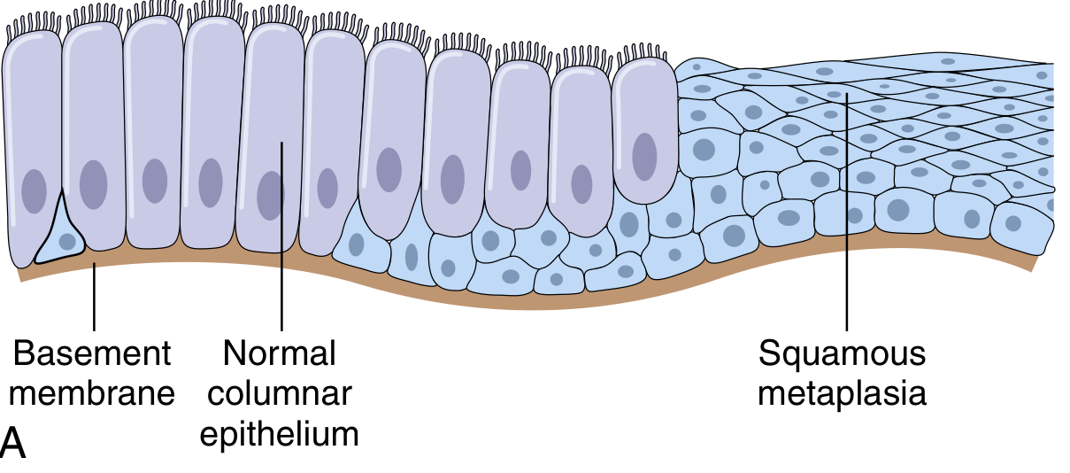
Schematic: Normal columnar epithelium (left) transitioning to squamous metaplasia (right) in a bronchus. Basement membrane remains intact throughout.
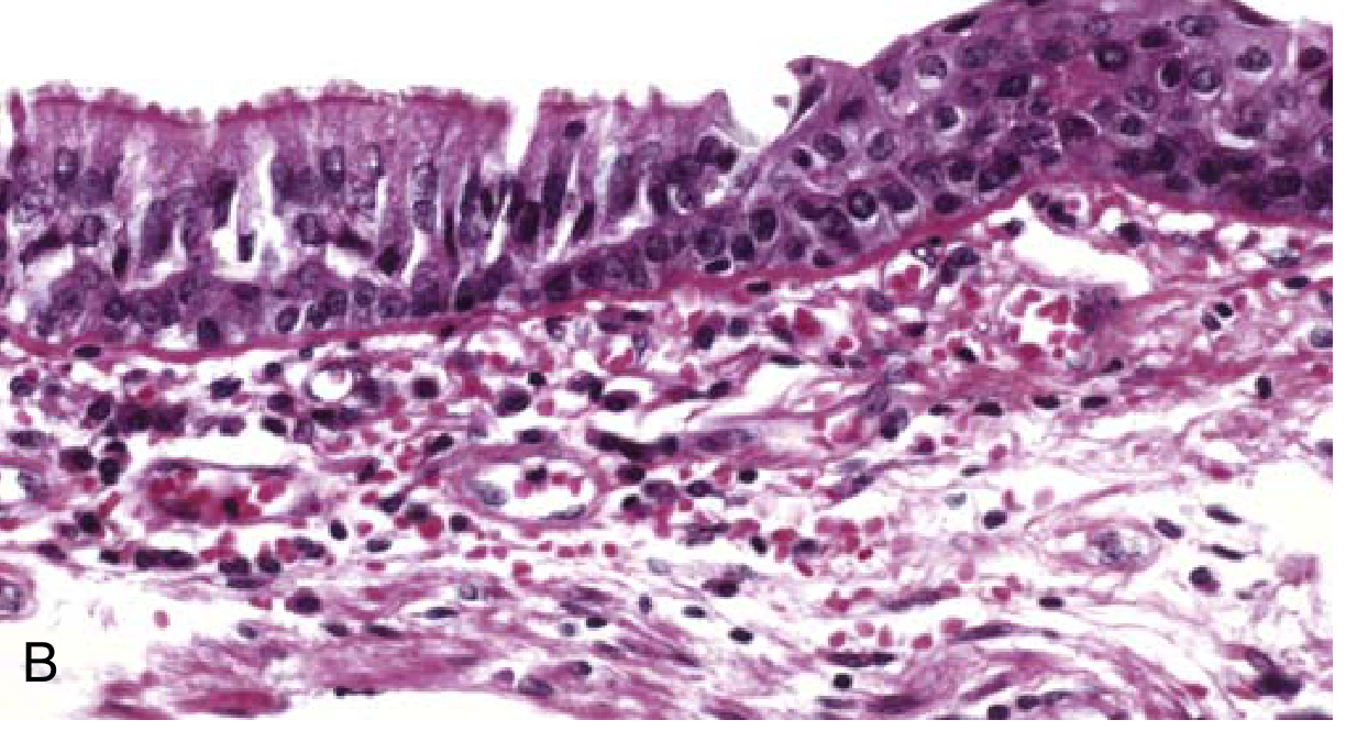
H&E histology: columnar ciliated epithelium (left) replaced by squamous epithelium (right) in smoking-related bronchial metaplasia.
Dysplasia - Detailed
Definition (Robbins): "Disordered growth, encountered principally in epithelial cells, characterized by cellular pleomorphism, large hyperchromatic nuclei with high N:C ratio, architectural disarray, and loss of orderly differentiation."
Morphological Features of Dysplasia:
| Feature | Description |
|---|---|
| Pleomorphism | Variation in cell size and shape |
| Nuclear enlargement | Large, dark (hyperchromatic) nuclei |
| High N:C ratio | Nucleus takes up more of cell volume |
| Loss of polarity | Cells lose their orderly orientation |
| Architectural disarray | Normal layered organisation disrupted |
| Increased mitoses | Mitotic figures abundant, found at all levels (not just basal) |
| Abnormal mitoses | Tripolar or multipolar mitotic figures |
| Maturation failure | Basal-type immature cells extend toward surface |
Spectrum of Dysplasia (cervix as model):
Normal epithelium
      ↓ (persistent HPV / irritant)
Mild dysplasia (CIN I) - lower 1/3 involved
      ↓
Moderate dysplasia (CIN II) - lower 2/3 involved
      ↓
Severe dysplasia (CIN III) - full thickness involved
      ↓
Carcinoma in Situ (CIS) - full thickness, NO basement membrane breach
      ↓
Invasive Carcinoma - basement membrane BREACHED
Key rule: As long as the basement membrane is intact, it is dysplasia or CIS - not invasive cancer.
The Relationship Between the Two
These are not mutually exclusive - they exist on a continuum:
Chronic Injury / Irritant
        ↓
   METAPLASIA  ──────────────────→  Metaplastic epithelium (survives better)
        ↓ (if stimulus persists)
   DYSPLASIA  ────────────────────→  Premalignant change (mutations accumulate)
        ↓
  Carcinoma in Situ
        ↓
  Invasive Carcinoma
Important: Not all metaplasia becomes dysplastic, and not all dysplasia progresses to cancer. Mild-moderate dysplasia can completely reverse if the causative stimulus is removed.
- Bronchial squamous metaplasia (smoker) → can develop squamous cell carcinoma
- Barrett's oesophagus (intestinal metaplasia) → can develop dysplasia → adenocarcinoma
- Gastric intestinal metaplasia → risk factor for gastric adenocarcinoma
Critical distinction from Robbins: "While dysplasia often occurs in metaplastic epithelium, not all metaplastic epithelium is dysplastic."
One-Line Memory Hooks
- Metaplasia = "Wrong cell, right behaviour" - adaptive, orderly, reversible
- Dysplasia = "Right place, wrong behaviour" - disordered, abnormal, premalignant
- Carcinoma in situ = dysplasia so severe it fills the full epithelial thickness - but hasn't crossed the basement membrane yet
(Robbins & Kumar Basic Pathology; Robbins, Cotran & Kumar Pathologic Basis of Disease)
This is a shared conversation. Sign in to Orris to start your own chat.