
What is Alzheimer disease? Classified drugs used in Alzheimer disease explain mechanism of action therapeutic uses and adverse effect of cholinergic activators
It's for 10 marks. Don't give too long, not too short also. Give according to that, how you have given previously for Parkinson's disease. Okay?
Alzheimer disease amyloid plaques neurofibrillary tangles pathology classification drugs
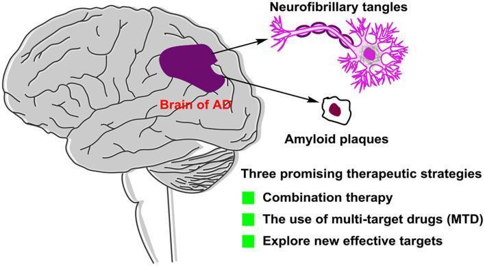
This medical infographic illustrates the pathology and future treatment strategies for Alzheimer's disease (AD). On the left, a sagittal view of a human brain labeled 'Brain of AD' highlights a central region in dark purple, indicating localized neurodegeneration or pathology. Arrows extend from this region to magnified cellular hallmarks: 'Neurofibrillary tangles' and 'Amyloid plaques.' The neurofibrillary tangles are depicted as purple, chain-like strands intertwined with a neuron's axonal and somatic structure. The amyloid plaque is shown as an extracellular deposit with a dark maroon core and a tan periphery. Below the anatomical illustrations, the graphic lists 'Three promising therapeutic strategies' for AD management: combination therapy, the use of multi-target drugs (MTD), and the exploration of new effective targets. This diagram serves as an educational summary of AD pathogenesis—specifically tau hyperphosphorylation and beta-amyloid accumulation—and the evolution of pharmacological approaches beyond traditional single-target inhibitors.
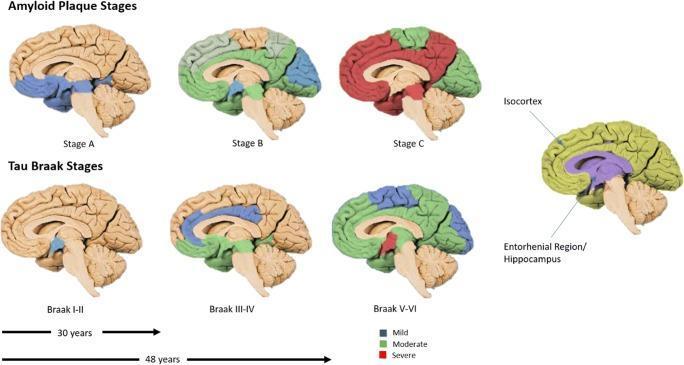
This medical schematic illustrates the neuroanatomical progression of Alzheimer’s disease pathology across different brain stages, focusing on amyloid-beta (Aβ) plaques and tau protein neurofibrillary tangles. The top row depicts Amyloid Plaque Stages A through C, showing a progression from localized mild density (blue) in the basal neocortex to moderate (green) and severe (red) densities as plaques spread throughout the isocortex. The bottom row displays the Tau Braak Stages (I-VI). Stages I-II show initial mild tau accumulation in the entorhinal region and hippocampus. Stages III-IV visualize moderate density spreading to the limbic system, while Stages V-VI demonstrate severe, widespread tau pathology throughout the isocortex. A color-coded legend indicates pathology density: blue for mild, green for moderate, and red for severe. An anatomical reference on the right identifies key regions like the Isocortex and Entorhinal Region/Hippocampus. Timelines below suggest a clinical progression spanning 30 to 48 years, highlighting the long-term nature of neurodegeneration.
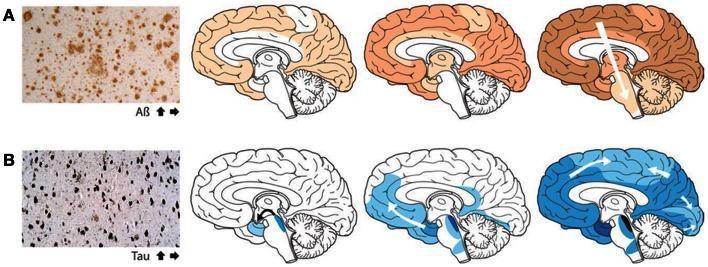
This medical illustration depicts the characteristic spatial progression of protein aggregation in Alzheimer's disease. The figure is divided into two primary rows: Row A focuses on ̢-amyloid (A̢) plaques, and Row B focuses on Tau-associated neurofibrillary tangles. Each row begins with a histological micrograph showing the morphology of the protein deposits, followed by a sequence of three sagittal brain diagrams illustrating staging. In Row A (orange shading), A̢ plaques are shown to initiate in the neocortex, subsequently spreading to the allocortex and deeper subcortical structures, following a top-down and outer-to-inner progression. In Row B (blue shading), neurofibrillary tangles are shown starting in the locus coeruleus and transentorhinal regions of the brainstem and medial temporal lobe, then progressing through the limbic system to the wider neocortex. White arrows indicate the direction of spread, emphasizing the stereotyped spatiotemporal evolution of AD pathology. This diagram serves as a pathophysiology resource for understanding Braak staging and the involvement of neuronal transport in the spread of proteopathic seeds.
Alzheimer Disease (AD)
Definition
- Bradley and Daroff's Neurology in Clinical Practice
Pathology
- Extracellular deposits of beta-amyloid (Aβ) protein derived from abnormal processing of amyloid precursor protein (APP)
- Start in the neocortex, then spread to hippocampus, basal ganglia, thalamus, and brainstem
- Neuritic plaques have a dense Aβ core surrounded by dystrophic neurites, microglia, and astrocytes
- Intraneuronal accumulations of hyperphosphorylated tau protein (normally a microtubule-stabilizing protein)
- Follow the Braak staging pattern: earliest involvement in locus coeruleus and entorhinal cortex, spreading to hippocampus and ultimately the neocortex
- When neurons die, tangles remain extracellularly as "ghost tangles"
Cholinergic Deficit - The Basis of Drug Therapy
Classification of Drugs Used in Alzheimer Disease
| Class | Drugs |
|---|---|
| Cholinesterase (AChE) Inhibitors | Donepezil, Rivastigmine, Galantamine, Tacrine (withdrawn) |
| NMDA Receptor Antagonist | Memantine |
| Anti-amyloid monoclonal antibodies | Lecanemab, Aducanumab (newer agents) |
| Vitamin E / Antioxidants | Adjunctive use only |
Note: Cholinesterase inhibitors are indicated for mild-to-moderate AD; memantine is indicated for moderate-to-severe AD. They are often combined for additive benefit.
Cholinergic Activators - Cholinesterase Inhibitors
Mechanism of Action
| Drug | Selectivity | Reversibility |
|---|---|---|
| Donepezil | AChE-selective; CNS-selective (minimal peripheral) | Reversible, non-competitive |
| Rivastigmine | Both AChE + BuChE (pseudoirreversible) | Pseudoirreversible (slow dissociation) |
| Galantamine | AChE inhibitor + allosteric nicotinic receptor modulator | Reversible, competitive |
| Tacrine | Non-selective AChE (all forms); significant peripheral activity | Reversible |
- Kaplan and Sadock's Synopsis of Psychiatry; Kaplan & Sadock's Comprehensive Textbook of Psychiatry
Pharmacokinetics (Key Points)
| Drug | Half-life | Dosing | Notes |
|---|---|---|---|
| Donepezil | 70 hrs | Once daily | 100% oral bioavailability; metabolized by CYP2D6, 3A4 |
| Rivastigmine | ~1 hr (plasma), 10 hrs (effect) | Twice daily (oral); Patch once daily | Degraded by esterases; patch reduces GI side effects |
| Galantamine | ~6 hrs | Twice daily | Alkaloid from Galanthus nivalis (daffodil); food reduces peak by 25% |
| Tacrine | 2-4 hrs | Four times daily | Largely withdrawn due to hepatotoxicity |
Therapeutic Uses
- Mild to moderate Alzheimer dementia - all three marketed agents (donepezil, rivastigmine, galantamine) are FDA-approved; donepezil is also approved for severe AD
- Slowing disease progression - slow progression of memory loss and reduce behavioral symptoms (apathy, depression, hallucinations, anxiety, purposeless motor behaviors)
- Lewy body dementia and Parkinson disease dementia - donepezil and rivastigmine are beneficial
- Vascular dementia - may respond to AChE inhibitors
- Traumatic brain injury - cognitive deficits may benefit from donepezil/rivastigmine
- Practical benefit: delay or reduction of need for nursing home placement
Donepezil is the only cholinesterase inhibitor approved for severe AD.
Adverse Effects
Donepezil
- Generally best tolerated
- Nausea, diarrhea, vomiting (<3% at 5 mg; higher at 10 mg) - typically resolve after 3 weeks
- Bradycardia (especially with underlying cardiac disease); syncope (rare)
- Weight loss
- Insomnia, muscle cramps
Rivastigmine
- More peripheral activity than donepezil → higher GI side effects
- Nausea, vomiting, dizziness, headache, diarrhea, abdominal pain, anorexia, fatigue, somnolence
- Adverse effects more common at doses >6 mg/day; reduced with transdermal patch
- Does not cause hepatic, renal, or hematologic abnormalities
Galantamine
- Dizziness, headache, nausea, vomiting, diarrhea, anorexia - generally mild and transient
- GI effects less severe than rivastigmine
Tacrine (withdrawn/historical)
- Hepatotoxicity (most significant): ALT/AST elevation in 25-30% of patients; requires weekly LFT monitoring for 18 weeks
- Nausea, vomiting, myalgia, anorexia, rash
- Requires four-times-daily dosing - impractical
- Contraindicated in jaundice; discontinued clinically due to these issues
| Drug | Dose (mg/day) | Nausea | Vomiting | Diarrhea | Dizziness |
|---|---|---|---|---|---|
| Donepezil | 5 | 4% | 3% | 9% | 15% |
| Donepezil | 10 | 17% | 10% | 17% | 13% |
| Rivastigmine | 1-4 | 14% | 7% | 10% | 15% |
| Rivastigmine | 6-12 | 48% | 27% | 17% | 24% |
| Galantamine | 16 | 13% | 6% | 12% | - |
- Kaplan and Sadock's Synopsis of Psychiatry, Table 21-56
Contraindications and Precautions
- Peptic ulcer disease - increased gastric acid secretion via vagal cholinergic pathways
- Sick sinus syndrome / cardiac conduction defects - risk of bradycardia/heart block
- Asthma / COPD - bronchoconstriction risk
- Seizure disorders - cholinergic activation can lower seizure threshold
- Idiosyncratic reaction: rare catastrophic reaction with grief and agitation (self-limited on discontinuation)
Quick Summary Flowchart
Alzheimer Disease
↓
Cholinergic deficit (↓ACh in hippocampus/cortex)
↓
Cholinesterase Inhibitors block AChE → ↑ACh in synapses
↓
Agents: Donepezil → Rivastigmine → Galantamine (Tacrine withdrawn)
↓
Indications: Mild-Moderate AD (all); Severe AD (donepezil only)
↓
ADRs: GI (nausea/vomiting/diarrhea), Bradycardia, Hepatotoxicity (tacrine only)
It's too long bro concise for 10 to 11 Marks
Alzheimer Disease
Definition
Pathology (Hallmarks)
Basis of Drug Therapy - Cholinergic Hypothesis
Classification of Drugs
| Class | Drugs |
|---|---|
| Cholinesterase Inhibitors | Donepezil, Rivastigmine, Galantamine, Tacrine (withdrawn) |
| NMDA Receptor Antagonist | Memantine |
| Anti-amyloid antibodies | Lecanemab, Aducanumab |
ChEIs → mild to moderate AD | Memantine → moderate to severe AD
Cholinesterase Inhibitors (Cholinergic Activators)
Mechanism of Action
| Drug | Selectivity |
|---|---|
| Donepezil | AChE-selective; CNS-selective (minimal peripheral) |
| Rivastigmine | AChE + BuChE (pseudoirreversible); more peripheral activity |
| Galantamine | AChE inhibitor + allosteric nicotinic receptor modulator |
| Tacrine | Non-selective; withdrawn |
Therapeutic Uses
- Mild to moderate AD - all three agents
- Severe AD - donepezil only (only FDA-approved ChEI for severe stage)
- Lewy body dementia and Parkinson disease dementia (donepezil, rivastigmine)
- Vascular dementia
- Slow progression of memory loss; reduce behavioral symptoms (apathy, depression, hallucinations, anxiety)
- Delay need for nursing home placement
Adverse Effects
- Nausea, vomiting, diarrhea, anorexia
- Dizziness, headache
- Weight loss
- Donepezil - Best tolerated; bradycardia/syncope (cardiac patients); insomnia, muscle cramps
- Rivastigmine - Most GI side effects (nausea 48% at high dose); reduced with transdermal patch; no hepatotoxicity
- Galantamine - Mild and transient GI effects; dizziness
- Tacrine - Hepatotoxicity (ALT/AST elevation in 25-30%); requires weekly LFT monitoring; 4× daily dosing - now withdrawn
- Peptic ulcer disease (↑ gastric acid)
- Sick sinus syndrome / heart block (bradycardia risk)
- Asthma/COPD (bronchoconstriction)
- Seizure disorders
Definition
Which definition do you need?
Definition, very short note on pathology,causes ,symptoms , classification, adverse effect,uses and most imp necessary information
Alzheimer Disease - Complete Notes
Definition
Pathology (Short Note)
- Amyloid Plaques - Extracellular deposits of beta-amyloid (Aβ) protein from abnormal cleavage of Amyloid Precursor Protein (APP). Start in neocortex → spread to hippocampus → brainstem.
- Neurofibrillary Tangles (NFTs) - Intraneuronal deposits of hyperphosphorylated tau protein (normally stabilizes microtubules). Follow Braak stages: locus coeruleus/entorhinal cortex → hippocampus → neocortex.
- Macroscopy: Diffuse cortical atrophy, especially hippocampus and association areas. Primary motor/sensory cortex is relatively spared.
Causes / Risk Factors
| Factor | Details |
|---|---|
| Age | Most important risk factor; prevalence doubles every 5 years after 65 |
| Genetics | APOE ε4 allele (sporadic AD); mutations in APP, Presenilin-1, Presenilin-2 (familial early-onset AD) |
| Gender | Women > Men (longer lifespan + hormonal factors) |
| Head trauma | Increases risk |
| Down syndrome | APP gene on chromosome 21; almost all develop AD by age 40 |
| Vascular risk factors | Hypertension, diabetes, obesity |
Symptoms (Clinical Features)
- Recent memory loss (short-term memory first affected)
- Difficulty with language (word-finding problems)
- Disorientation to time and place
- Confusion, poor judgment
- Behavioral changes: agitation, depression, anxiety, hallucinations
- Difficulty with daily activities (dressing, cooking)
- Complete loss of speech (aphasia)
- Inability to perform motor tasks (apraxia)
- Incontinence, bed-bound state
- Death (usually from aspiration pneumonia or infection)
Classification of Drugs
| Class | Drugs | Stage |
|---|---|---|
| Cholinesterase Inhibitors | Donepezil, Rivastigmine, Galantamine, Tacrine (withdrawn) | Mild - Moderate |
| NMDA Receptor Antagonist | Memantine | Moderate - Severe |
| Anti-amyloid antibodies | Lecanemab, Aducanumab | Early AD |
| Combination | Donepezil + Memantine | Moderate - Severe |
Mechanism of Action (ChEIs)
Therapeutic Uses
- Mild to moderate AD - donepezil, rivastigmine, galantamine
- Severe AD - donepezil only
- Lewy body and Parkinson disease dementia - donepezil, rivastigmine
- Vascular dementia
- Reduce behavioral symptoms (apathy, depression, hallucinations)
- Delay nursing home placement
Adverse Effects
- Nausea, vomiting, diarrhea, anorexia (most common - due to peripheral cholinergic excess)
- Dizziness, headache, weight loss
- Bradycardia/syncope - especially donepezil in cardiac patients
- Insomnia, muscle cramps - donepezil
- Hepatotoxicity - tacrine only (ALT/AST elevation in 25-30%); reason for withdrawal
- Dizziness, confusion, headache, constipation - generally well tolerated
- Peptic ulcer (↑ gastric acid)
- Sick sinus syndrome / heart block
- Asthma / COPD
- Seizure disorders
Key Points to Remember
| Point | Fact |
|---|---|
| Most common dementia | Alzheimer disease (70%) |
| Hallmark lesions | Amyloid plaques + NFTs (tau) |
| Drug basis | Cholinergic hypothesis |
| Only ChEI for severe AD | Donepezil |
| Withdrawn ChEI | Tacrine (hepatotoxicity) |
| NMDA antagonist | Memantine (moderate-severe AD) |
| Rivastigmine advantage | Transdermal patch - fewer GI effects |
| Galantamine unique action | AChE inhibition + nicotinic receptor modulation |
The information which you have given just now can I write these for the question What is Alzheimer disease classified drug used in Alzheimer disease explain the mechanism of action adverse effect and uses of cholinergic activators
Tips for writing in exam:
- Start with a one-line definition, then briefly cover pathology/causes/symptoms
- Draw the classification table - examiners love tables, gives a neat look
- For mechanism, write a short flowchart-style explanation (App → Aβ → plaque; AChE inhibition → ↑ACh)
- For adverse effects, mention tacrine hepatotoxicity separately - it's a high-yield point
- Mention donepezil is the only ChEI for severe AD - easy scoring point
Can you give again according to that question? Not too long not too short the length which we have given just now that much only I want
Alzheimer Disease
Definition
Pathology
- Amyloid Plaques - Extracellular deposits of beta-amyloid (Aβ) from abnormal APP processing
- Neurofibrillary Tangles (NFTs) - Intraneuronal hyperphosphorylated tau protein
- Macroscopy: Diffuse cortical atrophy, especially hippocampus and association areas
Causes / Risk Factors
- Age - Most important risk factor
- Genetics - APOE ε4 allele; APP, Presenilin-1, Presenilin-2 mutations (familial AD)
- Down syndrome - APP gene on chromosome 21
- Gender - Women > Men
- Vascular risk factors - Hypertension, diabetes
Symptoms
- Early: Recent memory loss, word-finding difficulty, disorientation
- Moderate: Confusion, behavioral changes (agitation, depression, hallucinations), impaired daily activities
- Late: Aphasia, apraxia, incontinence, bed-bound, death (usually from aspiration pneumonia)
Classification of Drugs
| Class | Drugs | Stage |
|---|---|---|
| Cholinesterase Inhibitors | Donepezil, Rivastigmine, Galantamine, Tacrine (withdrawn) | Mild - Moderate |
| NMDA Receptor Antagonist | Memantine | Moderate - Severe |
| Anti-amyloid antibodies | Lecanemab, Aducanumab | Early AD |
Cholinergic Activators - Cholinesterase Inhibitors
Mechanism of Action
| Drug | Selectivity |
|---|---|
| Donepezil | AChE-selective; CNS-selective |
| Rivastigmine | AChE + BuChE; pseudoirreversible |
| Galantamine | AChE inhibition + nicotinic receptor modulation |
| Tacrine | Non-selective; withdrawn |
Therapeutic Uses
- Mild to moderate AD - donepezil, rivastigmine, galantamine
- Severe AD - donepezil only (only FDA-approved ChEI)
- Lewy body dementia and Parkinson disease dementia
- Vascular dementia
- Reduces behavioral symptoms - apathy, depression, hallucinations
- Delays need for nursing home placement
Adverse Effects
- Nausea, vomiting, diarrhea, anorexia
- Dizziness, headache, weight loss
- Donepezil - Bradycardia, syncope (cardiac patients); insomnia, muscle cramps
- Rivastigmine - Most GI side effects; reduced with transdermal patch
- Galantamine - Mild, transient GI effects
- Tacrine - Hepatotoxicity (ALT/AST elevation in 25-30%); reason for withdrawal
- Peptic ulcer disease
- Sick sinus syndrome / heart block
- Asthma / COPD
- Seizure disorders
Mechanism of action, adverse effect, and therapeutic uses of cholinergic activators for four marks.
Cholinergic Activators (Cholinesterase Inhibitors)
Mechanism of Action
- Donepezil - AChE selective; CNS-selective (minimal peripheral effects)
- Rivastigmine - Inhibits both AChE + BuChE (pseudoirreversible)
- Galantamine - AChE inhibition + allosteric nicotinic receptor modulation
- Tacrine - Non-selective AChE inhibitor (withdrawn)
Therapeutic Uses
- Mild to moderate Alzheimer disease (all three agents)
- Severe AD - donepezil only
- Lewy body dementia and Parkinson disease dementia
- Vascular dementia
- Reduces behavioral symptoms - apathy, depression, hallucinations, anxiety
- Delays need for nursing home placement
Adverse Effects
- Nausea, vomiting, diarrhea, anorexia
- Dizziness, headache, weight loss
- Donepezil - Bradycardia, syncope; insomnia, muscle cramps
- Rivastigmine - Most GI side effects; reduced with transdermal patch
- Galantamine - Mild, transient GI effects
- Tacrine - Hepatotoxicity (25-30%); withdrawn from clinical use
Define receptors classify receptor and discuss about the signal transduction mechanism of Trans membrane enzyme linked receptors for 10 marks not too long
transmembrane enzyme linked receptor tyrosine kinase signal transduction mechanism
Receptors - Definition, Classification & Signal Transduction of Transmembrane Enzyme-Linked Receptors
Definition of Receptor
- Specificity - binds only specific ligands
- Affinity - strength of drug-receptor binding
- Sensitivity - response even at low ligand concentrations
- Reversibility - binding is usually reversible
Classification of Receptors
Type 1 - Ligand-Gated Ion Channels (Ionotropic Receptors)
- Located on cell membrane
- Ligand binding directly opens an ion channel
- Fastest response (milliseconds)
- Examples: Nicotinic ACh receptor (Na⁺/K⁺), GABA-A receptor (Cl⁻), Glutamate (NMDA) receptor
Type 2 - G-Protein Coupled Receptors (GPCRs / Metabotropic)
- 7-transmembrane spanning domain receptors
- Ligand → activates G-protein → activates/inhibits second messengers (cAMP, IP3, DAG)
- Slower response (seconds)
- Examples: Muscarinic, Adrenergic (α, β), Dopamine, Opioid receptors
Type 3 - Transmembrane Enzyme-Linked Receptors
- Single membrane-spanning domain with intrinsic enzyme activity (usually tyrosine kinase)
- Ligand binding activates enzyme domain intracellularly
- Examples: Insulin receptor, EGF, PDGF, VEGF receptors
- Also includes: Receptor Guanylyl Cyclase (ANP receptor), JAK-STAT linked receptors
Type 4 - Intracellular (Nuclear) Receptors
- Located in cytoplasm or nucleus
- Only activated by lipid-soluble ligands (steroids, thyroid hormone, Vit D)
- Hormone-receptor complex binds DNA → regulates gene transcription
- Slowest response (hours to days)
- Examples: Glucocorticoid, Estrogen, Androgen, Thyroid hormone receptors
Signal Transduction of Transmembrane Enzyme-Linked Receptors
Structure
- Single transmembrane-spanning protein
- Extracellular domain - ligand binding site
- Transmembrane domain - single alpha helix
- Intracellular domain - intrinsic tyrosine kinase catalytic domain (C-terminal)
Subtypes
| Subtype | Example Ligands |
|---|---|
| Receptor Tyrosine Kinase (RTK) | Insulin, EGF, PDGF, FGF, VEGF |
| Receptor Serine/Threonine Kinase | TGF-β |
| Receptor Guanylyl Cyclase | ANP (Atrial Natriuretic Peptide) |
| JAK-STAT linked receptors | Cytokines, Interferons, Growth hormone |
Mechanism - Step by Step (RTK)
- Grb2 (adaptor protein)
- PI3-Kinase (phosphatidylinositol 3-kinase)
- PLCγ (Phospholipase C-gamma)
| Pathway | Outcome |
|---|---|
| Grb2 → Ras → MAP Kinase pathway | Gene activation, cell proliferation, differentiation |
| PI3K → PIP3 → Akt/PKB pathway | Cell survival, glucose uptake, protein synthesis |
| PLCγ → IP3 + DAG | Ca²⁺ release, Protein Kinase C activation |
Insulin Receptor - Classic Example
- Preformed dimer in membrane (α2β2 tetrameric structure)
- Insulin binds α-subunits → β-subunits autophosphorylate → phosphorylates IRS (Insulin Receptor Substrate)
- IRS activates: Grb2-Ras-MAPK pathway (mitogenic effects) + PI3K-Akt pathway (metabolic effects: glucose uptake via GLUT4)
Clinical Significance
- RTKs and downstream signaling components (Ras, PI3K) are frequently mutated in cancer cells → constitutively active receptors → uncontrolled cell proliferation
- PTEN (tumor suppressor) dephosphorylates PIP3 → terminates PI3K signal; PTEN loss → cancer
- Drugs like imatinib (Gleevec) inhibit tyrosine kinase receptors → used in cancer therapy


Can I write these notes for the question which I have said just now? I will give the definition at starting,
Can I write these notes for the question which I have said just now? I will give the definition at starting,
- Type 1 - Ligand-gated Ion Channels
- Type 2 - G-Protein Coupled Receptors (GPCR)
- Type 3 - Transmembrane Enzyme-Linked Receptors
- Type 4 - Intracellular/Nuclear Receptors
- Extracellular ligand binding domain + intracellular tyrosine kinase domain
- Ligand binding → receptor dimerization → autophosphorylation → downstream signaling (MAPK, PI3K/Akt)
- Examples: Insulin receptor, VEGF-R, EGFR
- Draw the diagram you drew (the RTK diagram with ECF/ICF and enzyme receptors)
- Step-by-step mechanism (SH2 domains, effector proteins)
- MAP Kinase pathway → cell proliferation
- PI3K-Akt pathway → cell survival, glucose uptake
- PLCγ → IP3 + DAG → Ca²⁺ release
- Clinical significance: RTK mutations in cancer; imatinib targets tyrosine kinase
- GPCR (G-protein table with Gs, Gi, Gq, G12/13)
- Ion Channel Receptors
- JAK-STAT Receptors
- Nuclear/Transcription Factor Receptors
- Regulation table (up/down regulation, desensitization)
Some of the whole answer from my notes and your notes, and I only want to draw one diagram, not more than that. And give me the whole answer for the question which I have provided. And remember, it's for 10 marks, not too long, not too short.
Receptors - Definition, Classification & Signal Transduction of Transmembrane Enzyme-Linked Receptors
Definition
Classification of Receptors
Type 1 - Ligand-Gated Ion Channels (Ionotropic)
- Drug binds → direct opening/closing of ion channels
- Fast synaptic transmission (milliseconds)
- Examples: Nicotinic ACh receptor (Na⁺/K⁺), GABA-A (Cl⁻), Glycine (Cl⁻)
- Benzodiazepines and barbiturates modulate GABA-A channel
Type 2 - G-Protein Coupled Receptors (GPCR)
- 7-transmembrane domain; coupled to guanine nucleotide-binding protein
- Ligand → activates G-protein → second messenger
| G-Protein | Effect | 2nd Messenger | Examples |
|---|---|---|---|
| Gs | Stimulates adenylyl cyclase | ↑ cAMP | β-adrenergic, Glucagon, H₂ |
| Gi | Inhibits adenylyl cyclase | ↓ cAMP | α₂-adrenergic, M₂/M₄ muscarinic, Opioid |
| Gq | Activates phospholipase C | ↑ IP₃ + DAG → ↑Ca²⁺, PKC | α₁-adrenergic, M₁/M₃ muscarinic |
| G₁₂/₁₃ | Activates Rho kinase | Cytoskeletal changes | Thromboxane receptors |
- cAMP pathway: G-protein → adenylyl cyclase → cAMP → PKA → phosphorylation of target protein
- IP₃/DAG pathway: G-protein → PLC-β → PIP₂ → IP₃ (releases Ca²⁺ from ER) + DAG (activates PKC)
Type 3 - Transmembrane Enzyme-Linked Receptors (discussed in detail below)
Type 4 - Nuclear/Transcription Factor Receptors
- Drug/hormone enters cell → binds intracellular receptor → receptor-hormone complex enters nucleus → binds DNA response element → alters gene transcription
- Slowest mechanism (hours to days)
- Examples: Glucocorticoids, mineralocorticoids, estrogen, androgen, thyroid hormone, vitamin D, retinoic acid
Regulation of Receptors
| Type | Definition | Clinical Effect |
|---|---|---|
| Down regulation | ↓ receptor numbers/sensitivity with prolonged agonist exposure | Tolerance (e.g., β-agonist in asthma) |
| Up regulation | ↑ receptor numbers with prolonged antagonist use | Rebound effect on withdrawal (e.g., propranolol withdrawal → tachycardia) |
| Desensitization | Receptor uncoupling from G-protein, rapid onset | Acute tolerance |
Signal Transduction of Transmembrane Enzyme-Linked Receptors (RTK)
Structure
- Extracellular domain - ligand binding site
- Single transmembrane domain - spans membrane once
- Intracellular domain - intrinsic tyrosine kinase catalytic domain
Examples
Mechanism - Step by Step
- Grb2 (adaptor protein)
- PI3-Kinase
- PLCγ
| Pathway | Outcome |
|---|---|
| Grb2 → Ras → MAP Kinase pathway | Gene activation, cell proliferation, differentiation |
| PI3K → PIP3 → Akt/PKB pathway | Cell survival, glucose uptake (insulin) |
| PLCγ → IP₃ + DAG | Ca²⁺ release, PKC activation |
Diagram
Ligand
↓
[Extracellular - Binding Site]
══════════════════════════════ ← Cell Membrane
[Intracellular - Tyrosine Kinase Domain]
↓ Dimerization + Autophosphorylation
P-Tyr ←——→ P-Tyr (SH2 docking)
↓
Grb2 → MAPK → Cell Proliferation
PI3K → Akt → Cell Survival
PLCγ → IP₃ → Ca²⁺ Release
Clinical Significance
- RTK mutations → constitutively active receptors → uncontrolled cell proliferation → cancer
- Imatinib, erlotinib - tyrosine kinase inhibitors used in cancer therapy
- PTEN (tumor suppressor) terminates PI3K signaling; PTEN loss → cancer
Yes, I don't write that regulation receptor part. It's... Will be enough, right?
- Define receptors ✅
- Classify receptors ✅
- Signal transduction of transmembrane enzyme-linked receptors ✅
- Definition
- Classification (all 4 types with brief points)
- GPCR table (Gs, Gi, Gq, G12/13) - since it's part of classification
- RTK mechanism (step by step)
- One diagram (RTK)
- Clinical significance (2-3 lines)
0k