
What’s the clinical examination in a patient with Genu Valgum or pea planus or CTEV or scoliosis
genu valgum knock knee clinical examination intermalleolar distance
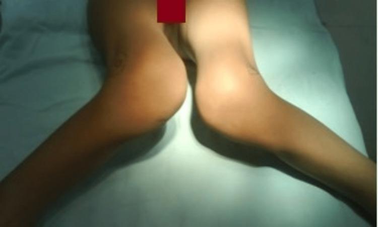
This clinical photograph displays a bilateral lower extremity deformity in a patient, likely within an orthopedic or pediatric context. The image illustrates a severe, persistent bilateral genu valgum (knock-knee) deformity, characterized by the inward angulation of the distal femur and proximal tibia. When the knees are brought into close proximity, there is a substantial intermalleolar distance, indicating significant lateral deviation of the lower legs. The visual evidence suggests a skeletal dysplasia or developmental condition, such as Ellis-van Creveld (EVC) syndrome, given the associated clinical context of dysplastic epiphyses. The skin appears intact without visible scarring or primary lesions in the visualized field. This image serves as an educational example for identifying pathological knee alignment and the clinical manifestation of severe valgus stress on the lower limbs.
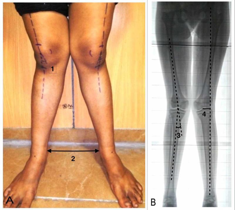
This dual-panel image demonstrates the preoperative assessment of a lower limb deformity, specifically genu valgum (knock-knees). Panel A is a clinical photograph of a patient standing in an anterior view. It displays surface anatomy and clinical measurements. Marked on the skin are the anatomical axes of the femurs and tibiae. Label (1) indicates the clinical tibiofemoral angle, while label (2) highlights the intermalleolar distance (IMD), measured between the medial malleoli while the knees are touching. Panel B is a corresponding standing anteroposterior (AP) long-limb X-ray scanogram showing the skeletal structure from the hips to the ankles. This radiographic view allows for precise angular measurements. Label (3) indicates the radiological tibiofemoral angle, formed by the intersection of the femoral and tibial anatomical axes. Label (4) denotes the mechanical axis deviation (MAD), which is the distance between the center of the knee joint and the mechanical axis line (drawn from the center of the femoral head to the center of the ankle). This comparison illustrates the correlation between physical examination findings and radiographic evidence in orthopedic surgical planning.
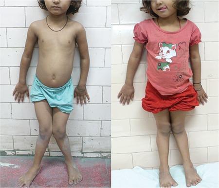
A comparison of two clinical photographs demonstrating the progression and partial resolution of genu valgum (knock-knees) in a pediatric patient. The left image shows a child with significant bilateral genu valgum; when the knees are adducted and touching, there is a wide intermalleolar distance between the ankles. The right image, taken after a 1-year follow-up following medical intervention (stosstherapy for nutritional rickets), shows a marked reduction in the angular deformity. The mechanical axis of the lower limbs appears more aligned, with a substantially narrower gap between the ankles when the knees are in proximity. These images illustrate the clinical manifestation of metabolic bone disease in children and the potential for lower limb remodeling with appropriate treatment. The visual focus is on the orthopedic alignment of the femurs and tibiae relative to the midline.
scoliosis Adams forward bend test rib hump
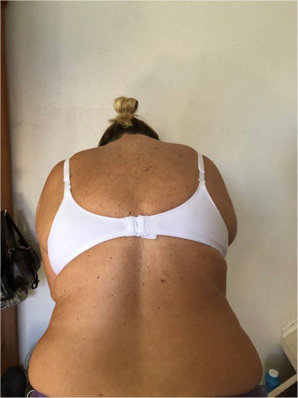
Clinical photograph demonstrating a posterior view of a patient performing the Adams Forward Bend Test, a clinical screening tool for scoliosis. The patient is leaning forward at the waist, which accentuates truncal asymmetry. The image clearly shows a prominent right-sided thoracic rib hump, where the ribs on the convex side of the spinal curvature protrude posteriorly due to vertebral rotation. This results in a noticeable dorsal tilt and uneven contour of the back compared to the contralateral side. Additional findings include asymmetrical shoulder heights and uneven soft tissue folds at the waistline. The photograph serves to illustrate the physical manifestation of structural scoliosis, highlighting how forward flexion makes rotational spinal deformities more visible to the clinician. The skin also displays numerous benign-appearing melanocytic nevi across the dorsal surface, though these are secondary to the primary orthopedic assessment.
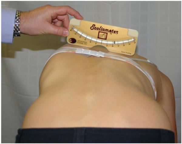
A clinical photograph demonstrating a procedural assessment for scoliosis using the Adams forward bend test. The patient is positioned in forward flexion, which highlights a prominent right-sided rib hump, indicative of thoracic vertebral rotation and spinal convexity. An examiner is shown holding a Scoliometer (a specialized inclinometer) across the patient's mid-thoracic spine to quantify the Angle of Trunk Rotation (ATR). The device features a curved scale and a ball bearing level to provide a numerical degree of asymmetry. This clinical procedure is a standard screening and monitoring tool used in orthopedics and physical therapy to evaluate the severity and progression of adolescent idiopathic scoliosis and other structural spinal deformities. The image clearly illustrates the anatomical relationship between spinal curvature and visible thoracic cage distortion.
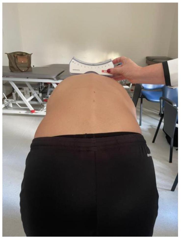
This clinical photograph demonstrates a procedural assessment for scoliosis using a scoliometer during an Adams Forward Bend Test. The image shows the posterior view of a patient's torso in a flexed position, revealing a prominent rib hump and paraspinal muscle asymmetry on the left side of the upper back. A clinician, wearing a white lab coat, is holding a white scoliometer horizontally across the apex of the spinal curvature to measure the Angle of Trunk Rotation (ATR). The scoliometer features a curved base designed to fit over the spinous processes and a graduated scale with black markings and a red indicator to quantify the degree of vertebral rotation. This diagnostic procedure is essential for identifying adolescent idiopathic scoliosis and determining the need for further radiological imaging. The clinical significance of this visual is to illustrate the quantification of trunk asymmetry and postural changes associated with lateral spinal curvature and rotational deformity.
CTEV clubfoot talipes equinovarus clinical features hindfoot varus forefoot adduction
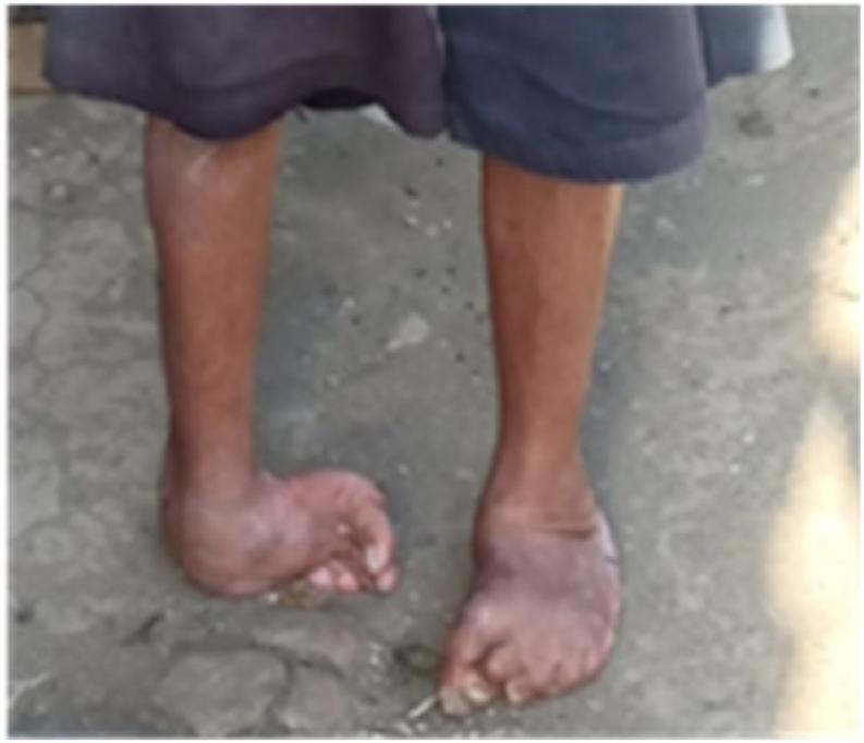
This clinical photograph shows a bilateral lower limb presentation of congenital talipes equinovarus (CTEV), commonly known as clubfoot, in an adult patient. The image illustrates severe structural deformities characterized by forefoot adduction, midfoot cavus, and hindfoot varus. The left foot exhibits more profound involvement, with the foot severely rotated medially (inward) and the weight-bearing surface appearing to be the lateral border of the foot rather than the sole. The right foot shows similar inward angulation and equinus, though less pronounced than the left. Associated muscle atrophy of the lower calves is visible, a common finding in untreated or chronic CTEV. The presentation demonstrates the long-term musculoskeletal impact and abnormal gait mechanics associated with uncorrected congenital foot deformities, highlighting the clinical need for specialized orthopedic intervention or orthotic management to improve stability and weight distribution.
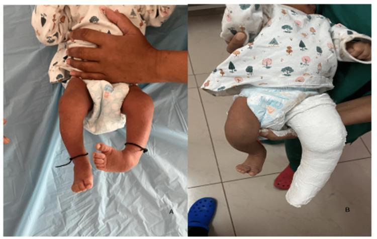
Clinical photographs depicting the management of Congenital Talipes Equinovarus (CTEV), commonly known as clubfoot, using the Ponseti technique. Figure A shows an infant with bilateral clubfoot exhibiting classic deformities: forefoot adduction, hindfoot varus, and equinus. A prominent medial longitudinal arch is visible on the right foot, signifying cavus deformity. Figure B demonstrates the initial phase of serial casting. The left lower extremity is encased in a white, above-knee plaster cast. This first cast is specifically designed to correct the cavus component by supinating the forefoot to align it with the hindfoot while maintaining some abduction. The cast extends from the upper thigh to the toes, which remain visible for neurovascular monitoring. This visual documentation illustrates the step-wise orthopedic correction of neonatal foot deformities through manual manipulation and rigid immobilization.
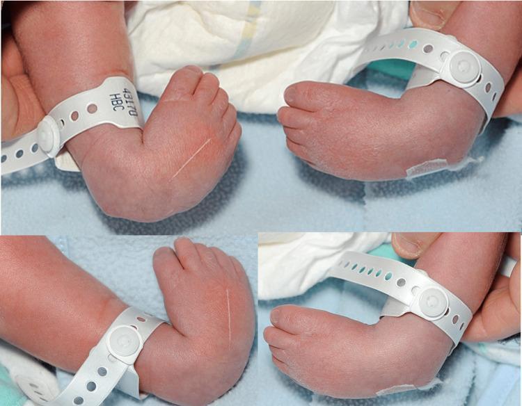
This composite clinical photograph displays a newborn's feet from multiple angles, demonstrating bilateral congenital talipes equinovarus (CTEV), commonly known as clubfoot. The visual features exhibit the characteristic four components of the deformity: hindfoot equinus (downward pointing of the foot at the ankle), hindfoot varus (inward tilting of the heel), midfoot adductus (medial deviation of the forefoot), and cavus (high medial longitudinal arch). Notable clinical signs include deep skin creases on the medial aspect of the midfoot and posterior aspect of the ankle. The infant is wearing white hospital identification bands around both lower legs. This image serves as an educational tool in pediatric orthopedics to illustrate the physical presentation of idiopathic clubfoot at birth, which is typically assessed using the Pirani or Dimeglio scoring systems for severity before initiating Ponseti method manipulative treatment.
pes planus flat foot clinical examination arch
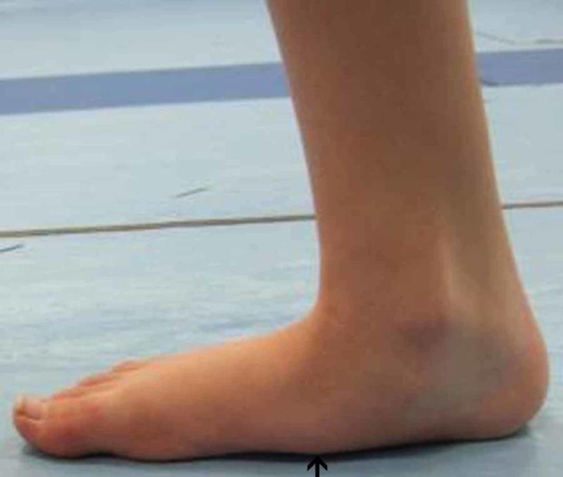
This clinical photograph displays a medial-side view of a right foot during a pre-operative gait analysis, illustrating significant musculoskeletal deformity. The primary visible feature is the complete flattening of the medial longitudinal arch, a condition known as pes planus (flat foot). The medial border of the foot shows direct contact with the weight-bearing surface, indicated by a black arrow pointing to the collapsed arch region. Additionally, there is a visible inward tilting of the ankle and outward displacement of the heel, characteristic of a valgus hindfoot deformity (pes planovalgus). This image serves as a clinical representation of pediatric or adult acquired flatfoot deformity, demonstrating the associated pronated posture and the loss of normal anatomical curvature. It is intended for educational use in podiatry, orthopedics, and physical therapy to identify the visual markers of arch collapse and hindfoot malalignment before surgical intervention.
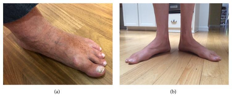
This comparative clinical photograph displays two different presentations of pes planus (flat feet). Panel (a) is a lateral-oblique view of a right foot showing a significantly collapsed medial longitudinal arch with the midfoot in contact with the floor. The skin exhibits mottling, venous prominence, and hyperpigmentation, with thickening and discoloration of the hallux nail. Panel (b) is an anterior view of both feet in a weight-bearing position, demonstrating bilateral flattening of the arches accompanied by a valgus alignment at the ankles and abduction of the forefeet. While both cases illustrate characteristic arch collapse, panel (a) shows additional dermatological and vascular changes, whereas panel (b) highlights postural and structural malalignment associated with the condition. These images are used in medical education to illustrate the musculoskeletal morphology of flat feet and their potential association with hereditary neuropathies like Charcot-Marie-Tooth disease (CMT).
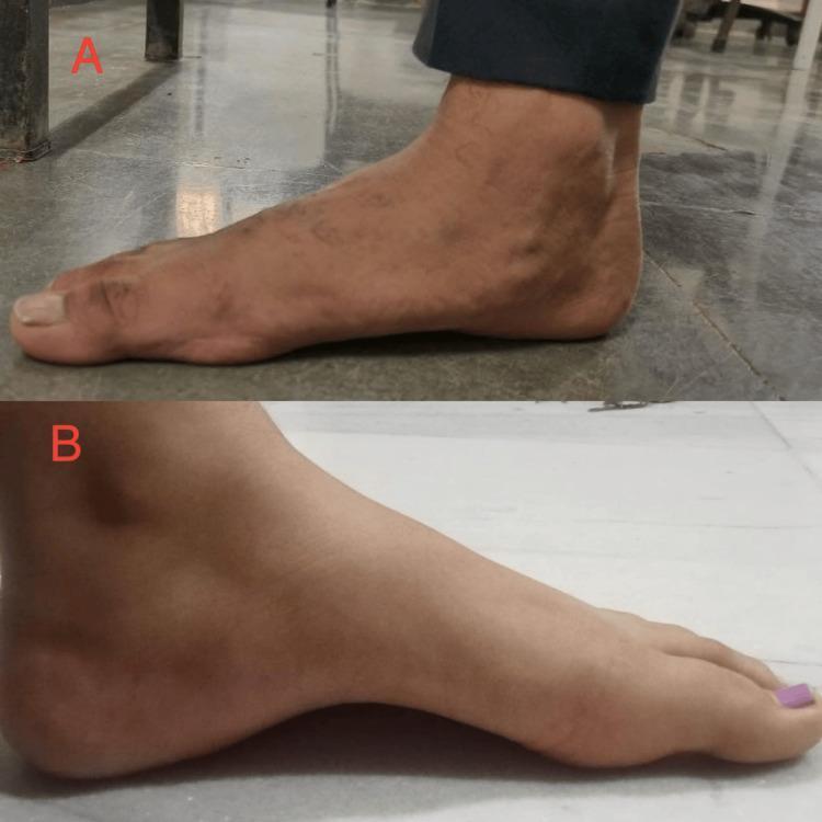
This clinical photograph provides a side-by-side comparison of two distinct foot arch morphologies in a musculoskeletal context. Image A illustrates Pes Planus (flat foot), characterized by a collapsed medial longitudinal arch where the plantar surface of the midfoot makes near-complete contact with the ground. Image B illustrates Pes Cavus (high-arched foot), showing a markedly exaggerated medial longitudinal arch with a significant visible gap between the midfoot and the floor. The comparison demonstrates common orthopedic variations of the foot's structural alignment. These images serve as educational visual aids for identifying common foot deformities associated with gait abnormalities, plantar fascia tension, and muscle imbalances in physiotherapy and orthopedic assessments. The contrast highlights the difference between an absent arch (A) and an abnormally high arch (B), both of which can impact biomechanical stability and result in pain or instability.
Clinical Examination in Orthopaedic Deformities
1. GENU VALGUM (Knock Knees)
Definition & Measurement
History
- Age of onset (physiologic vs. pathologic)
- Pain, limp, difficulty walking
- Family history
- Nutritional history (rickets)
Inspection (Standing)
- Gait: observe for awkward waddling, toe-in gait
- Alignment: observe in both frontal and sagittal planes - knees touch but ankles are apart
- Skin changes: look for bowing, skin over medial knee
- Limb length discrepancy: assess pelvic tilt
Measurement (Key Clinical Measure)
- Intermalleolar Distance (IMD): With the patient standing and medial femoral condyles (knees) touching, measure the distance between the medial malleoli. IMD > 10 cm is pathological and an indication for surgical consideration.
- Tibiofemoral angle: normally 5-7° of valgus in adults; >10° is pathological
Special Tests
- Assess for compensatory foot changes: secondary pes planus
- Check for tibial torsion: external tibial torsion often accompanies genu valgum
- Patellar tracking: assess for lateral patellar subluxation tendency
- Ligament assessment: medial collateral ligament laxity
Causes to Exclude
- Renal osteodystrophy (most common pathological cause if bilateral)
- Osteochondromas
- Rickets (check metaphyseal flaring, wrist signs)
- Post-infectious asymmetric tibial overgrowth
2. PES PLANUS (Flat Foot)
Definition
- Flexible (physiologic in young children, arch reappears on toe standing)
- Rigid (structural - always flat, may be painful)
History
- Age, bilateral vs. unilateral
- Pain (under arch, heel, calf)
- Family history
- Any associated neurological symptoms
Inspection
- Observe the arch - is it present when non-weight-bearing?
- Look for valgus hindfoot (heel tilted outward = hindfoot valgus)
- Skin calluses over medial midfoot
- Arch collapses completely
- "Too many toes sign": viewed from behind, more than 2 toes visible lateral to the heel (due to forefoot abduction and hindfoot valgus)
- Medial border of foot contacts the ground
Key Differentiating Tests
- Jack's toe dorsiflexion (windlass) test: Passively dorsiflexing the big toe - if the arch reconstitutes, it is a flexible flat foot (normal plantar fascia mechanism)
- Single heel rise test: Ask patient to stand on one leg and rise on tiptoes - in flexible flat foot the heel inverts to varus; in rigid flat foot or tibialis posterior dysfunction, the heel stays in valgus or the patient cannot perform the test
- Subtalar movement: Assess subtalar joint range of motion - rigid flat foot has restricted subtalar motion
Palpation
- Tibialis posterior tendon (medial to medial malleolus) - tenderness suggests tibialis posterior tendon dysfunction
- Plantar fascia origin at calcaneum
Neurological Check
- Exclude underlying neurological cause (e.g., cerebral palsy, spina bifida)
3. CTEV - Congenital Talipes Equinovarus (Clubfoot)
Definition
- C - Cavus (high-arched medial longitudinal arch)
- A - Adductus (forefoot medial deviation)
- V - Varus (hindfoot inversion/varus)
- E - Equinus (plantarflexion at ankle, inability to dorsiflex)
Clinical Examination at Birth
- Both feet should be examined systematically
- Foot is small, with shortened calf muscles
- Foot turned inward and downward
- Lateral border of foot facing upward in severe cases
- Deep skin creases on the medial side of the foot and posteriorly at the ankle
- Calf muscle hypoplasia (the affected calf is thinner)
| Component | What to Assess |
|---|---|
| Equinus | Dorsiflexion of ankle restricted (cannot reach neutral/90°) |
| Varus | Heel inverted inward |
| Adductus | Forefoot deviated medially |
| Cavus | High medial arch - check under the foot |
- Talar head palpable prominently on dorsolateral surface
- Check for skin crease depth (medial and posterior creases)
- Assess for bony prominences on lateral foot
- Attempt gentle dorsiflexion and external rotation - documents whether the deformity is correctable (flexible vs. rigid)
- Document the maximum achievable correction - baseline for Ponseti scoring
- Pirani Scoring (0-6): Grades 3 hindfoot signs (posterior crease, empty heel, equinus) and 3 midfoot signs (medial crease, curved lateral border, talar head coverage) - score out of 6; higher = more severe
- Dimeglio Classification: Grades by reducibility of each deformity component (Grades I-IV)
- Hip: examine both hips for developmental dysplasia (DDH) - associated in ~2%
- Spine: check for neural tube defects (spina bifida - may cause secondary CTEV)
- Opposite foot: 50% are bilateral
- Check for arthrogryposis (multiple joint contractures)
- Neurological: rule out spinal muscular atrophy, meningomyelocele
4. SCOLIOSIS
Definition
History
- Age of onset, progression
- Pain (unusual in idiopathic scoliosis; red flag if present in a child)
- Family history
- Respiratory symptoms (severe curves >70° can impair breathing)
- Menarcheal status in girls (growth remaining)
Inspection - Patient Undressed, Standing
- Shoulder asymmetry (one shoulder higher)
- Scapular prominence on convex side
- Waist asymmetry - unequal skin folds
- Pelvic tilt / limb length discrepancy - must be excluded as a cause of apparent scoliosis
- Spinal curve - note direction and level (thoracic, thoracolumbar, lumbar)
- Assess for kyphosis or lordosis changes - is the sagittal alignment normal?
- Café-au-lait spots + cutaneous neurofibromas + axillary freckling = Neurofibromatosis Type 1
- Midline hair patches or skin dimples = Spina bifida occulta or tethered cord
Adams Forward Bend Test (KEY TEST)
- Positive test: Rib hump (posteriorly prominent ribs) on the convex side of the curve - this is due to vertebral rotation causing ribs to rotate backward
- Loin prominence instead if lumbar curve
- This test differentiates structural (fixed rotation - hump persists) from postural scoliosis (hump absent)
Scoliometer (Angle of Trunk Rotation - ATR)
- Placed at the apex of the curve during Adams forward bend test
- ATR >7° warrants radiological evaluation
- Used to monitor progression without repeated X-rays
Measurement of Curve (on X-ray - Cobb Angle)
- <10°: normal variation
- 10-25°: mild (observe)
- 25-40°: moderate (brace)
-
40-45°: severe (surgical consideration)
Limb Length Assessment
- Measure true leg length (ASIS to medial malleolus) and apparent leg length (umbilicus/xiphisternum to medial malleolus)
- LLD causes compensatory spinal tilt that can mimic scoliosis ("functional scoliosis") - a block under the short leg corrects it
Neurological Examination (Mandatory)
- Abdominal reflexes: absent or asymmetrical reflexes suggest intraspinal pathology (syringomyelia, spinal cord anomaly)
- Power, tone, reflexes and sensation in upper and lower limbs
- Gait assessment
- In kyphosis: also assess sagittal alignment and forward gaze
Quick Comparison Summary
| Feature | Genu Valgum | Pes Planus | CTEV | Scoliosis |
|---|---|---|---|---|
| Key measurement | Intermalleolar distance | Arch height, heel alignment | CAVE components, Pirani score | Cobb angle (X-ray), ATR (scoliometer) |
| Key clinical test | IMD with knees touching | Jack's test, single heel rise | Flexibility of deformity | Adams forward bend test |
| Pathological threshold | IMD >10 cm, angle >15° | Rigid + painful | Fixed equinovarus | Cobb angle >10° |
| Key associated findings | Rickets, renal osteodystrophy | Tibialis posterior dysfunction | DDH, spina bifida | Neurofibromatosis, LLD |
Treatment of avulsed hamstring tendon in a 67 year old lady post bilateral TKR surgery
proximal hamstring avulsion repair elderly treatment
proximal hamstring avulsion treatment elderly post TKR surgical conservative management
hamstring avulsion total knee replacement complication
proximal hamstring repair outcomes older patients conservative versus surgical
proximal hamstring avulsion repair suture anchor ischial tuberosity surgery
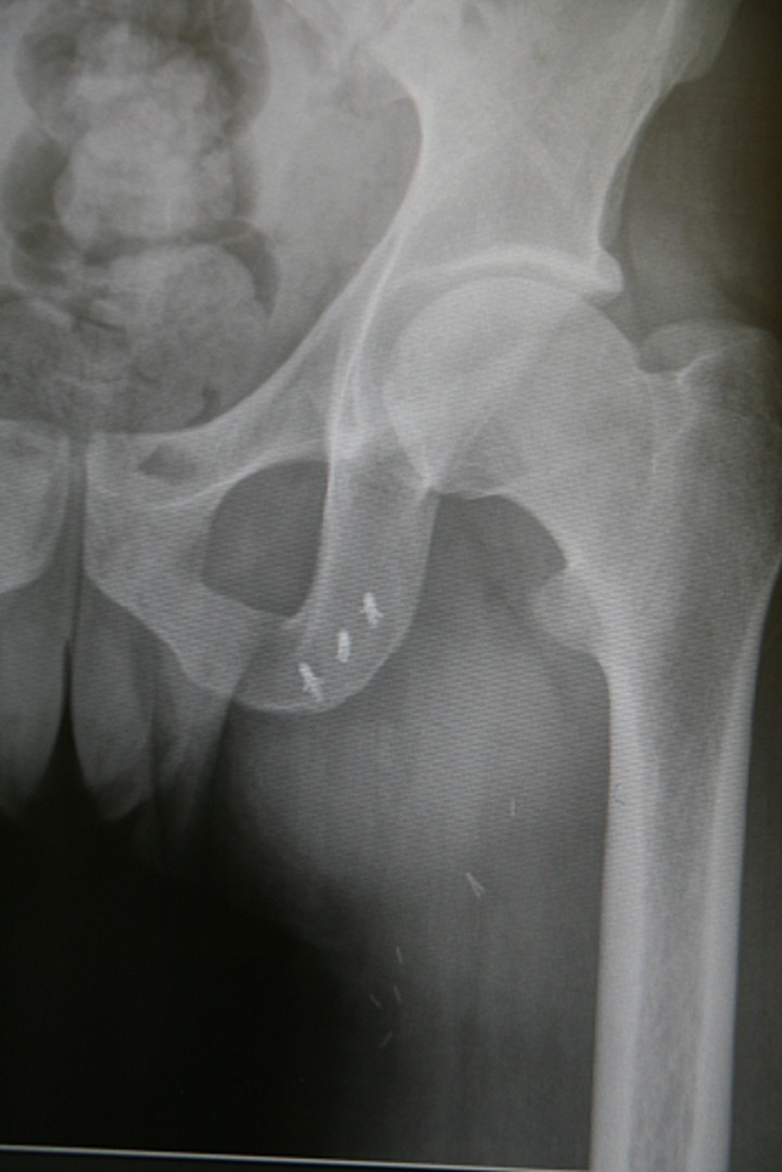
**Imaging Modality:** Anteroposterior (AP) projection radiograph (X-ray). **Anatomical Region:** Left hip and pelvis, including the proximal femur, acetabulum, and pubic rami. **Observed Pathology/Postoperative Status:** Postoperative imaging demonstrating the surgical repair of a proximal hamstring avulsion. The hip joint appears congruous with no acute fractures or dislocations of the femoral head or neck. **Characteristic Visual Features:** - **Suture Anchors:** Three radiopaque metallic suture anchors are visualized embedded within the inferior pubic ramus/ischial tuberosity region. - **Hardware Distribution:** The anchors are arranged in a linear pattern along the anatomical origin of the hamstring muscle group. - **Soft Tissue:** Several small, faint radiopaque markers or surgical clips are visible in the proximal soft tissues of the medial thigh, consistent with suture reinforcement or local hemostasis. **Key Diagnostic Features:** The presence of specific orthopedic hardware at the ischial tuberosity confirms a surgical reattachment procedure (suture anchor fixation) for a high-grade proximal hamstring tendon tear or avulsion. No secondary degenerative changes or heterotopic ossification are noted in the immediate field of view.
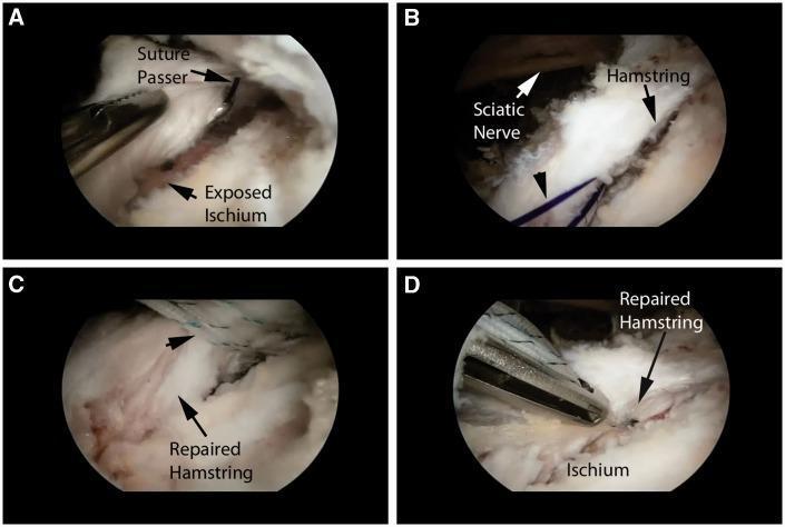
This four-panel (A-D) figure demonstrates the endoscopic repair of a proximal hamstring tendinous avulsion from the ischial tuberosity. (A) Shows the preparation of the bleeding corticocancellous bed on the exposed ischium, with a suture passer ready to penetrate the tendon tissue. (B) Highlights a shuttle suture (black arrowhead) placed through the hamstring tendon. Crucially, the image illustrates the immediate anatomical proximity of the sciatic nerve (white arrow) to the surgical site, emphasizing the need for nerve protection during suture management. (C) Displays multiple mattress sutures secured within the substance of the hamstring tendon to reapproximate it to the bone. (D) Shows the completed repair with the hamstring tendon firmly anchored back to the ischium. The series illustrates an endoscopic orthopedic procedure utilizing suture anchors and arthroscopic techniques to treat proximal hamstring ruptures while managing critical neurovascular structures like the sciatic nerve.
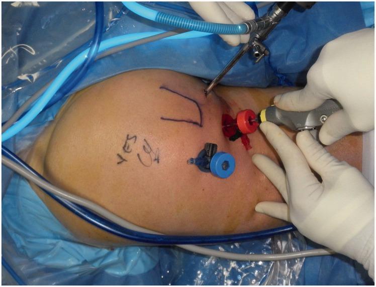
This clinical photograph demonstrates the positioning and portal placement for an endoscopic repair of a proximal hamstring avulsion. The patient is in a prone position, with the posterior hip region surgically prepared and draped. Visible on the skin are preoperative markings, including a 'U' shape likely delineating the ischial tuberosity and site verification markers. Two primary surgical portals are identified by color-coded cannulas: a superior medial portal with a red cannula containing an arthroscope for visualization, and an inferior distal portal with a blue cannula through which an oscillating shaver is being maneuvered by a gloved hand. This setup allows for sub-muscular dissection, debridement of devitalized tissue from the ischial tuberosity, and subsequent suture anchor placement for tendon reattachment. The procedure is orthopaedic in nature, focusing on sports medicine and minimally invasive techniques for managing deep gluteal space pathology and hamstring origin injuries.
Treatment of Avulsed Hamstring Tendon in a 67-Year-Old Post-Bilateral TKR
Understanding the Anatomy and Injury
- Number of tendons involved (1, 2, or all 3)
- Degree of retraction (< 2 cm vs. 2-3 cm vs. > 3 cm)
- Acuity: acute (<4 weeks), subacute (4-12 weeks), chronic (>3 months)
Pre-Treatment Workup
Imaging
- X-ray (AP pelvis, proximal femur): identifies bony avulsion fragment from the ischial tuberosity; also checks TKR hardware for any loosening
- MRI (non-contrast, fat-suppressed inversion recovery + proton density sequences in multiple planes): defines number of tendons torn, degree of retraction, and muscle degeneration - this is the key diagnostic tool per Campbell's Operative Orthopaedics
- Ultrasound: a useful bedside alternative to assess retraction and partial vs. complete tear
Clinical Signs to Confirm
- Bowstring sign: absence of palpable tension in the distal hamstrings with patient prone and knee flexed to 90° - positive in complete 3-tendon avulsion
- Ecchymosis over the posterior thigh
- Palpable proximal defect
- Weakness in prone knee flexion
- Sitting pain over the ischium (a hallmark complaint)
Decision Framework: Conservative vs. Operative
| Factor | Favors Conservative | Favors Operative |
|---|---|---|
| Number of tendons torn | Single tendon | All 3 tendons (complete avulsion) |
| Retraction | < 2 cm | ≥ 3 cm (2 tendons) or any 3-tendon complete |
| Time since injury | >3 months (chronic - repair difficult) | Acute (<4 weeks, ideally <3 months) |
| Functional demand | Very low | Moderate-high (post-TKR stability needs) |
| Age | Advanced age alone is NOT a contraindication | Age >50 does NOT worsen surgical outcomes (ISAKOS data) |
| Comorbidities | High anaesthetic risk | Otherwise fit |
| Patient preference | Avoids surgery | Wants optimal functional recovery |
Key Evidence Point
Conservative Treatment
- Single-tendon tears (regardless of retraction)
- Two-tendon tears with retraction < 2 cm
- Chronic injuries (>3 months) where scar has formed and acute repair is no longer feasible
- Patients unfit for anaesthesia
Phase 1 - Acute (0-6 weeks)
- Rest, cryotherapy, analgesia (paracetamol, NSAIDs with caution in elderly)
- Progressive weight-bearing with crutches
- Positioning: avoid combined hip flexion + knee extension (the position that stretches the repair zone); patient sits with hip slightly extended, or uses a wedge cushion
- Brace (if used): locked knee brace in extension or hip orthosis in slight extension
- Avoid prolonged sitting on hard surfaces (ischial pressure aggravates symptoms)
Phase 2 - Subacute (6-12 weeks)
- Gentle passive range of motion of hip and knee
- Isometric hamstring contractions (pain-free range)
- Hydrotherapy / pool walking (reduces load while maintaining mobility)
Phase 3 - Active Rehabilitation (3-6 months)
- Progressive hamstring strengthening: prone leg curls, Nordic curls (graded)
- Core and gluteal strengthening
- Proprioception training - particularly important for TKR stability
- Functional activities: stair training, sit-to-stand (especially important for this patient to maintain independence post-bilateral TKR)
Operative Treatment
- Complete three-tendon tears with or without displacement
- Two-tendon tears with ≥ 3 cm of distal retraction
- Partial tears failing conservative management
- Acute presentation (<3-4 months from injury) - once chronic, scar fibrosis makes mobilisation of the retracted tendon extremely difficult
Surgical Approach Options
1. Open Repair (Technique 53.28 - Campbell's)
- Patient prone
- Longitudinal incision at edge of gluteus maximus (or horizontal incision in gluteal crease - more cosmetic but less extensible)
- Gluteus maximus retracted proximally; fascia opened protecting the posterior femoral cutaneous nerve and inferior cluneal nerve
- Tendons identified; large hematoma evacuated in acute injuries
- Traction sutures placed in tendon stumps; sciatic nerve identified and protected
- Ischial tuberosity exposed with retractors
- 2-3 suture anchors placed within the proximal hamstring footprint on the ischium; suture sets from each anchor passed through tendons in Krackow or modified Mason-Allen fashion
- Knotless technique using SwiveLock anchors is preferred by many surgeons
- If repair is performed >3 weeks post-injury, the knee must be flexed to allow tendon approximation to the tuberosity
2. Endoscopic/Arthroscopic Repair (Technique 53.29 - Campbell's)
- 4-cm incision in gluteal crease; 30° arthroscope into space between gluteus maximus and ischial tuberosity
- Ischial bursectomy via accessory portal
- Sciatic nerve identified, neurolysed, and protected (critical step)
- Torn tendons debrided; 4-mm burr to prepare ischial bone
- 1-2 suture anchors placed under direct vision; sutures passed through tendons in mattress fashion
- Tension-slide technique to bring tendons to bone; arthroscopic knot tied; lateral-row SwiveLock anchor placed
- Well-suited for partial tears or cases with less retraction
Postoperative Rehabilitation (Critical Modification for Bilateral TKR Patient)
Bracing
- Custom pelvic-thigh-hip spica orthosis: maintains hip in extension, knee left free - preferred option
- OR locked hinged knee brace in full extension
- Bracing continued for 6 weeks
Weight-bearing
- Full weight-bearing with crutches while wearing hip orthosis
- Non-weight-bearing if using knee brace in flexion
Special Considerations for Bilateral TKR Patient:
- Bracing conflict: A knee brace in extension post-hamstring repair conflicts with normal TKR rehabilitation which requires knee flexion. The hip spica orthosis (keeping the hip extended, knee free) is therefore the preferred choice in this patient to protect the repair without restricting the already-replaced knee joints.
- Crutch use: Standard for 2 weeks minimum, but this patient needs to be assessed for upper limb strength and balance - elderly patients post-bilateral TKR may have compromised balance and risk falls.
- DVT prophylaxis: Mandatory - this patient has had bilateral TKR (already at elevated thrombosis risk) and is now post-hip surgery with restricted mobility. Anticoagulation (LMWH or rivaroxaban) is essential.
- Hamstring strength is critical for TKR outcome: The hamstrings protect the posterior capsule and PCL mechanism of a TKR. Inadequate hamstring function can lead to posterior instability of the TKR. Functional rehabilitation in this patient is therefore more pressing than in a patient with native knees.
Rehabilitation Milestones
| Phase | Week | Goals |
|---|---|---|
| Protection | 0-6 wks | Pain control, protected WB, prevent contracture, brace |
| Early mobilisation | 6-12 wks | Hip ROM, isometric hamstring activation, pool therapy |
| Strengthening | 3-6 months | Progressive hamstring + quadriceps loading, sit-to-stand training |
| Functional | 6-12 months | Stair climbing, gait normalisation, return to daily activities |
Summary of Treatment Algorithm for This Patient
67-year-old, post-bilateral TKR, proximal hamstring avulsion
       ↓
Confirm with MRI: retraction? number of tendons? acuity?
       ↓
Single tendon or <2cm retraction → Conservative (PT + protection + analgesia)
       ↓
Complete 3-tendon or ≥3cm retraction:
       ↓
Acute (<3 months) → OPERATIVE: open or endoscopic repair with suture anchors
                    + hip spica orthosis post-op (NOT knee brace - protects TKR)
                    + DVT prophylaxis
                    + staged bilateral rehab coordination
       ↓
Chronic (>3 months) → Conservative preferred (scar tissue prevents easy reduction)
                      Consider ultrasound-guided steroid injection if recalcitrant
Key Takeaways for This Specific Patient
- Age is NOT a contraindication to surgical repair - patients >50 years have equal or better pain relief from surgery without increased complication rates.
- Bilateral TKR makes hamstring function more important, not less - these muscles are the dynamic posterior stabilisers of the replaced knees.
- Hip orthosis is preferred over knee brace post-repair in this patient to avoid conflicting with TKR biomechanics.
- DVT prophylaxis is mandatory given the combined risk profile.
- Timing is critical: acute repair (<3 months) yields significantly better outcomes than delayed repair; if chronic, conservative management is preferred.
- The sciatic nerve is at direct surgical risk during repair and must be identified and protected in all approaches.
Write a reflective essay on “automation in the operating room: How do I tackle robotic-assisted surgery?” Reflect on the emergence of robotic-assisted surgery and the use of machine learning to learn expert gestures. How does AI impact surgical performance, safety and outcomes? How do visual enhancements like photo acoustic guidance , influence surgical decision-making? What role should AI play in high-stakes procedures and what safeguards are needed to build clinician and patient trust ? While writing the essay, don’t just celebrate the tech, but reflect on how AI improves precision and reduces fatigue, but also raises concerns like equipment errors or over-reliance on machines. What are the safeguards ( Human oversight, fail safes, ethical review) which are needed to balance performance with trust? Think in terms of tradeoffs like precision vs trust and show how risks can be managed responsibly. Talk about how patients can start trusting AI , by transparency , validation , clear communication with patients and by keeping clinicians in the loop. Show nuance in how AI reshapes roles , rather than just outcomes. Tie every technical point back to patient care and clinician experience. Recognise that adoption of AI isn’t only about accuracy , it’s about ethics , oversight and system readiness. Apply AI concepts accurately and appropriately within a healthcare context. Provide a meaningful and insightful analysis of the case study. Use proper APA citations while listing references, when needed and finish the essay within 2500-3000 words.
robotic surgery machine learning surgical gestures performance outcomes
artificial intelligence surgical safety patient outcomes operating room
photoacoustic imaging surgical guidance decision making AI operating room 2023 2024
PMID: 36550203
PMID: 36997732
photoacoustic imaging intraoperative guidance surgery clinical
AI robotic surgery surgeon fatigue trust ethics oversight fail-safe patient trust 2024 2025
PMID: 39999009
PMID: 40985285
PMID: 37224375