
Close Reduction, poss. Orif plating radius
distal radius fracture ORIF volar locking plate X-ray
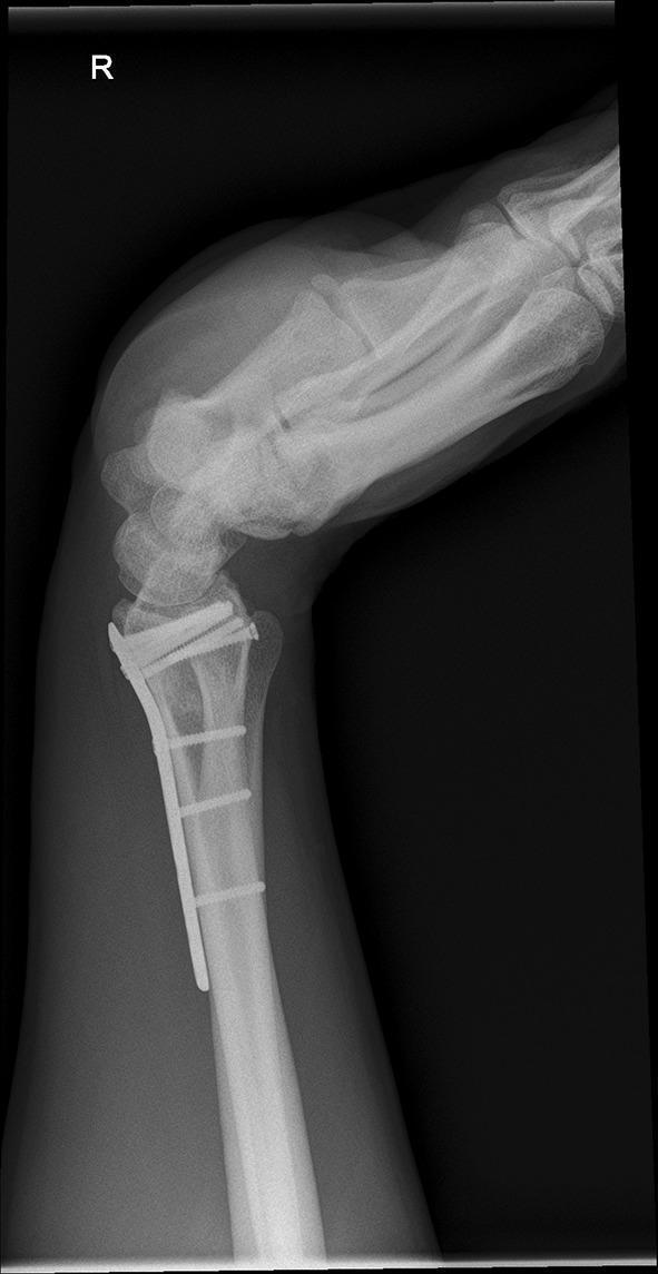
This diagnostic image is a lateral X-ray radiograph of a right wrist in a position of flexion. The image demonstrates a distal radius fracture that has undergone open reduction and internal fixation (ORIF). A volar locking plate is visible along the anterior (volar) aspect of the distal radius, secured with multiple cortical and subchondral locking screws. The screws are positioned near the radiocarpal joint surface to provide stability to the distal fragments. The carpal bones, including the scaphoid and lunate, are visualized in an articulated position with the distal radius, showing a clear flexed orientation. The distal radioulnar joint (DRUJ) and the proximal carpal row are included in the view. There is no evidence of hardware failure, such as plate fracture or screw loosening, and the bone density appears within normal limits. This radiographic view is utilized in orthopedic surgery to assess the range of motion (ROM) and the stability of internal fixation before planned implant removal.
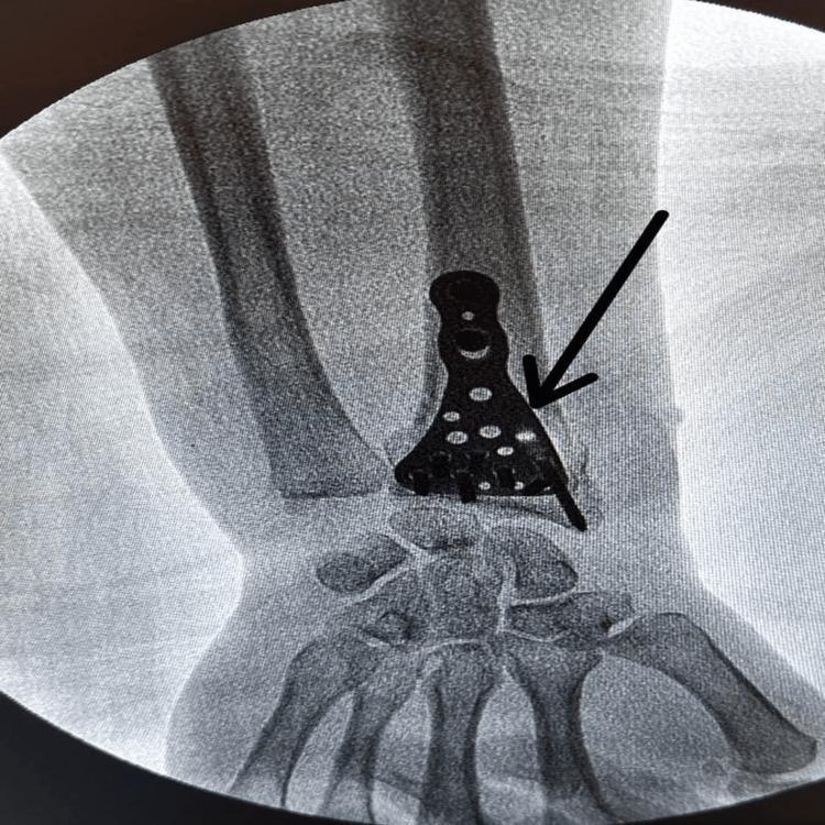
This diagnostic fluoroscopic X-ray image demonstrates a post-operative view of the distal forearm and wrist, specifically illustrating open reduction and internal fixation (ORIF) for a distal radius fracture. Centered on the distal radius is an anatomically shaped, metallic volar-locked distal radius plate. The implant features a characteristic triangular distal head that conforms to the radial metaphysis and an elongated proximal shaft. The distal portion of the plate contains multiple circular radiopaque densities representing locking screws or screw holes, arranged in a configuration designed to stabilize subchondral bone and articular fragments. The proximal shaft contains several elongated screw holes for diaphyseal fixation. A black arrow points specifically to the volar plate at the fracture site. The carpal bones and distal radioulnar joint are partially visible, demonstrating the plate's proximity to the radiocarpal joint surface. This image serves as a clinical example of orthopedic osteosynthesis in the management of high-energy wrist trauma.
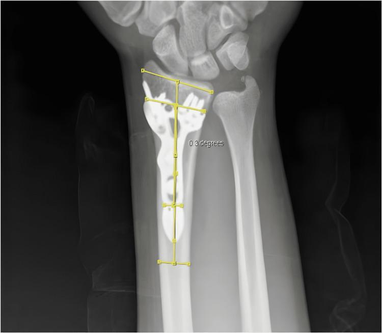
This diagnostic image is an anteroposterior (AP) X-ray of the wrist and distal forearm, demonstrating a distal radius fracture status post-open reduction and internal fixation (ORIF). A radiopaque locking plate is visualized along the volar aspect of the distal radius, secured with multiple cortical and locking screws. Yellow digital annotations illustrate a clinical measurement for coronal malposition known as the 'AYE Angle.' This measurement is obtained by calculating the angle between the vertical midline axis of the distal radius shaft and the longitudinal midline axis of the implanted hardware. In this specific case, the measurement is labeled as 0.3 degrees, indicating minimal coronal malalignment. The surrounding anatomy including the distal ulna, carpal bones, and radiocarpal joint space are well-visualized. This diagnostic tool is used in orthopedic surgery to assess the precision of hardware placement and post-operative alignment, which is critical for preventing complications like tendon irritation or restricted range of motion.
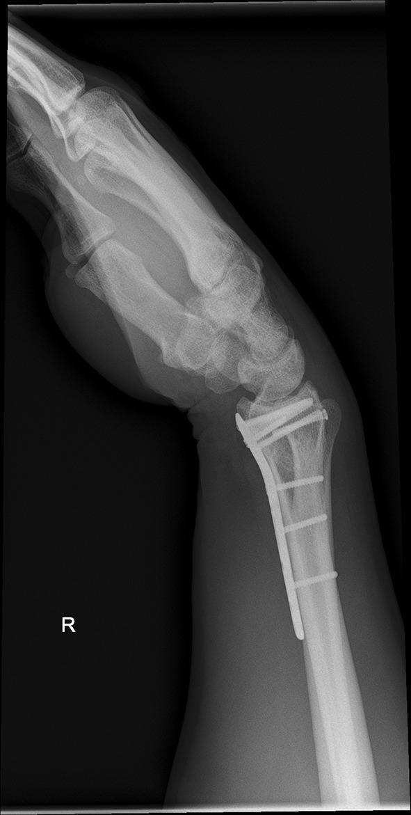
This diagnostic X-ray illustrates a lateral view of a right wrist in a flexed position, serving as an assessment of range of motion prior to hardware removal. The radiograph displays a distal radius fracture previously managed with open reduction and internal fixation (ORIF). A volar locking plate is visible, secured to the distal radial metaphysis and shaft with multiple screws. The distal screws are positioned in a subchondral fashion to support the articular surface. Anatomical structures clearly visualized include the distal radius, distal ulna, carpal bones (including the scaphoid and lunate), and the bases of the metacarpals. The image evaluates the relationship between the hardware and the carpal alignment during joint movement, highlighting potential mechanical impingement or soft tissue restrictions, such as the palmar and dorsal scar formation mentioned in the clinical context. This imaging is characteristic of orthopedic follow-up for complex intra-articular fractures.
Closed Reduction +/- ORIF with Plate: Distal Radius Fracture
Overview
Closed Reduction Technique
- Dorsal hematoma block with local anaesthetic OR regional/general anaesthesia
- Finger traps with ~10 lb longitudinal traction applied to thumb and index finger
- Re-create the deformity briefly
- Apply traction and ulnar deviation
- Translate the distal fragment volarly (hook the volar cortex)
- Apply a sugar-tong splint with a three-point mold to maintain reduction
- MCP and IP joints kept free for movement
- Radial shortening < 5 mm
- Dorsal articular tilt < 5-10 degrees
- Intraarticular step-off < 2 mm
Decision to Proceed to ORIF
- Patient age > 60 years
- Dorsal metaphyseal comminution
- Intraarticular involvement with > 2 mm step-off
- Associated ulnar fracture
- Severe osteoporosis
- Volar cortex not intact / cannot be hooked
- Soft tissue compromise
Operative Options
| Technique | Indications |
|---|---|
| Closed Reduction + Percutaneous Pinning (CRPP) | Extraarticular fractures in younger patients without osteoporosis; metaphyseal instability with simple articular displacement |
| Volar Locking Plate (VLP) - ORIF | Most common; volarly or dorsally displaced fractures, >2 mm articular displacement, metaphyseal comminution, combined radius/ulna fractures |
| Dorsal Plating | Dorsally displaced fractures with dorsal bony defects; direct articular visualization |
| Distraction/Bridge Plating | Highly comminuted, unstable, or elderly patients with severe osteoporosis; multiple-trauma patients needing weight bearing |
| External Fixation | Unstable fractures with soft tissue compromise; adjunct to internal fixation |
Volar Locking Plate (VLP) - Standard Technique
- Approach: Henry approach between FCR tendon and radial artery (or through floor of FCR sheath)
- Plate placed at or proximal to the watershed line (distal to watershed = FPL rupture risk)
- Reduction confirmed under fluoroscopic guidance (articular surface not directly visualized)
- Fixed-angle and variable-angle locking screws available
- Pronator quadratus repair over the plate at closure
- Can be combined with dorsal bone grafting for dorsal comminution
Key Complications to Know
| Complication | Details |
|---|---|
| FPL rupture | Most common flexor tendon injury; from plate placed distal to watershed line |
| EPL rupture | Most common with volar plates due to over-length dorsal screw penetration |
| Acute carpal tunnel syndrome | Progressive paresthesias + disproportionate pain - requires urgent decompression |
| CRPS | Risk increases with overdistraction (especially external fixation) |
| Pin site infection / tendon tethering | With percutaneous pinning |
| DRUJ instability | Concurrent ulnar styloid fracture treatment generally not needed if DRUJ is stable after radius fixation |
ORIF X-ray Appearances
Key Points to Remember
- Closed reduction is always attempted first in displaced fractures unless there is an obvious contraindication
- Loss of reduction is more common in patients >50 years old
- VLP has largely replaced percutaneous pinning but one high-level study showed no clear advantage of VLP over K-wire pinning
- The controversy around the "large increase in ORIF" remains: evidence for superior long-term outcomes from volar locked plating vs. non-operative treatment is not conclusive for all fracture types
- Post-operative early vs. late motion: no evidence supports early motion offering advantage in functional recovery

420, verano 22 M
- Lacerated Wound, 4 cm - Mentum (chin)
- Multiple Abrasions - Right Posterior Forearm and Bilateral Knees
- Mechanism: Fall from 12 feet
- Background: Intellectual Disability
- 1d S/P Wound Suturing, Mentum (8/5)
"MPI sec to MVA 1 Fracture, Close, Comminuted, Displaced; Multiple Abrasions L Shoulder R Forearm L Knee S/P Wound Debridement and Repair, Right Forearm 8/3 - Plan: Close Reduction poss ORIF plating, proximal radius."
Yes
Which patient's clinical summary would you like?
Clinical Summary - 420 VERANO 22/M
- Lacerated wound, 4 cm - Mentum (chin)
- Multiple abrasions - Right posterior forearm and bilateral knees Post-op Status: 1 day post wound suturing of mentum (done 8/5)
Current Management Plan
1. Wound Care (Mentum Laceration - Post-Suturing Day 1)
- Cleanse daily with dilute hydrogen peroxide to remove crusting and debris (stop once scab separates - toxic to new epithelium)
- Keep wound clean and dry; safe to bathe after 24 hours post-repair
- Apply clean dressing as needed
- Facial sutures: 5 days from repair (i.e., around 8/10)
- Replace with Steri-Strips after removal to reinforce the healing wound and minimize scarring
2. Abrasion Care (Right Posterior Forearm, Bilateral Knees)
- Clean with saline irrigation or gentle soap and water
- Non-adherent dressing or moist wound care
- Monitor for signs of infection
- Elevate affected extremity (right forearm) above heart level to minimize edema
3. Infection Surveillance
- Redness, increasing pain, swelling around wound
- Fever
- Red streaks progressing up the extremity
- Pus or discharge from wound
4. Tetanus Prophylaxis
- Verify immunization history
- If < 3 doses or uncertain: give Tetanus Toxoid + TIG (since this is not a clean minor wound - fall + abrasion)
- If ≥ 3 doses and last dose within 5 years: no additional immunization needed
- Consider Tdap if no prior dose received
5. Antibiotic Prophylaxis
| Scenario | Recommendation |
|---|---|
| Uncomplicated wound | 1st-generation cephalosporin (e.g., Cefalexin) |
| Grossly contaminated / retained foreign body | Amoxicillin-clavulanate or 2nd-gen cephalosporin |
- Fall injuries with ground contact carry contamination risk - consider short course antibiotic cover
6. Special Considerations - Intellectual Disability
- Patient may have limited ability to report pain or new symptoms - rely on caregiver/guardian observation
- Ensure guardian is fully briefed on wound care steps and return precautions
- Consider restraint precautions if patient may disturb sutures; chin/mentum location is at risk of self-manipulation
- Low threshold for follow-up if compliance with wound care is uncertain
- Assess for any undetected injuries from the 12-ft fall - consider imaging of cervical spine and chest if not already done on admission, given fall height
Additional: Surveillance for Hidden Injuries (12-ft Fall)
- Cervical spine - must be clinically cleared or imaged
- Thoracolumbar spine - compression fractures
- Pelvis and lower extremities - given bilateral knee abrasions
- Head CT - if any LOC, altered GCS, or cognitive baseline change (complicated by baseline intellectual disability)
426 jorquina
Clinical Summary - 426 JORQUIA 22/M
Post-Op Day 5 Management
1. Vitals & Monitoring
- Monitor temperature, HR, BP, RR, SpO2 each shift
- Watch for fever spikes (early sign of surgical site infection or intra-abdominal collection)
- Monitor bowel sounds and return of bowel function
2. Diet & Ambulation
- Diet: Should be tolerating regular/solid diet by POD 1-2 after uncomplicated laparoscopic appendectomy; by POD 5, full diet expected
- Ensure adequate oral fluid intake
- Ambulation: Full ambulation expected at POD 5 - early mobilization reduces ileus and DVT risk
- Encourage walking several times daily
3. Wound Care (Port Sites)
- 3 laparoscopic port sites (umbilicus + 2 lower abdomen)
- Closed with dissolvable sutures - no formal suture removal needed
- Inspect port sites daily for:
- Redness, swelling, discharge (signs of surgical site infection - most common complication after appendectomy)
- Dehiscence
- Keep clean and dry; light dressing if needed
4. Analgesia
- By POD 5, should be transitioning to oral analgesia only
- Paracetamol 500-1000 mg q6-8h PRN
- NSAIDs (e.g., Ibuprofen 400 mg q8h with food) if no contraindications
- Step down from IV analgesia if still ongoing
5. Antibiotics
- For uncomplicated appendicitis: perioperative antibiotics only (single pre-op dose of 2nd-generation cephalosporin, e.g., Cefoxitin or Cefazolin + Metronidazole)
- No prolonged post-op antibiotics indicated for uncomplicated, non-perforated appendicitis
- If antibiotics still running - review and discontinue if uncomplicated case confirmed intraoperatively
6. Discharge Planning (POD 5 - near discharge)
- Afebrile for >24 hours
- Tolerating full oral diet
- Pain controlled on oral medications
- Bowel function returned (flatus/stool)
- Port sites clean, no signs of infection
- Port site care - keep clean, monitor for infection signs
- Activity: Light activity for 1 week; avoid strenuous exercise and heavy lifting for 2-3 weeks
- Return to school/work: ~1 week post-op
- Full physical activity: 2-3 weeks post-op
- Return to ER if: fever >38°C, worsening abdominal pain, vomiting, port site redness/pus, inability to tolerate food
7. Complications to Watch (POD 5)
| Complication | Features |
|---|---|
| Surgical site infection | Most common; port site redness, warmth, discharge, fever |
| Intra-abdominal abscess | Persistent fever, abdominal pain, leukocytosis post-op |
| Ileus | Absent bowel sounds, distension, no flatus - less common by POD 5 |
| DVT/PE | Rare in young patient but monitor for calf pain, dyspnea |