

8091e051-2ff7-4704-958e-7d6889f778a8.jpeg
Show images
Here is the image you shared:
analyse_media
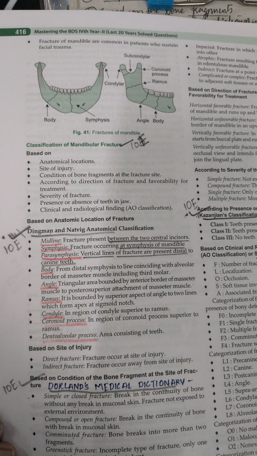
This is page 416 from Mastering the BDS IVth Year - II (Last 20 Years Solved Questions), covering the Classification of Mandibular Fractures.
Key Content on the Page
Diagram (Fig. 41): Two views of the mandible illustrating fracture sites - Symphysis, Body, Condylar, Subcondylar, Coronoid process, Ramus, and Angle.
Classification of Mandibular Fracture - Based on:
- Anatomical locations
- Site of injury
- Condition of bone fragments at the fracture site
- Direction of fracture and favorability for treatment
- Severity of fracture
- Presence or absence of teeth in jaw
- Clinical and radiological finding (AO classification)
Dingman & Natvig Anatomical Classification (underlined in red):
| Region | Description |
|---|---|
| Midline | Between the two central incisors |
| Symphysis | At symphysis of mandible |
| Parasymphysis | Distal to canine teeth |
| Body | Distal symphysis to alveolar border of masseter, including 3rd molar |
| Angle | Bounded by anterior to posterosuperior attachment of masseter |
| Ramus | Superior aspect of angle to sigmoid notch apex |
| Condyle | Superior to ramus in condyle region |
| Coronoid process | Superior to ramus in coronoid region |
| Dentoalveolar process | Area consisting of teeth |
Fracture Types by Bone Condition (Dorland's Medical Dictionary):
- Simple/Closed: No break in mucosal skin
- Compound/Open: Break in mucosal skin
- Comminuted: Bone breaks into >2 fragments
- Greenstick: Incomplete fracture (text cuts off)
The right column (partially visible) also covers: direction-based classification (favorable vs. unfavorable), severity classification, Kazanjian's classification (Classes I-III based on tooth presence), and the AO classification (F, L, O, S, A parameters).
This is a shared conversation. Sign in to Orris to start your own chat.