
Teach thyroid gland in detail 1st yr mbbs anatomy
Reading File
Now let me search the library for thyroid anatomy content in parallel:
Finding Sources
Finding Sources
Gray's Anatomy for Students is the ideal 1st yr MBBS source. Let me read the thyroid section from it plus search for related anatomy topics in parallel:
Reading File
Finding Sources
Finding Sources
Excellent content found. Let me now read the remaining sections - arterial supply continued, venous drainage, nerve supply, lymphatics, and pathology:
Reading File
Reading File
I have thorough content. Let me now fetch key images to include in the response:
Loading Image
Loading Image
Reading File
I now have all the content needed from Gray's Anatomy for Students to deliver a complete 1st year MBBS anatomy lecture on the thyroid gland. Let me compile the comprehensive response.
Thyroid Gland - Anatomy (1st Year MBBS)
Source: Gray's Anatomy for Students
1. Introduction and Overview
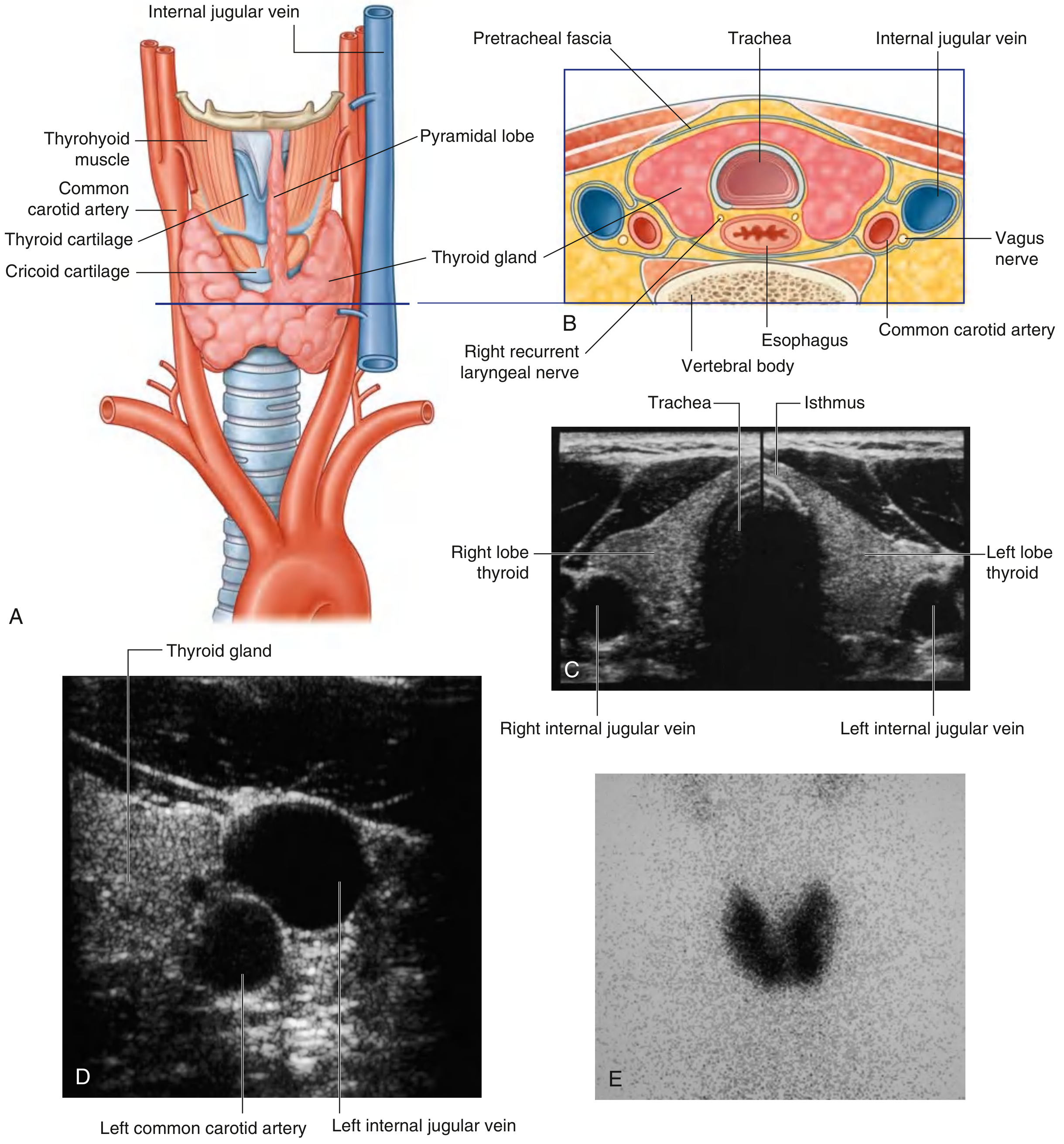
The thyroid gland is the largest endocrine gland in the body. It is a large, unpaired gland situated anteriorly in the neck, below and lateral to the thyroid cartilage. It lies in the visceral compartment of the neck, surrounded by the pretracheal layer of cervical fascia - the same compartment that contains the pharynx, trachea, and esophagus.
2. Gross Anatomy and Shape
The thyroid consists of:
- Two lateral lobes - these cover the anterolateral surfaces of the trachea, the cricoid cartilage, and the lower part of the thyroid cartilage
- An isthmus - connects the two lateral lobes and crosses the anterior surfaces of the 2nd and 3rd tracheal cartilages
- Pyramidal lobe (present in ~40-50% of individuals) - a small tongue of tissue extending upward from the isthmus, often from the left side, along the path of the thyroglossal duct
The gland lies deep to the sternohyoid, sternothyroid, and omohyoid muscles.
3. Relations (Clinically Important)
| Surface | Relations |
|---|---|
| Anterolateral | Sternohyoid, sternothyroid, omohyoid muscles; anterior border of sternocleidomastoid |
| Medial (deep) | Trachea, cricoid cartilage, thyroid cartilage, pharynx, esophagus |
| Posterior | Carotid sheath (common carotid artery, internal jugular vein, vagus nerve) |
| Posteromedial groove | Recurrent laryngeal nerve (in tracheo-oesophageal groove) |
| Posterior surface | Parathyroid glands (4 in number) |
4. Fascia and Capsule
The gland is enclosed in a true capsule of fibrous tissue that sends septa into the substance of the gland. Outside this, the pretracheal fascia forms a false capsule (surgical capsule). The gland is attached to the trachea and cricoid cartilage by a thickening of the pretracheal fascia called the ligament of Berry (posterior suspensory ligament) - this is why the thyroid moves up on swallowing (clinically useful in examination).
5. Arterial Supply
Two major pairs of arteries supply the thyroid gland:
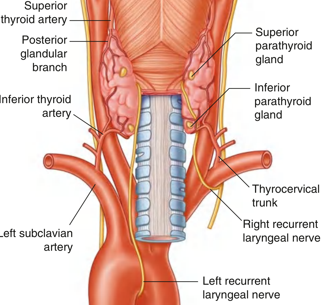
A. Superior Thyroid Artery
- First branch of the external carotid artery
- Descends along the lateral margin of the thyrohyoid muscle to reach the superior pole of the lateral lobe
- Divides into:
- Anterior glandular branch - runs along the superior border of the gland, anastomoses with its twin across the isthmus
- Posterior glandular branch - passes to the posterior side, may anastomose with the inferior thyroid artery
B. Inferior Thyroid Artery
- Branch of the thyrocervical trunk (from the 1st part of the subclavian artery)
- Ascends along the medial edge of the anterior scalene muscle
- Passes posteriorly to the carotid sheath
- Reaches the inferior pole of the lateral lobe and divides into:
- Inferior branch - supplies lower part of gland
- Ascending branch - supplies the parathyroid glands
C. Thyroid Ima Artery (occasional)
- Arises from the brachiocephalic trunk or arch of the aorta
- Ascends on the anterior surface of the trachea to supply the isthmus
- Important to know before tracheostomy - unexpected bleeding!
6. Venous Drainage
Three veins drain the thyroid (no valves - forms a venous plexus):
| Vein | Drains into |
|---|---|
| Superior thyroid vein | Internal jugular vein |
| Middle thyroid vein | Internal jugular vein |
| Inferior thyroid vein | Right and left brachiocephalic veins respectively |
The inferior thyroid veins are bilateral and descend in front of the trachea - important to ligate during thyroidectomy.
7. Lymphatic Drainage
Lymph drains to:
- Paratracheal nodes (nodes beside the trachea)
- Deep cervical nodes (along the internal jugular vein)
From deep cervical nodes, lymph flows into the thoracic duct (left) and right lymphatic duct.
8. Nerve Supply
The thyroid gland receives vasomotor (autonomic) nerve supply only - it has no somatic innervation:
- Sympathetic fibers - from the superior, middle, and inferior cervical ganglia via the cardiac plexus
- Parasympathetic fibers - from the vagus nerve [CN X]
These nerves regulate blood flow, not secretory activity (secretion is controlled by TSH from the pituitary).
9. Recurrent Laryngeal Nerve - Critical Relation
The recurrent laryngeal nerve (RLN) is the most important surgical relation of the thyroid gland:
- Arises from the vagus nerve (CN X)
- Right RLN: loops around the right subclavian artery
- Left RLN: loops around the arch of the aorta (longer course)
- Both ascend in the tracheo-oesophageal groove (groove between trachea and esophagus)
- Pass deep to the posteromedial surface of the lateral lobes of the thyroid
- Enter the larynx by passing deep to the inferior constrictor muscle
- The RLN passes very close to the inferior thyroid artery - they may cross each other (either artery anterior or posterior to nerve - highly variable)
- The nerves may also pass through the ligament of Berry
Clinical importance: Damage to the RLN during thyroidectomy causes hoarseness of voice (unilateral) or stridor and respiratory compromise (bilateral). Vocal cord assessment is mandatory before and after thyroid surgery.
10. Development (Embryology)
- Thyroid is the first endocrine gland to develop (appears in the 4th week of intrauterine life)
- Arises as a median outgrowth from the floor of the pharynx, near the base of the tongue (at what later becomes the foramen cecum of the tongue)
- Descends as the thyroglossal duct, passing anterior to the hyoid bone, to reach its final position at the front of the trachea
- The thyroglossal duct normally disappears early in development
Remnants and anomalies:
| Anomaly | Description |
|---|---|
| Thyroglossal duct cyst | Most common - midline neck swelling, moves up on swallowing and on tongue protrusion (unique sign). Treated by Sistrunk's operation (excision of cyst + mid-portion of hyoid bone) |
| Lingual thyroid | Failure to descend - gland remains at the tongue base. May be the only thyroid tissue present! |
| Pyramidal lobe | Remnant of the lower part of the thyroglossal duct |
| Ectopic thyroid | Anywhere along the path of migration |
| Thyroglossal fistula | Persistent connection from foramen cecum to skin |
11. Parathyroid Glands (Related Structure)
Four small, ovoid, yellowish glands on the posterior surface of the lateral lobes of the thyroid:
- Superior parathyroids (2) - derived from the 4th pharyngeal pouch; more constant in position
- Inferior parathyroids (2) - derived from the 3rd pharyngeal pouch; variable position (can be anywhere from carotid bifurcation to mediastinum)
Blood supply: Inferior thyroid arteries (mainly); superior thyroid artery may contribute to superior parathyroids.
These are at risk during thyroidectomy - accidental removal causes hypoparathyroidism with hypocalcemia and tetany.
12. Clinical Anatomy - High-Yield Points
1. Goiter (enlarged thyroid gland)
- Diffuse or nodular enlargement
- Causes: iodine deficiency (endemic), autoimmune (Graves' disease, Hashimoto's thyroiditis), multinodular goiter
- A large goiter can compress the trachea (stridor), esophagus (dysphagia), recurrent laryngeal nerve (hoarseness), and superior vena cava (SVC syndrome)
- A retrosternal goiter descends into the superior mediastinum
2. Thyroidectomy complications
- RLN injury → hoarseness (unilateral) or stridor (bilateral)
- Hypoparathyroidism → hypocalcemia → tetany, carpopedal spasm, Chvostek's and Trousseau's signs
- Thyroid storm → life-threatening hyperthyroidism post-op
- Thyroid ima artery injury → unexpected hemorrhage in the midline
3. Why thyroid moves on swallowing?
Because it is attached to the larynx and trachea by the ligament of Berry and enclosed in the pretracheal fascia, which is connected to the laryngeal cartilages. This distinguishes thyroid swellings from other neck lumps on clinical exam.
4. Tracheostomy warning
Thyroid ima artery (when present) runs in the midline in front of the trachea at the level of the 2nd and 3rd tracheal rings - exactly where tracheostomy is performed. Must be ligated first to avoid catastrophic hemorrhage.
13. Summary Table - Thyroid Anatomy at a Glance
| Feature | Detail |
|---|---|
| Type | Largest endocrine gland, bilobed with isthmus |
| Location | Visceral compartment of neck, C5-T1 vertebral level |
| Isthmus level | 2nd and 3rd tracheal cartilages |
| Arterial supply | Superior thyroid a. (ECA) + Inferior thyroid a. (thyrocervical trunk) |
| Venous drainage | Superior & middle → IJV; Inferior → Brachiocephalic veins |
| Lymphatics | Paratracheal nodes → deep cervical nodes |
| Nerve supply | Autonomic (sympathetic + parasympathetic) - vasomotor only |
| Key surgical relation | Recurrent laryngeal nerve (posteromedial) |
| Fascia | True capsule + pretracheal fascia (false capsule) |
| Embryological origin | Floor of pharynx, foramen cecum → via thyroglossal duct |
| Developmental week | 4th week of intrauterine life |
Sources: Gray's Anatomy for Students, pp. 1157-1160 (Thyroid gland, Arterial supply, Venous and lymphatic drainage, Recurrent laryngeal nerves, Parathyroid glands, Clinical correlations)
This is a shared conversation. Sign in to Orris to start your own chat.