
Hii
Hey there! How can I help you today?
Tell me about diabetic retinopathy
Diabetic retinopathy (DR) is a microvascular complication of diabetes affecting the retina - the leading cause of blindness in working-age adults worldwide. It results from chronic hyperglycemia damaging retinal blood vessels, leading to leakage, ischemia, and abnormal new vessel growth. Stages range from mild non-proliferative changes to sight-threatening proliferative disease and diabetic macular edema.
Searching sources...
Reading File
Now let me search the medical library and PubMed in parallel for comprehensive coverage.
Finding Sources
Searching PubMed
"diabetic retinopathy"[MeSH Terms] AND management
Good results. Now let me read the key sections from Kanski's Ophthalmology and Wills Eye Manual in parallel:
Reading File
Reading File
Let me now read the detailed DR content from Kanski's and the treatment section from Wills Eye in parallel:
Reading File
Reading File
Let me now read the pathogenesis, classification, and treatment sections from Kanski's:
Reading File
Reading File
Let me get the pathogenesis and classification content from the earlier Kanski block:
Reading File
Finding Sources
The block3.md file ends at 7566. Let me look for the pathogenesis/classification content in the next block:
Finding Sources
Finding Sources
Let me use FTS paragraph search to find the specific DR pathogenesis and signs content:
Finding Sources
Finding Sources
Let me get the Harrison's DR content and the relevant Kanski content:
Reading File
Finding Sources
Reading File
I now have enough comprehensive information to write a detailed answer. Let me also fetch the retinal capillary image from Kanski's:
Loading Image
I now have comprehensive information from multiple authoritative sources. Here is a complete overview of diabetic retinopathy:
Diabetic Retinopathy
Overview
Diabetic retinopathy (DR) is the most common cause of new blindness in most industrialized countries, primarily affecting working-age adults. With effective screening and implementation of evidence from major clinical trials (ETDRS, DCCT, UKPDS), the risk of severe visual loss can be reduced to less than 5%. - Kanski's Clinical Ophthalmology, 10th Ed.
Pathogenesis
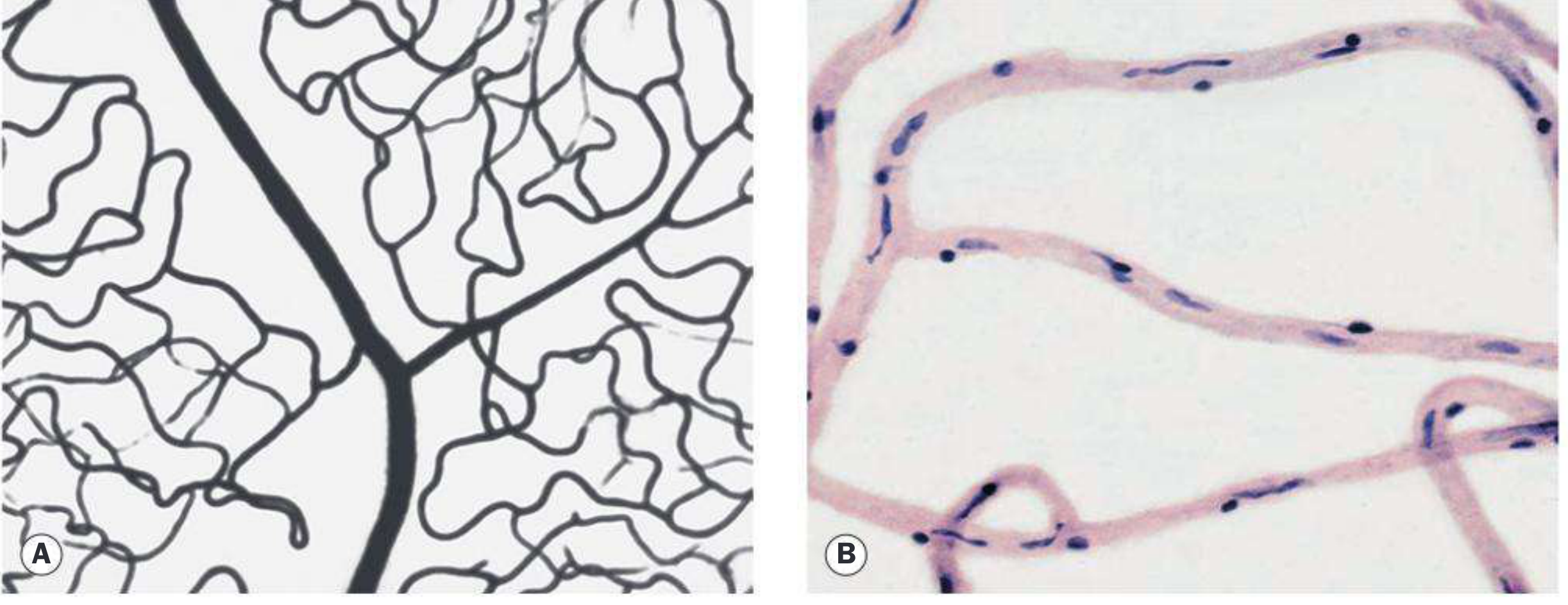
The primary target tissue is the retinal capillary. Chronic hyperglycemia damages capillary endothelial cells and pericytes - the structural support cells of retinal vessels. This leads to:
- Capillary leakage - breakdown of the blood-retinal barrier, causing fluid and protein extravasation
- Capillary occlusion - non-perfusion zones develop, causing localized retinal ischemia
- VEGF upregulation - ischemic retina releases vascular endothelial growth factor (VEGF), driving pathological neovascularization
- Neovascularization - fragile, leaky new vessels grow on the disc, retina, or iris, prone to hemorrhage and fibrous contraction
Risk Factors
| Risk Factor | Details |
|---|---|
| Duration of diabetes | Most important predictor - DR rarely occurs within the first few years |
| Poor glycemic control | Higher HbA1c = greater risk; tight control (DCCT, UKPDS) delays progression |
| Hypertension | Must be kept <140/80 mmHg; especially important in Type 2 with maculopathy |
| Nephropathy | Severe nephropathy worsens DR; renal transplantation may improve it |
| Pregnancy | ~5% of mild DR and ~33% of moderate DR progress to PDR during pregnancy |
| Hyperlipidaemia | Associated with hard exudates and macular disease |
| Drugs | Pioglitazone raises risk of worsening diabetic macular oedema |
| GLP-1 agonists | When associated with marked glycemic improvement, can transiently worsen DR |
Kanski's Clinical Ophthalmology, 10th Ed.; Harrison's Principles of Internal Medicine, 22nd Ed.
Prevalence: ~40% of all people with diabetes. In Type 2 diabetes, 67% develop DR within 10 years of diagnosis, with 10% developing proliferative disease.
Classification - Disease Severity Scale
(International Clinical Diabetic Retinopathy Severity Scale)
1. No Apparent Retinopathy
No visible abnormalities on dilated fundoscopy.
2. Mild Non-Proliferative DR (NPDR)
- Microaneurysms only - the earliest visible sign; small saccular outpouchings of capillary walls
3. Moderate NPDR
- More than mild, less than severe
- May have cotton-wool spots (CWS) and venous beading
- Hard exudates (lipid deposits), dot-and-blot hemorrhages
4. Severe NPDR (4-2-1 Rule)
Any ONE of the following in the absence of proliferative disease:
- >20 intraretinal hemorrhages in all 4 quadrants
- Venous beading in 2 or more quadrants
- Prominent intraretinal microvascular abnormalities (IRMA) in 1 or more quadrants
5. Proliferative DR (PDR)
Neovascularization of:
- NVD - new vessels at the disc
- NVE - new vessels elsewhere on retina
- NVI - neovascularization of iris (rubeosis iridis)
- NVA - neovascularization of angle (leads to neovascular glaucoma)
- Vitreous/preretinal hemorrhage
Diabetic Macular Edema (DME)
- Can occur at any stage of DR
- DME affecting or threatening the fovea is an independent indication for treatment
- Leading cause of visual impairment in diabetic patients
Wills Eye Manual, 6th Ed.; Kanski's Clinical Ophthalmology, 10th Ed.
Clinical Signs Summary
| Stage | Key Signs |
|---|---|
| Mild NPDR | Microaneurysms |
| Moderate NPDR | Dot/blot hemorrhages, hard exudates, CWS, venous beading |
| Severe NPDR | 4-2-1 rule features, IRMA |
| PDR | NVD/NVE, vitreous hemorrhage, tractional retinal detachment |
| DME | Retinal thickening ± hard exudate rings at/near macula |
Differential Diagnosis
| Condition | Distinguishing Feature |
|---|---|
| CRVO | Optic disc swelling, dilated/tortuous veins, splinter hemorrhages, unilateral |
| BRVO | Hemorrhages along a vein, not crossing horizontal raphe |
| Hypertensive retinopathy | Flame-shaped hemorrhages, AV nicking, microaneurysms rare |
| OIS | Midperipheral hemorrhages, pain, absent exudates |
| Radiation retinopathy | History of radiation, microaneurysms rare |
| Sickle cell | Peripheral "sea fan" neovascularization |
Wills Eye Manual
Treatment
1. Systemic Management (Primary Prevention)
- Tight glycemic control - HbA1c optimization; reduces development and progression (DCCT/UKPDS evidence)
- Caution: rapid improvement in control can transiently worsen DR in the first 6-12 months
- Blood pressure control - target <140/80 mmHg
- Fenofibrate - may reduce progression of DR by lowering triglycerides
- Aspirin - does NOT alter the natural history of DR
2. Laser Photocoagulation
Panretinal Photocoagulation (PRP) - indicated for high-risk PDR features:
- NVD > 1/4 to 1/3 disc area in size
- Any NVD with preretinal hemorrhage or vitreous hemorrhage (VH)
- NVE > 1/2 disc area with preretinal hemorrhage or VH
- Any NVI or NVA
Focal/grid laser - historically used for clinically significant DME; now largely replaced by anti-VEGF.
3. Anti-VEGF Therapy (Intravitreal Injections)
- First-line treatment for center-involving DME
- Alternative to PRP for PDR (preferred if DME coexists or if vitreous hemorrhage limits peripheral retinal view)
- Agents: ranibizumab, aflibercept, bevacizumab
- A 2025 meta-analysis (PMID 39128789) confirmed anti-VEGF is at least equivalent to PRP for PDR with better preservation of visual field; however, patients lost to follow-up have worse anatomic and visual outcomes without PRP backup
4. Vitrectomy
Indicated for:
- Dense, non-clearing or recurrent vitreous hemorrhage causing significant vision loss
- Tractional retinal detachment involving or progressing toward the macula
- Macular epiretinal membranes (ERMs) or vitreomacular traction
- Dense premacular hemorrhage
- Chronic DME unresponsive to other treatments
- Severe fibrovascular proliferation unresponsive to laser/anti-VEGF
Note: Young Type 1 diabetics tend to have more aggressive PDR and may benefit from earlier intervention.
Screening Guidelines
| Patient Group | First Exam | Follow-up |
|---|---|---|
| Type 1 DM | 5 years after diagnosis | Annually |
| Type 2 DM | At time of diagnosis | Annually |
| Pregnancy in DM | Before conception or early 1st trimester | Based on severity |
| No/minimal NPDR | Gestational course - minimal risk of progression | Annual |
| PDR or high-risk NPDR | Immediate treatment + close follow-up | Every 2-3 months |
All diabetic patients require dilated fundus examination by an ophthalmologist or retinal photography with remote reading - routine non-dilated exams by primary care are inadequate. - Harrison's Internal Medicine, 22nd Ed.
Ophthalmic Complications of Diabetes (Full Spectrum)
Common: Maculopathy (DME, macular ischaemia), retinopathy, unstable refraction
Uncommon: Recurrent styes, xanthelasma, accelerated cataract, tractional retinal detachment, neovascular glaucoma, ocular motor nerve palsies, reduced corneal sensitivity
Rare: Papillopathy, Wolfram syndrome, acute-onset cataract, rhino-orbital mucormycosis
Recent Evidence Updates (2024-2026)
- A 2025 systematic review and meta-analysis (PMID 39128789) confirms anti-VEGF injections are a valid alternative to PRP for PDR, with advantages in preserving peripheral visual field.
- A 2026 systematic review on gut-eye axis (PMID 41484843) highlights emerging evidence for gut microbiota's role in DR pathophysiology - a potentially new therapeutic avenue.
- A 2025 systematic review on risk prediction models (PMID 40717809) reports advances in AI-based risk stratification tools for DR screening.
Sources: Kanski's Clinical Ophthalmology 10th Ed. | Wills Eye Manual | Harrison's Principles of Internal Medicine 22nd Ed. | Textbook of Family Medicine 9th Ed.
Abnormal uterine bleeding
Abnormal uterine bleeding (AUB) refers to any bleeding from the uterus that differs from normal menstruation in frequency, duration, volume, or regularity. It affects up to 30% of women of reproductive age and is one of the most common gynecological complaints. The PALM-COEIN classification system (2011, FIGO) organizes causes into structural and non-structural categories.
Searching sources...
Reading File
Now let me search the library and PubMed in parallel:
Finding Sources
Searching PubMed
"abnormal uterine bleeding"[MeSH Terms] AND management
Excellent sources found. Let me read from Berek & Novak's (gold standard gynecology text), Robbins, and Goldman-Cecil in parallel:
Reading File
Reading File
Reading File
Searching PubMed
"abnormal uterine bleeding" treatment
Now let me read the Goldman-Cecil treatment section and the Bailey & Love surgical section, plus the Sabiston content:
Reading File
Reading File
Reading File
Excellent content. Let me read the continuation of the Sabiston section and the Bailey & Love tables for the structural/non-structural causes:
Reading File
Reading File
I now have comprehensive, authoritative content from multiple sources. Let me compile the full answer:
Abnormal Uterine Bleeding (AUB)
Definition
AUB is defined as bleeding from the uterus that is excessive or occurs outside of the normal menstrual cycle. A normal menstrual cycle should occur regularly between 24 and 38 days, with duration of flow between 4 and 8 days. Any deviation in frequency, regularity, duration, or volume constitutes AUB. - Sabiston Textbook of Surgery
Previously used terms (menorrhagia, metrorrhagia, menometrorrhagia, dysfunctional uterine bleeding) have been replaced by the PALM-COEIN classification system.
Epidemiology
- Affects 10-30% of women of reproductive age
- ~20% of all AUB cases are postmenarchal (adolescents with immature HPO axis)
- ~50% are perimenopausal (incipient ovarian failure)
- Postmenopausal bleeding is a red flag - must always be investigated to exclude endometrial carcinoma
- Most cases (~75%) have no demonstrable organic cause and are due to anovulation - Goldman-Cecil Medicine
PALM-COEIN Classification (FIGO 2011)
This system divides causes into structural (PALM) and non-structural (COEIN) categories:
PALM - Structural Causes (visible on imaging/histology)
| Letter | Cause | Key Features |
|---|---|---|
| P | Polyp (endometrial/endocervical) | Overgrowth of endometrial glands and stroma; sessile or pedunculated; up to 5% malignant transformation; inner feeder vessel on Doppler |
| A | Adenomyosis | Endometrial glands within myometrium; causes heavy, painful periods; diagnosed on USS or MRI |
| L | Leiomyoma (fibroids) | Most common benign gynecologic tumor; affects up to 70% by age 50; more prevalent and severe in Black women; classified by FIGO types 0-8 |
| M | Malignancy/Hyperplasia | Endometrial hyperplasia or carcinoma; most common gynecologic malignancy; must exclude in all postmenopausal bleeding and high-risk premenopausal patients |
COEIN - Non-Structural Causes
| Letter | Cause | Key Features |
|---|---|---|
| C | Coagulopathy | von Willebrand disease (most common), ITP, thrombocytopenia; suspect if heavy bleeding since menarche, family history, or systemic bleeding signs |
| O | Ovulatory dysfunction | PCOS (most common), hyperprolactinemia, thyroid disease, HPA axis dysfunction; unopposed estrogen causes irregular shedding |
| E | Endometrial | Endometritis (primary endometrial disorder); altered local hemostatic mechanisms |
| I | Iatrogenic | OCP breakthrough bleeding, IUD-related, tamoxifen (increases polyps, hyperplasia, cancer risk by 2x), anticoagulants |
| N | Not otherwise classified | AVM (arteriovenous malformations), chronic renal/hepatic disease |
Sabiston Textbook of Surgery; Bailey & Love's Short Practice of Surgery, 28th Ed.
Causes by Age Group (Robbins' Pathology)
| Age Group | Common Causes |
|---|---|
| Prepuberty | Precocious puberty (hypothalamic, pituitary, or ovarian origin) |
| Adolescence | Anovulatory cycles (immature HPO axis), coagulation disorders |
| Reproductive age | Complications of pregnancy (abortion, ectopic, trophoblastic disease), PCOS, fibroids, polyps, OCP use, coagulopathy |
| Perimenopausal | Anovulatory cycles due to incipient ovarian failure, endometrial hyperplasia |
| Postmenopausal | Endometrial atrophy (most common), endometrial hyperplasia, carcinoma, polyps, HRT-related |
Bleeding Patterns and Their Significance
| Pattern | Likely Associations |
|---|---|
| Heavy menstrual bleeding (HMB) | Fibroids, adenomyosis, coagulopathy, anovulation |
| Intermenstrual bleeding (IMB) | Cervical/endometrial polyp, cervical malignancy, endometritis |
| Postcoital bleeding (PCB) | Cervical ectropion, cervical polyp, cervical cancer |
| Postmenopausal bleeding (PMB) | Atrophy, endometrial cancer, polyps, HRT |
Bailey & Love's Short Practice of Surgery, 28th Ed.
Pathophysiology
The central mechanism in most non-structural AUB is anovulation:
- Without ovulation, no corpus luteum forms - no progesterone is produced
- Estrogen stimulates the endometrium unopposed, causing excessive proliferation
- The thickened, unstable endometrium sheds irregularly, causing unpredictable, often heavy bleeding
- This estrogen breakthrough or estrogen withdrawal bleeding is the basis of anovulatory AUB
Two specific subtypes:
- Estrogen withdrawal bleeding - drop in estrogen without progesterone support
- Estrogen breakthrough bleeding - prolonged unopposed estrogen stimulation with irregular shedding
Goldman-Cecil Medicine; Robbins & Kumar Basic Pathology
Diagnosis
History
- Menstrual cycle characterization (frequency, duration, volume, regularity)
- Prospective bleeding diary/chart if the pattern is unclear
- Sexual history, contraceptive use, medications (tamoxifen, anticoagulants, OCPs)
- Family history (coagulopathy, cancer)
- Symptoms of systemic disease (thyroid, bleeding elsewhere)
Physical Examination
- Pelvic exam - assess for uterine size, tenderness, masses
- Cervical exam - ectropion, polyps, contact bleeding
- Papanicolaou smear (cervical cancer screening)
Laboratory Investigations
- Pregnancy test (beta-hCG) - exclude pregnancy complications first
- Full blood count - assess severity/anemia
- Coagulation screen - PT, aPTT, VWF antigen/activity (if coagulopathy suspected)
- Thyroid function tests (TSH)
- Prolactin level
- Fasting blood glucose
- STI screening (chlamydia, gonorrhoea - endometritis)
- Cervical cancer screening if not up to date
Imaging
- Pelvic ultrasound (2D/3D or saline sonogram/SIS) - first-line imaging
- Evaluates endometrial thickness, uterine/ovarian pathology
- Endometrial thickness >4 mm in postmenopausal women is abnormal
- Doppler for polyp feeder vessels
Endometrial Sampling
Endometrial biopsy (Pipelle) or hysteroscopy + biopsy is indicated when:
- All women age ≥45 years with AUB
- Women <45 years with risk factors: obesity, unopposed estrogen exposure, PCOS, persistent AUB refractory to medical treatment, elevated familial cancer risk
- All postmenopausal women with bleeding
- Suspected endometrial pathology on USS
- Persistent intermenstrual bleeding
- Women on HRT with irregular bleeding after initial 3 months
- Endometrial thickness >4 mm (postmenopausal) or >7 mm (PCOS)
Note: Hysteroscopy combined with biopsy has superior sensitivity and specificity compared to either alone. - Bailey & Love's Short Practice of Surgery
Treatment
1. Medical Management (First Line)
For Anovulatory/Non-structural AUB:
Acute profuse bleeding (hemodynamically stable):
- Combined OCP - 1 pill every 6 hours for 5-7 days (off-label); bleeding should cease within 24 hours; warn patient of heavy withdrawal bleed 2-4 days after stopping
- IV conjugated estrogens - 25 mg IV every 4 hours (up to 3 doses) + concurrent progestin (medroxyprogesterone acetate 5-10 mg for 10 days)
- Blood transfusion if anemia is severe
Ongoing/Recurrent Bleeding:
- Combined OCP - cyclic dosing (if pregnancy not desired)
- Progestins - cyclic medroxyprogesterone acetate or norethindrone (if OCP not tolerated)
- Levonorgestrel IUD (LNG-IUS, Mirena) - highly effective for HMB, ovulatory AUB, adenomyosis-related bleeding
- NSAIDs - tranexamic acid + NSAIDs for ovulatory HMB
- Tranexamic acid - antifibrinolytic; effective for heavy menstrual bleeding
For Fibroids specifically:
- GnRH antagonists - elagolix 300 mg twice daily or relugolix 40 mg daily (oral medication options); a 2025 meta-analysis (PMID 39821450) confirms efficacy of GnRH antagonists for fibroid-related bleeding
- Progestins/OCP - symptom control
For ovulatory dysfunction/PCOS:
- OCP to regulate cycles
- Induce ovulation if pregnancy is desired
Fenofibrate Note:
- May reduce progression of related hormonal dysregulation, though primarily used for triglycerides
2. Surgical/Procedural Management
| Procedure | Indication | Notes |
|---|---|---|
| Endometrial ablation | Persistent AUB refractory to medical therapy; completed family | Not 100% effective; ~29% require hysterectomy by 60 months |
| Hysteroscopic polypectomy | Endometrial/endocervical polyps | First-line for polyps |
| Myomectomy | Symptomatic fibroids, fertility preservation desired | Hysteroscopic (types 0-2), laparoscopic/open (types 3-7) |
| Uterine artery embolization (UAE) | Symptomatic fibroids, alternative to hysterectomy | ~31% ultimately require hysterectomy; effective alternative per 2024 meta-analysis (PMID 39164326) |
| Dilation & Curettage (D&C) | Diagnostic; NOT effective as sole treatment for AUB | Not necessary before hysterectomy |
| Hysterectomy | Refractory AUB; malignancy found on biopsy; patient choice; failed all other options | Definitive treatment; should be considered only after medical/conservative options exhausted |
Berek & Novak's Gynecology; Sabiston Textbook of Surgery; Goldman-Cecil Medicine
Special Considerations
Postmenopausal Bleeding
- Any bleeding >12 months after last period (without hormonal contraception) is abnormal and requires immediate investigation
- Most common cause = endometrial atrophy (thinning due to estrogen deficiency)
- Must exclude endometrial carcinoma (most common gynecologic cancer)
- Investigations: USS (endometrial thickness), endometrial biopsy, hysteroscopy
Tamoxifen Users
- Tamoxifen (SERM used in breast cancer) causes uterine abnormalities in 10-40% of women
- Associated with endometrial polyps, hyperplasia, carcinoma, and uterine sarcomas
- Doubles the risk of endometrial cancer
- Aromatase inhibitors (AIs) are associated with fewer uterine complications than tamoxifen
Adolescents
- Most common cause is anovulation due to immaturity of the hypothalamic-pituitary-ovarian axis
- Always screen for coagulation disorders (von Willebrand disease) - especially if heavy bleeding since menarche
- 2025 systematic review (PMID 39617186) highlights coagulation disorders as a frequently missed cause of HMB in adolescents and adults
Pregnancy Must Always Be Excluded First
- Miscarriage, ectopic pregnancy, and trophoblastic disease all cause AUB
- Beta-hCG is mandatory in any woman of reproductive age presenting with AUB
Prognosis
The prognosis of AUB depends on its underlying cause. Most cases of anovulatory/functional AUB respond well to medical management. Structural causes (fibroids, polyps) are highly treatable with surgery. When malignancy is detected early, prognosis is good. Delayed diagnosis of endometrial carcinoma carries a significantly worse prognosis. - Goldman-Cecil Medicine
Sources: Sabiston Textbook of Surgery | Goldman-Cecil Medicine | Berek & Novak's Gynecology | Robbins & Kumar Basic Pathology | Bailey & Love's Short Practice of Surgery, 28th Ed.
This is a shared conversation. Sign in to Orris to start your own chat.